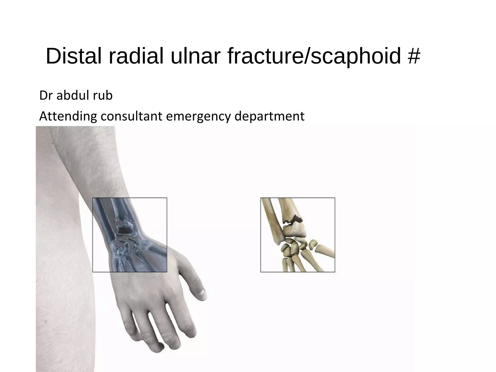
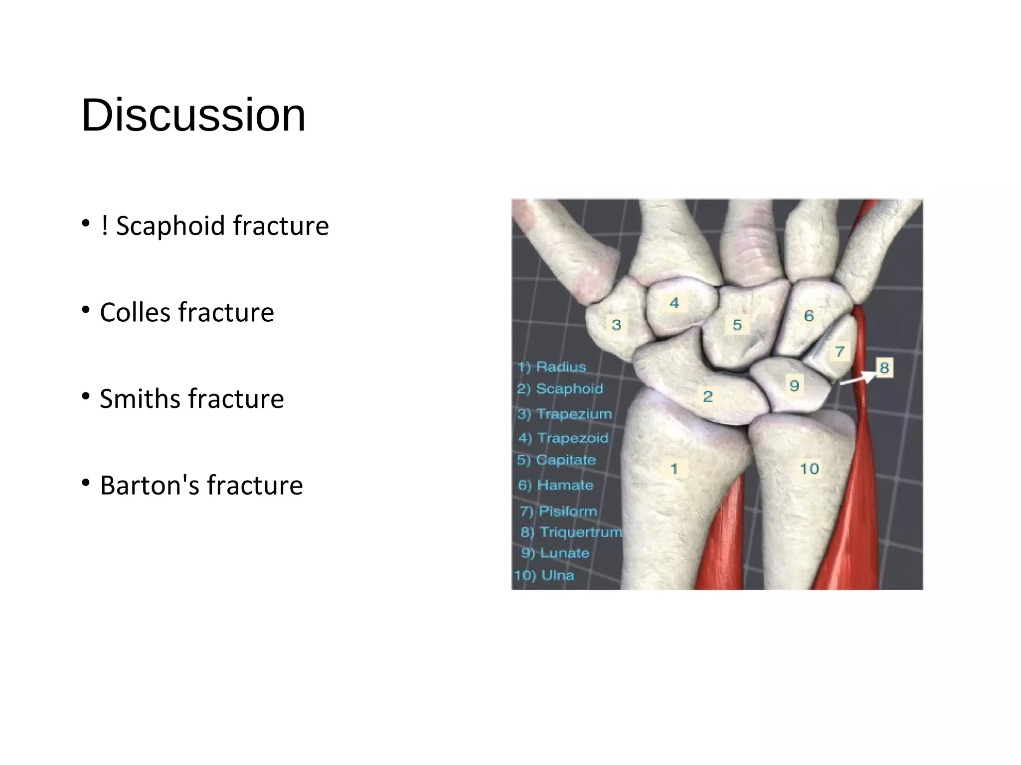
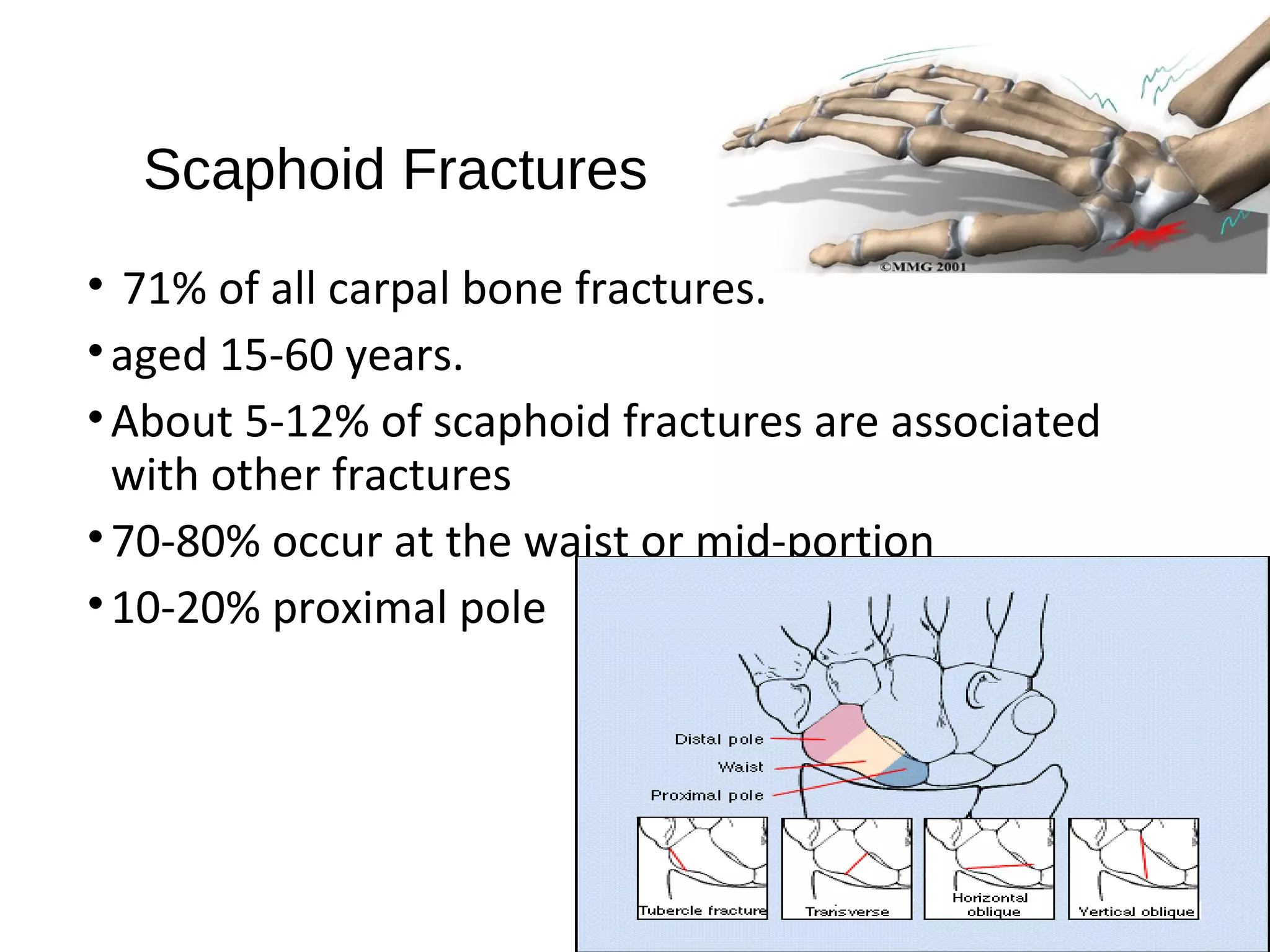
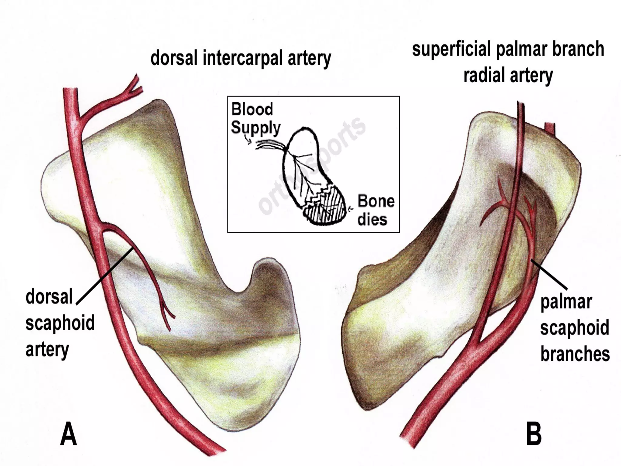
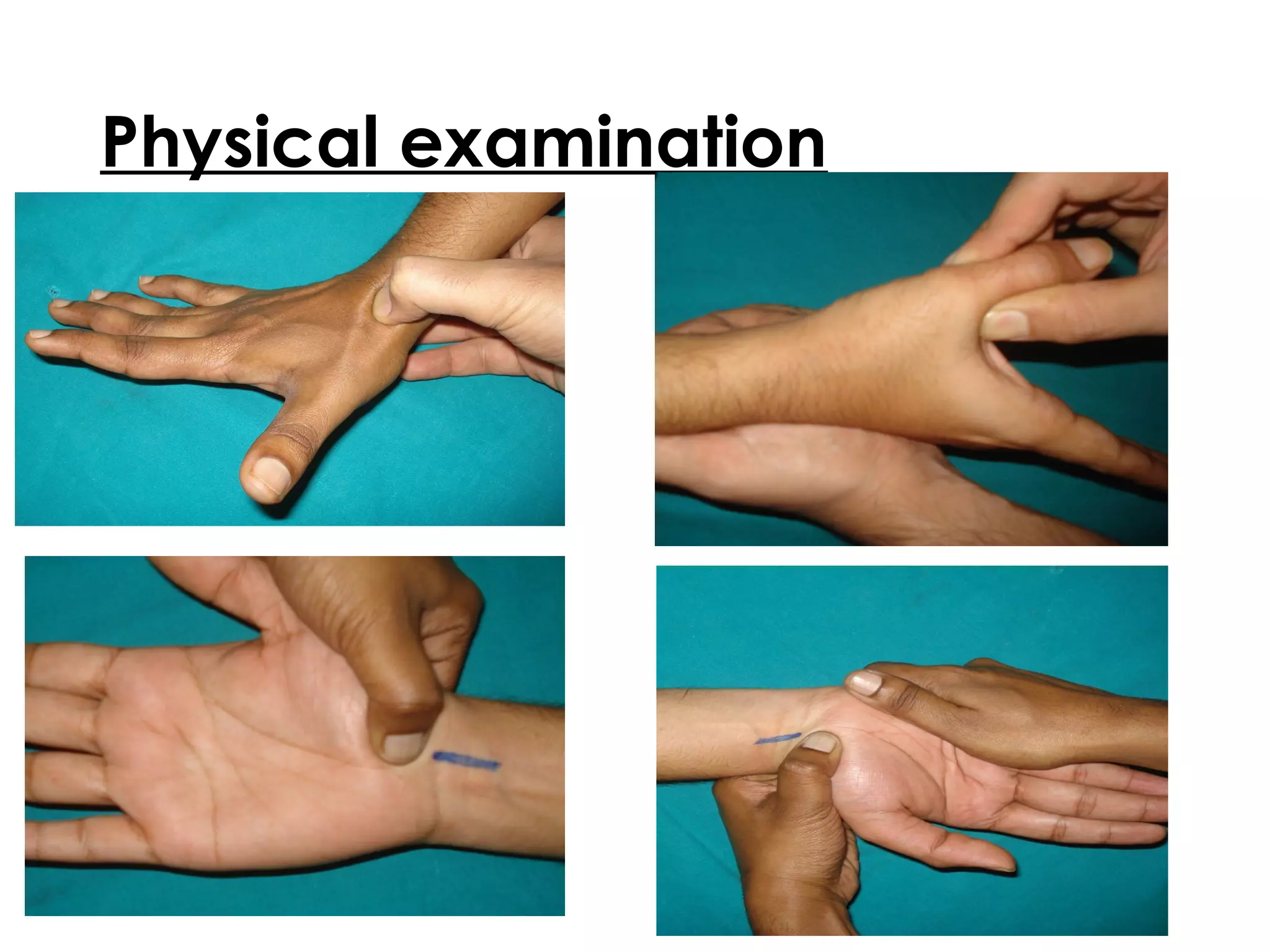
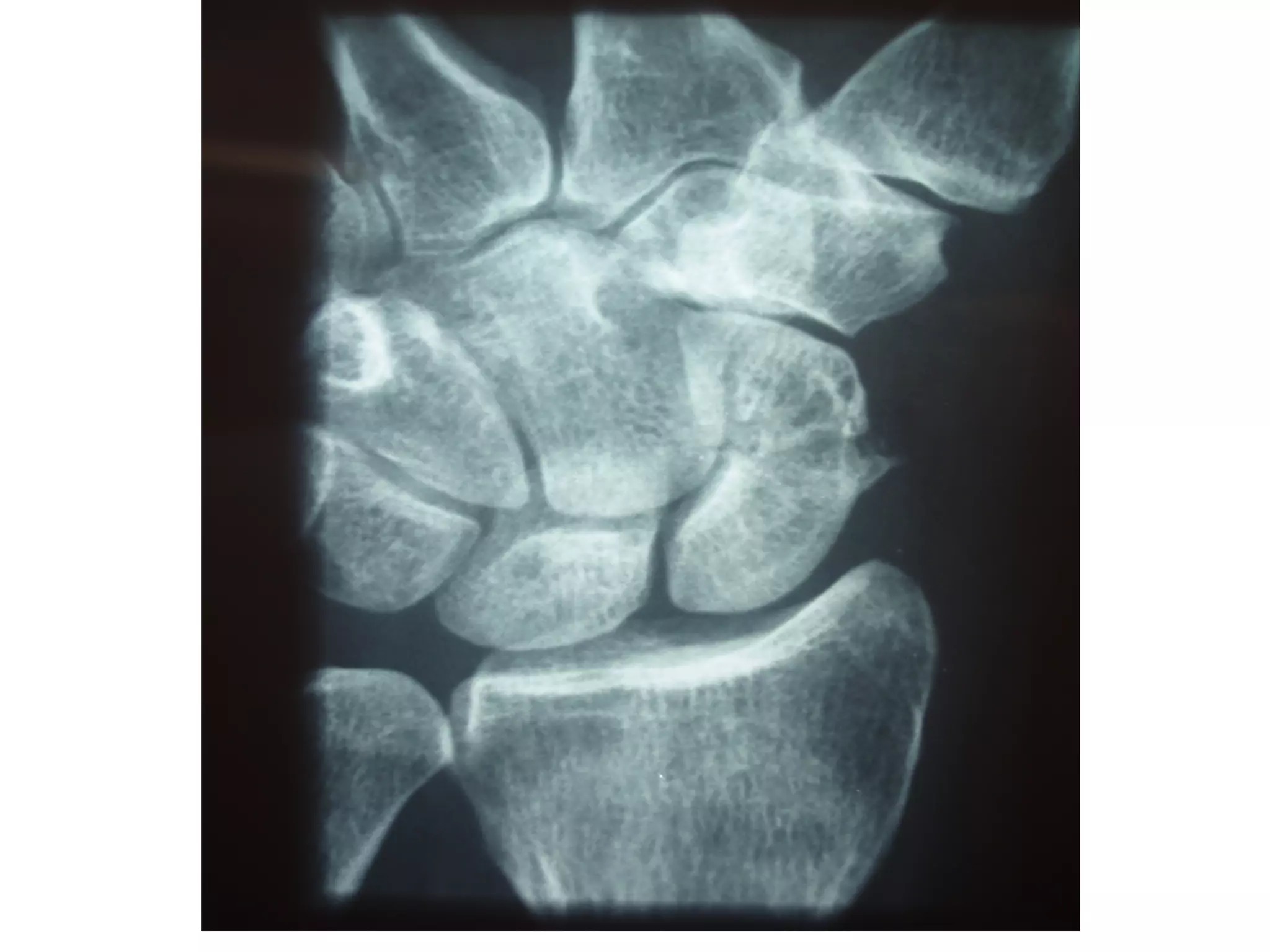
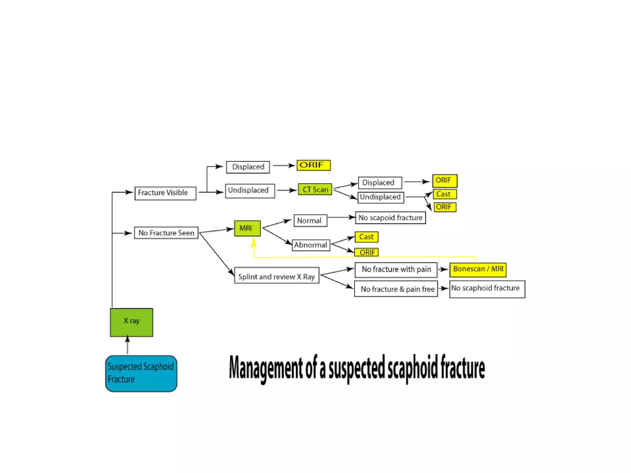
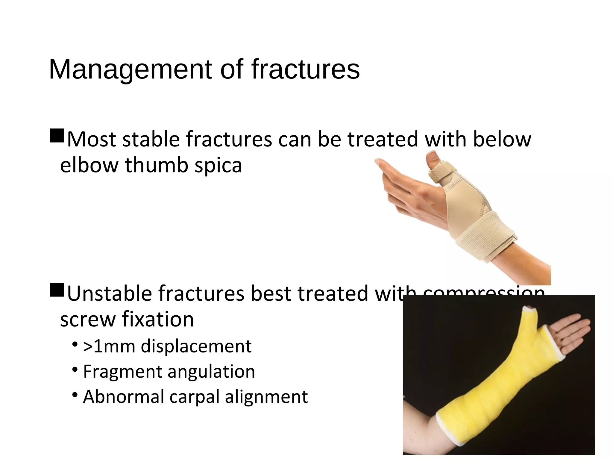
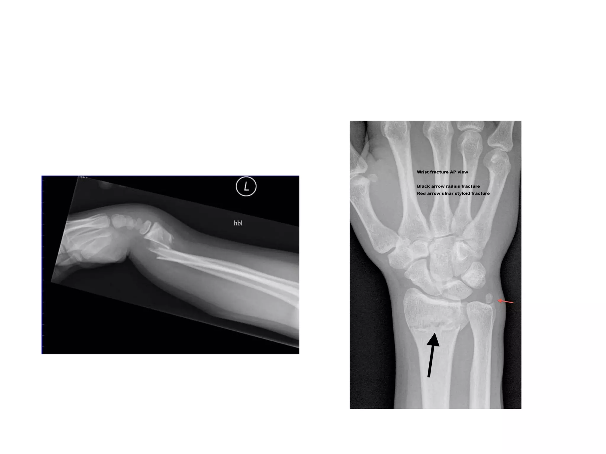
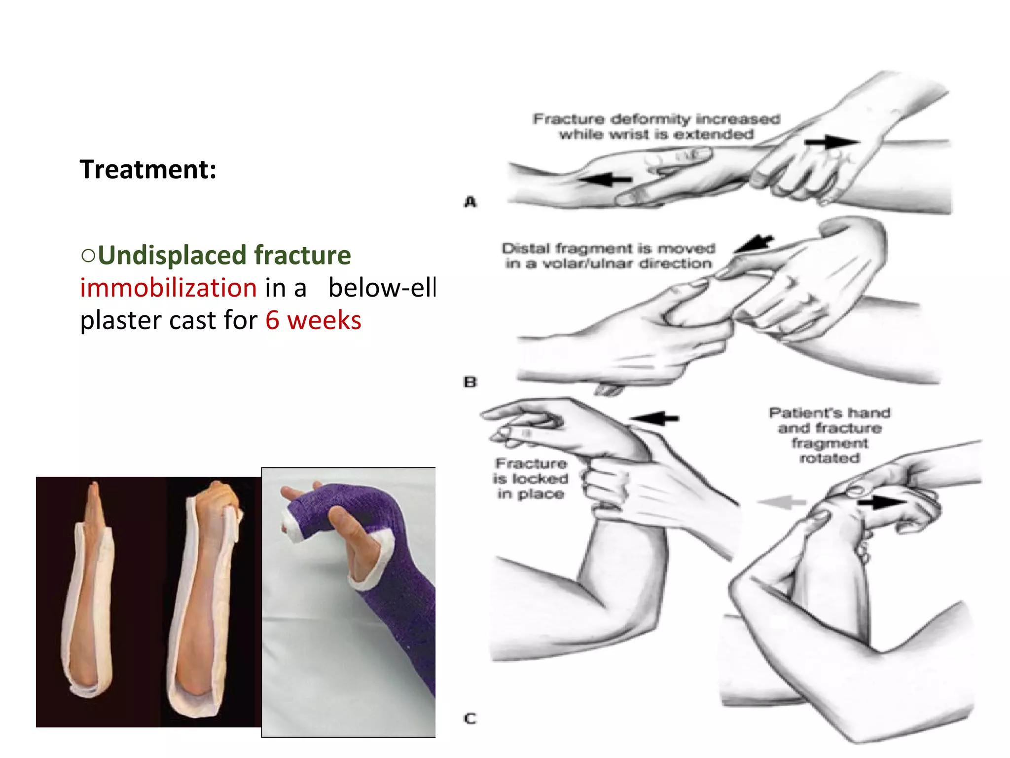
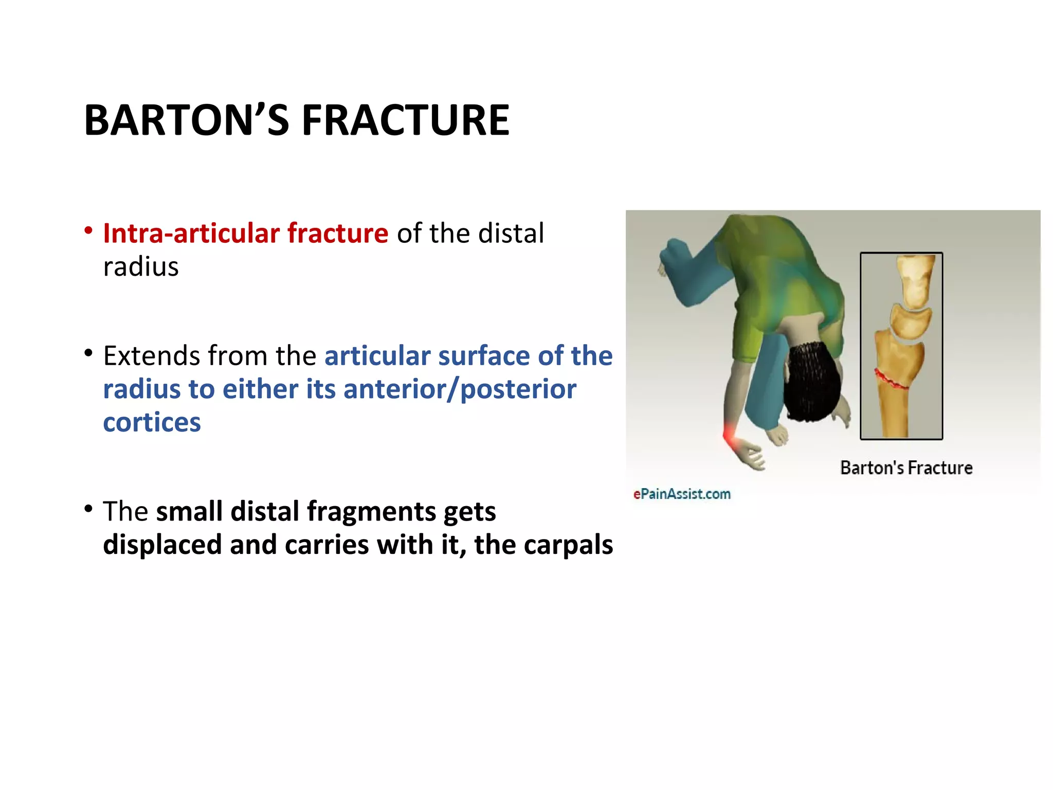
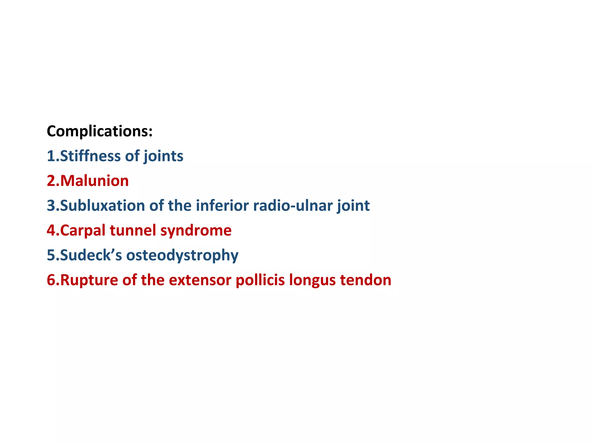
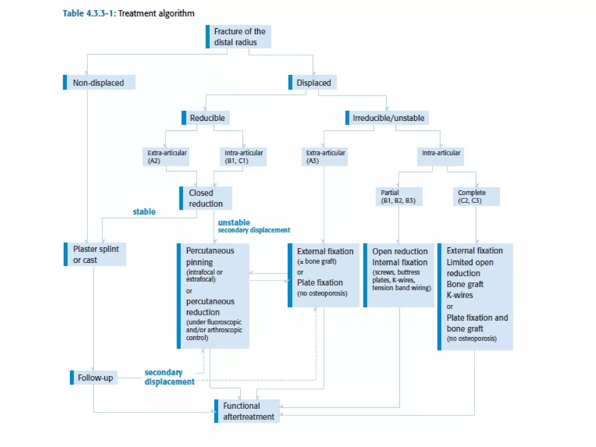
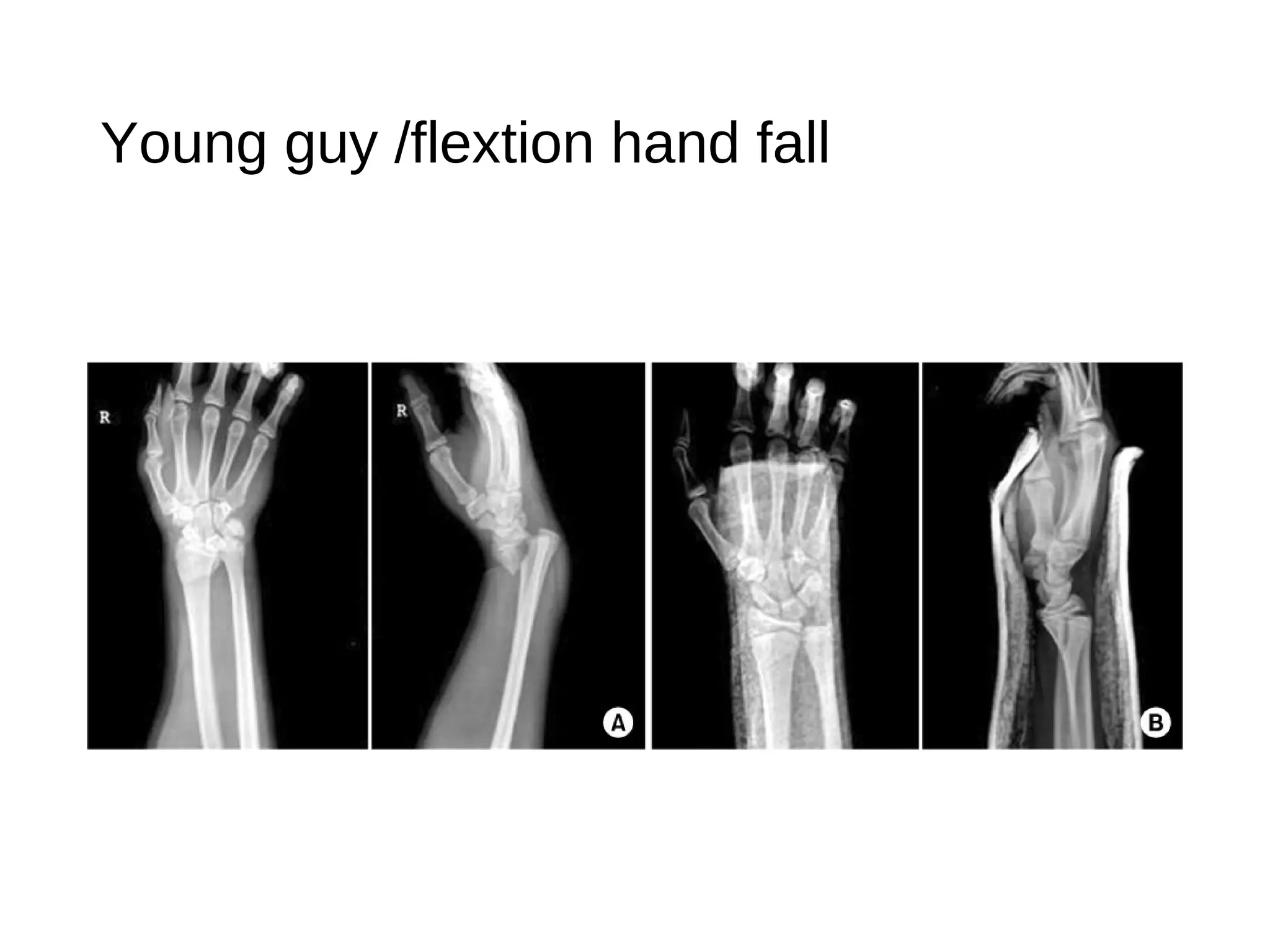
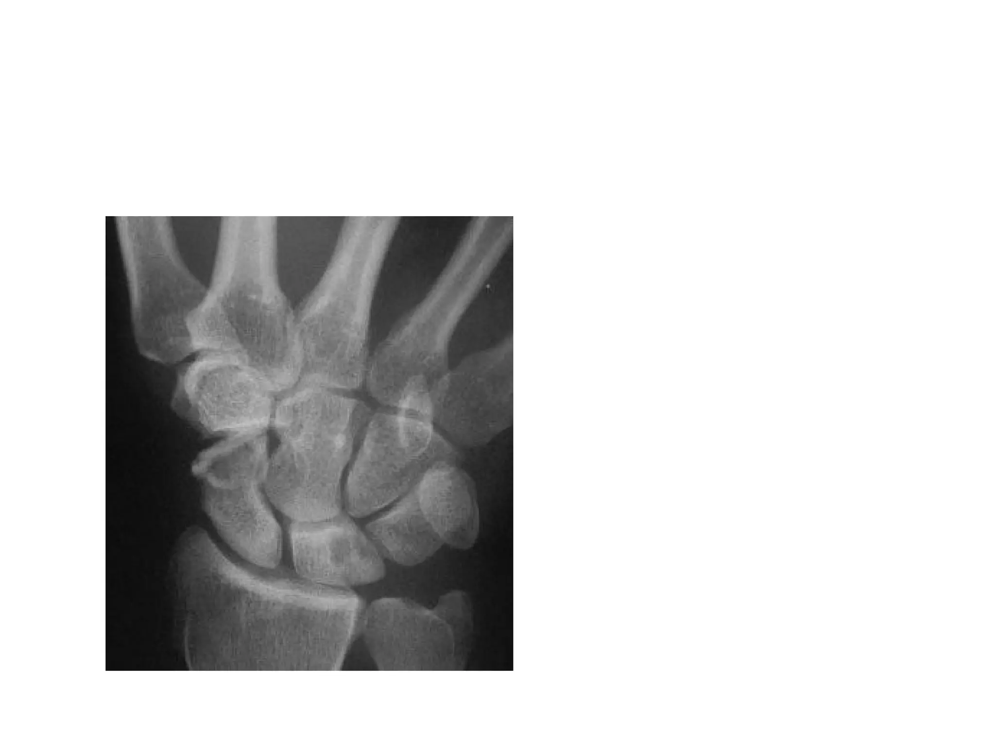
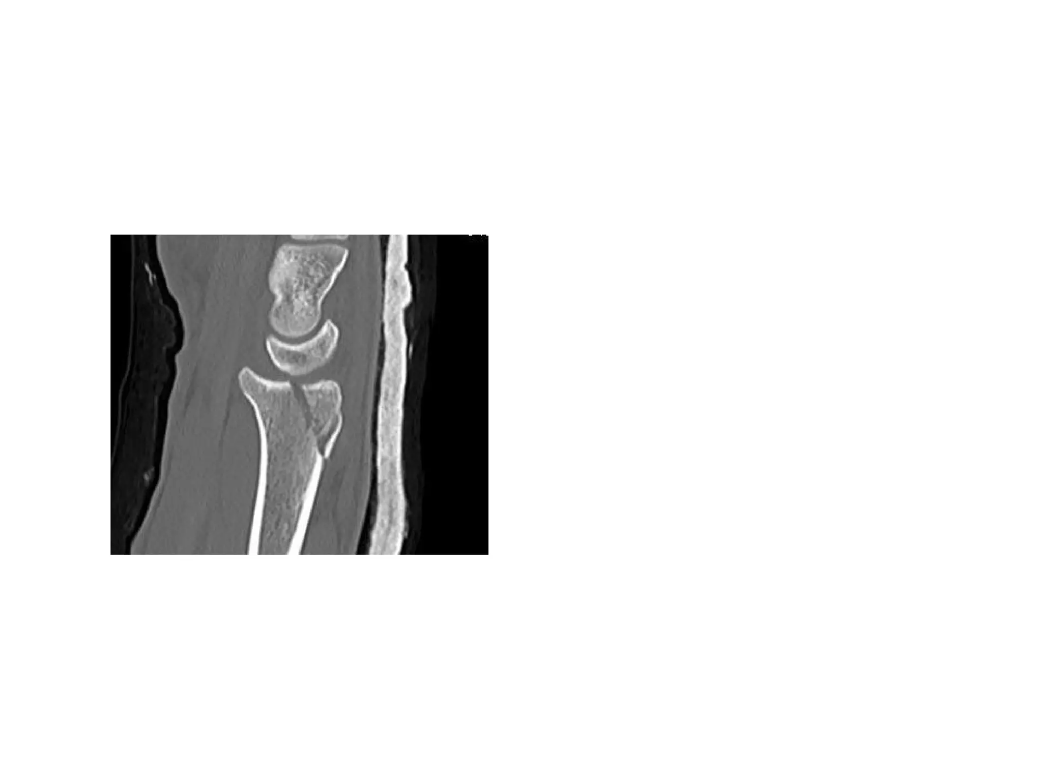
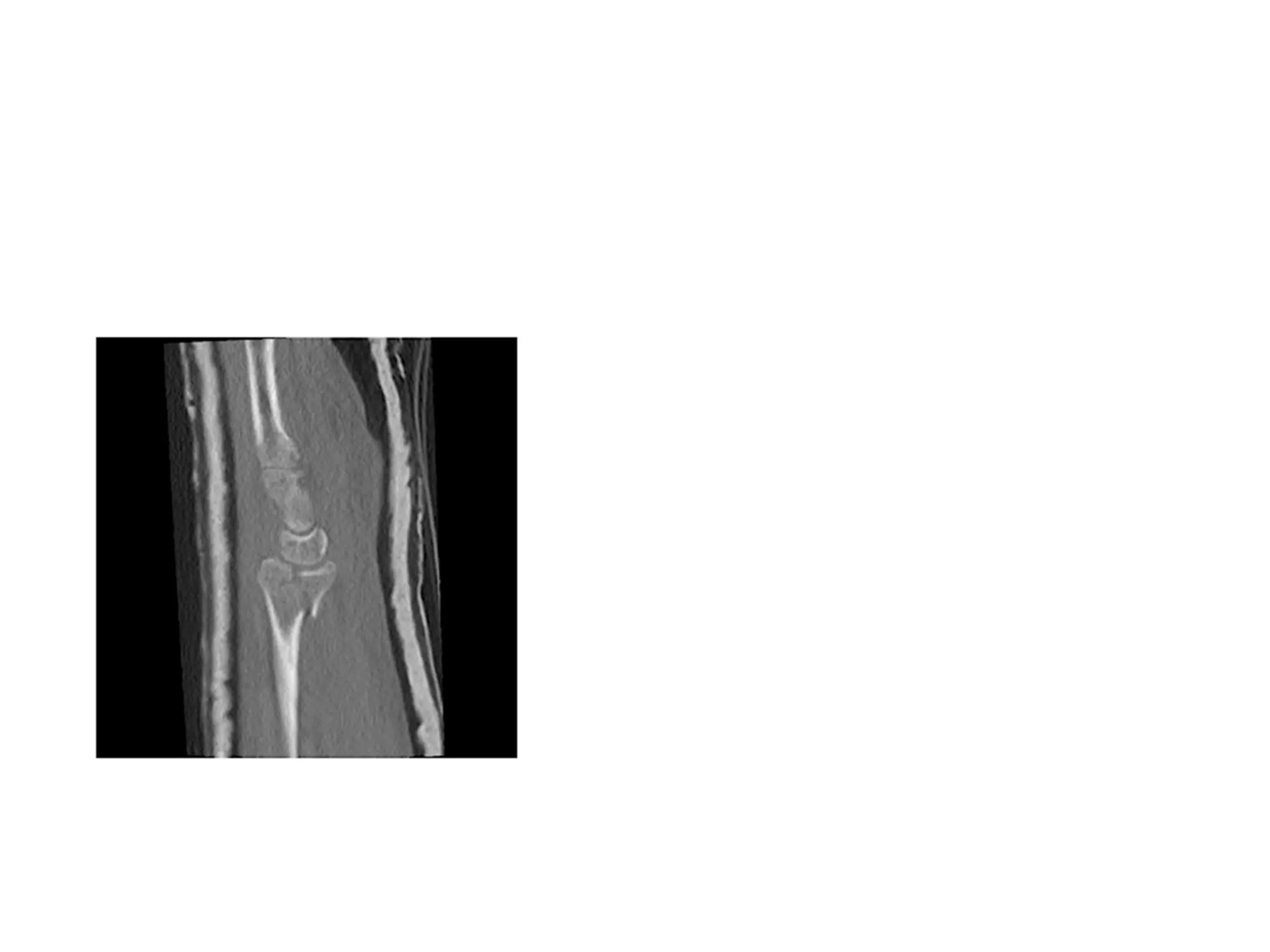
Distal radial ulnar fracture/scaphoid #
Dr abdul rub
Attending consultant emergency department
Discussion
• ! Scaphoid fracture
• Colles fracture
• Smiths fracture
• Barton's fracture
Scaphoid Fractures
• 71% of all carpal bone fractures.
•aged 15-60 years.
•About 5-12% of scaphoid fractures are associated
with other fractures
•70-80% occur at the waist or mid-portion
•10-20% proximal pole
Physical examination
•Snuff box tenderness 100% sensitivity
•Scaphoid tubercle tenderness 20% specific
•Adding Scaphoid compression test :
Specificity reaches 74% (Parvizi et al)
Suggested by:
• patient’s age,
• mechanism of injury
• signs and symptoms
Imaging
• Xray….Can miss 20%of #
• CT Scan-for staging & if plain flims normal
• MRI-most sensitive test(within 24hrs with pain)
• Bone Scan (3-4days)-100% SENSITIVE
Wrist PA
Management of fractures
Most stable fractures can be treated with below
elbow thumb spica
Unstable fractures best treated with compression
screw fixation
• >1mm displacement
• Fragment angulation
• Abnormal carpal alignment
Closed treatment
•Stable non displaced fractures
•Short arm for 6-8 weeks in tubercle or distal
pole fractures
•Upto 12 weeks in waist fractures
•Position- wrist in neutral position
Nigel R.Clay, Joseph J.Dias, P.S. Costigan, P.J. Gregg, N.J. Barton. Need
The Thumb To be Immobilized In Scaphoid Fractures.
Complication$$
• Malunion
• AVN 15-30%
Distal radio-ulnar #
1. COLLES’ FRACTURE
2. SMITH’S FRACTURE
3. BARTON’S FRACTURE
• Osteoporotic bone
• Extra articular fracture-metaphysis involvement
• 2cm from radio carpel joint
• Look for styloid process# ulna
Other displacements (1/> displacements below occur in majority of the cases;
although in few cases it may be crack fracture without displacement)
oImpaction of fragments
oDorsal displacement
oDorsal tilt
oLateral displacement
oLateral tilt
oSupination
Clinical features:
oPain
oSwelling
oDeformity of the wrist
oOn examination;
 Tenderness
 Irregularity of the lower end of
radius
Radiological features: (Differentiated from
other fractures at the same site by looking at
the displacements)
oDorsal tilt can be detected on a lateral
X-ray:
 normal  faces ventrally
 dorsal tilt  faces dorsally or becomes
neutral
oLateral tilt can be detected on an AP X-
ray:
 normal  faces medially
 lateral tilt  faces laterally or becomes
horizontal
Treatment:
oUndisplaced fracture
immobilization in a below-elbow
plaster cast for 6 weeks
oDisplaced fracture manipulative
reduction followed by immobilization
in Colles’ cast
SMITH’S FRACTURE
• Fracture at the distal end of the radius,
at its cortico-cancellous junction, with
ventral tilt and other displacements
• Uncommon
• Treatment:
• Closed reduction
• Plaster cast immobilization (6 weeks)
BARTON’S FRACTURE
• Intra-articular fracture of the distal
radius
• Extends from the articular surface of the
radius to either its anterior/posterior
cortices
• The small distal fragments gets
displaced and carries with it, the carpals
• Displacements:
• Volar Barton’s fracture (anterior
type)
• Dorsal Barton’s fracture
(posterior type)
• Treatment:
• Closed manipulation
• ORIF with plate in cases where
closed reduction fails
Complications:
1.Stiffness of joints
2.Malunion
3.Subluxation of the inferior radio-ulnar joint
4.Carpal tunnel syndrome
5.Sudeck’s osteodystrophy
6.Rupture of the extensor pollicis longus tendon
Young guy /flextion hand fall
Hyperextension & fall
Questions?

distal radius & scaphoid fracture

  • 1.
    Distal radial ulnarfracture/scaphoid # Dr abdul rub Attending consultant emergency department
  • 2.
    Discussion • ! Scaphoidfracture • Colles fracture • Smiths fracture • Barton's fracture
  • 3.
    Scaphoid Fractures • 71%of all carpal bone fractures. •aged 15-60 years. •About 5-12% of scaphoid fractures are associated with other fractures •70-80% occur at the waist or mid-portion •10-20% proximal pole
  • 5.
  • 6.
    Physical examination •Snuff boxtenderness 100% sensitivity •Scaphoid tubercle tenderness 20% specific •Adding Scaphoid compression test : Specificity reaches 74% (Parvizi et al)
  • 7.
    Suggested by: • patient’sage, • mechanism of injury • signs and symptoms Imaging • Xray….Can miss 20%of # • CT Scan-for staging & if plain flims normal • MRI-most sensitive test(within 24hrs with pain) • Bone Scan (3-4days)-100% SENSITIVE
  • 8.
  • 11.
    Management of fractures Moststable fractures can be treated with below elbow thumb spica Unstable fractures best treated with compression screw fixation • >1mm displacement • Fragment angulation • Abnormal carpal alignment
  • 12.
    Closed treatment •Stable nondisplaced fractures •Short arm for 6-8 weeks in tubercle or distal pole fractures •Upto 12 weeks in waist fractures •Position- wrist in neutral position Nigel R.Clay, Joseph J.Dias, P.S. Costigan, P.J. Gregg, N.J. Barton. Need The Thumb To be Immobilized In Scaphoid Fractures.
  • 13.
  • 14.
    Distal radio-ulnar # 1.COLLES’ FRACTURE 2. SMITH’S FRACTURE 3. BARTON’S FRACTURE
  • 15.
    • Osteoporotic bone •Extra articular fracture-metaphysis involvement • 2cm from radio carpel joint • Look for styloid process# ulna
  • 16.
    Other displacements (1/>displacements below occur in majority of the cases; although in few cases it may be crack fracture without displacement) oImpaction of fragments oDorsal displacement oDorsal tilt oLateral displacement oLateral tilt oSupination
  • 17.
    Clinical features: oPain oSwelling oDeformity ofthe wrist oOn examination;  Tenderness  Irregularity of the lower end of radius Radiological features: (Differentiated from other fractures at the same site by looking at the displacements) oDorsal tilt can be detected on a lateral X-ray:  normal  faces ventrally  dorsal tilt  faces dorsally or becomes neutral oLateral tilt can be detected on an AP X- ray:  normal  faces medially  lateral tilt  faces laterally or becomes horizontal
  • 19.
    Treatment: oUndisplaced fracture immobilization ina below-elbow plaster cast for 6 weeks oDisplaced fracture manipulative reduction followed by immobilization in Colles’ cast
  • 20.
    SMITH’S FRACTURE • Fractureat the distal end of the radius, at its cortico-cancellous junction, with ventral tilt and other displacements • Uncommon • Treatment: • Closed reduction • Plaster cast immobilization (6 weeks)
  • 22.
    BARTON’S FRACTURE • Intra-articularfracture of the distal radius • Extends from the articular surface of the radius to either its anterior/posterior cortices • The small distal fragments gets displaced and carries with it, the carpals
  • 23.
    • Displacements: • VolarBarton’s fracture (anterior type) • Dorsal Barton’s fracture (posterior type) • Treatment: • Closed manipulation • ORIF with plate in cases where closed reduction fails
  • 24.
    Complications: 1.Stiffness of joints 2.Malunion 3.Subluxationof the inferior radio-ulnar joint 4.Carpal tunnel syndrome 5.Sudeck’s osteodystrophy 6.Rupture of the extensor pollicis longus tendon
  • 26.
  • 28.
  • 32.

Editor's Notes

  • #5 Primary blood supply is dorsal branch of radial artery -70 to 85% other is volar scaphoid branch supplies distal portion of the bone 20 to 30 %
  • #6 1.Snuff box tenderness 2.scaphoid compression test 3.scaphoid tubercle tenderness 4.scaphoid shift test
  • #9 1.PA 2.LATERAL 3.SCAPHOID VIEW…WRIST EXTENSION 30 DEGREE & ULNAR DEVIATION 20 DEGREE 4.SUPINATED OBLIQUE OTHERS PRONATED OBLIQUE
  • #10 IMAGING OF CHOICE SCAPHOID VIEW ……
  • #15 Fall on extended hand colles…fall on flexed hand smiths
  • #19 Lateral view for dorsal displacement & AP view for lateral displacement
  • #20 Slight flextion,pronation & ulnar deviation…
  • #21 Fall on back-dorsum of hand
  • #27 Smiths fracture…………………extention hand fall
  • #28 bartons
  • #29 colles
  • #30 Scaphoid fracture
  • #31 Dorsal barton
  • #32 Volar bartons