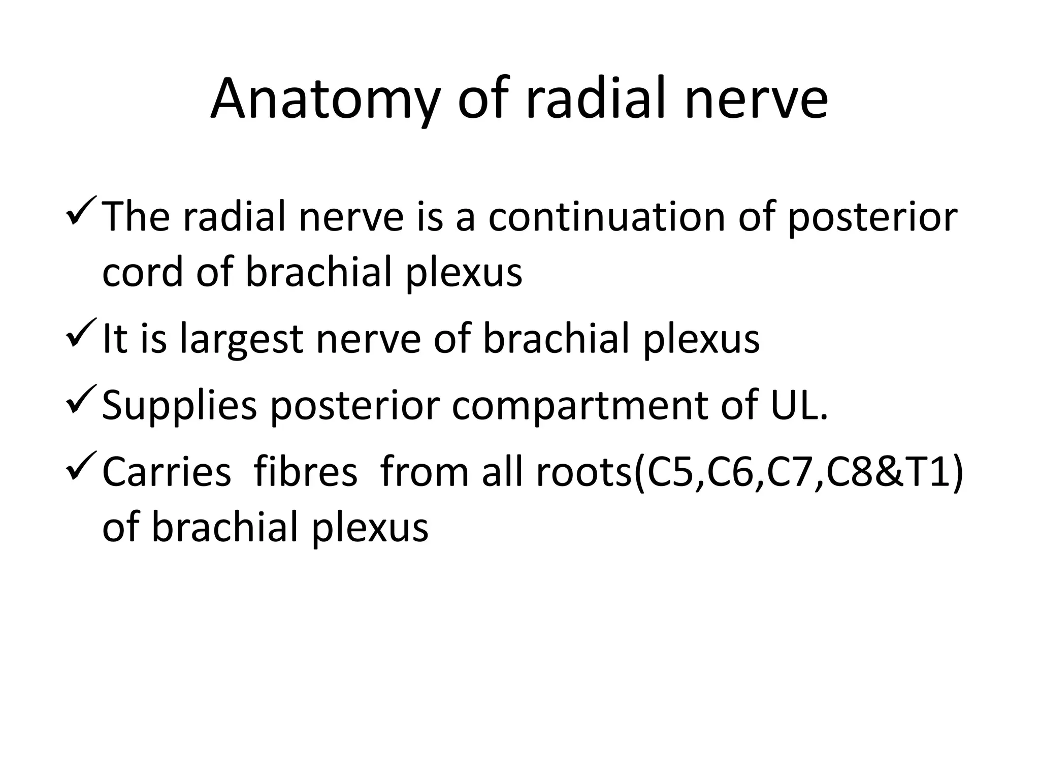
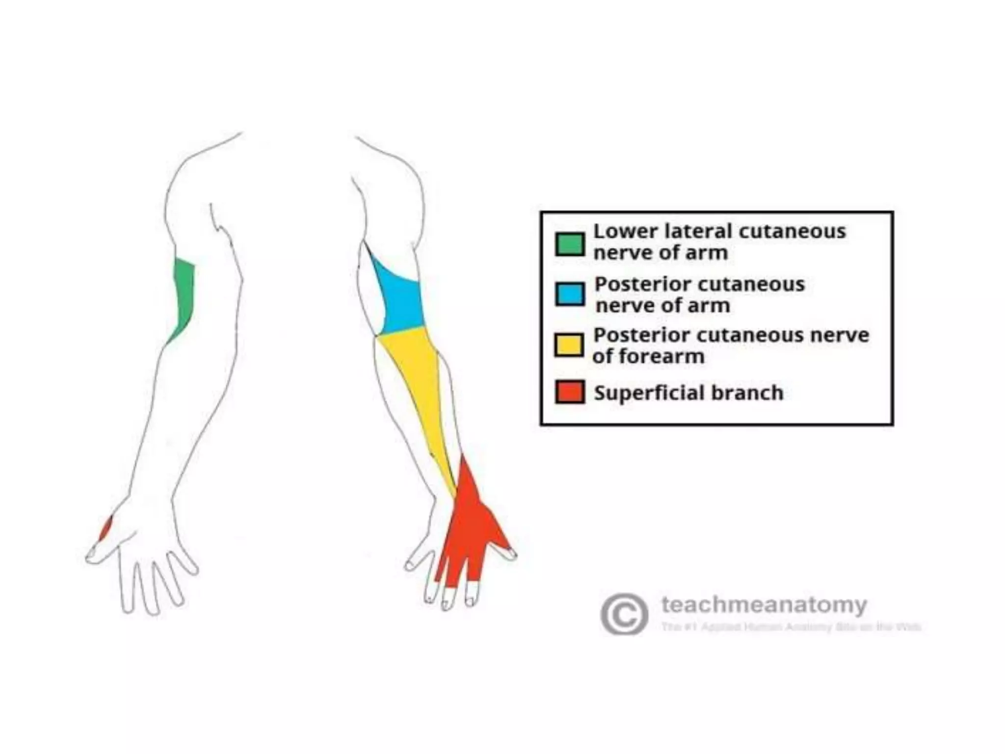
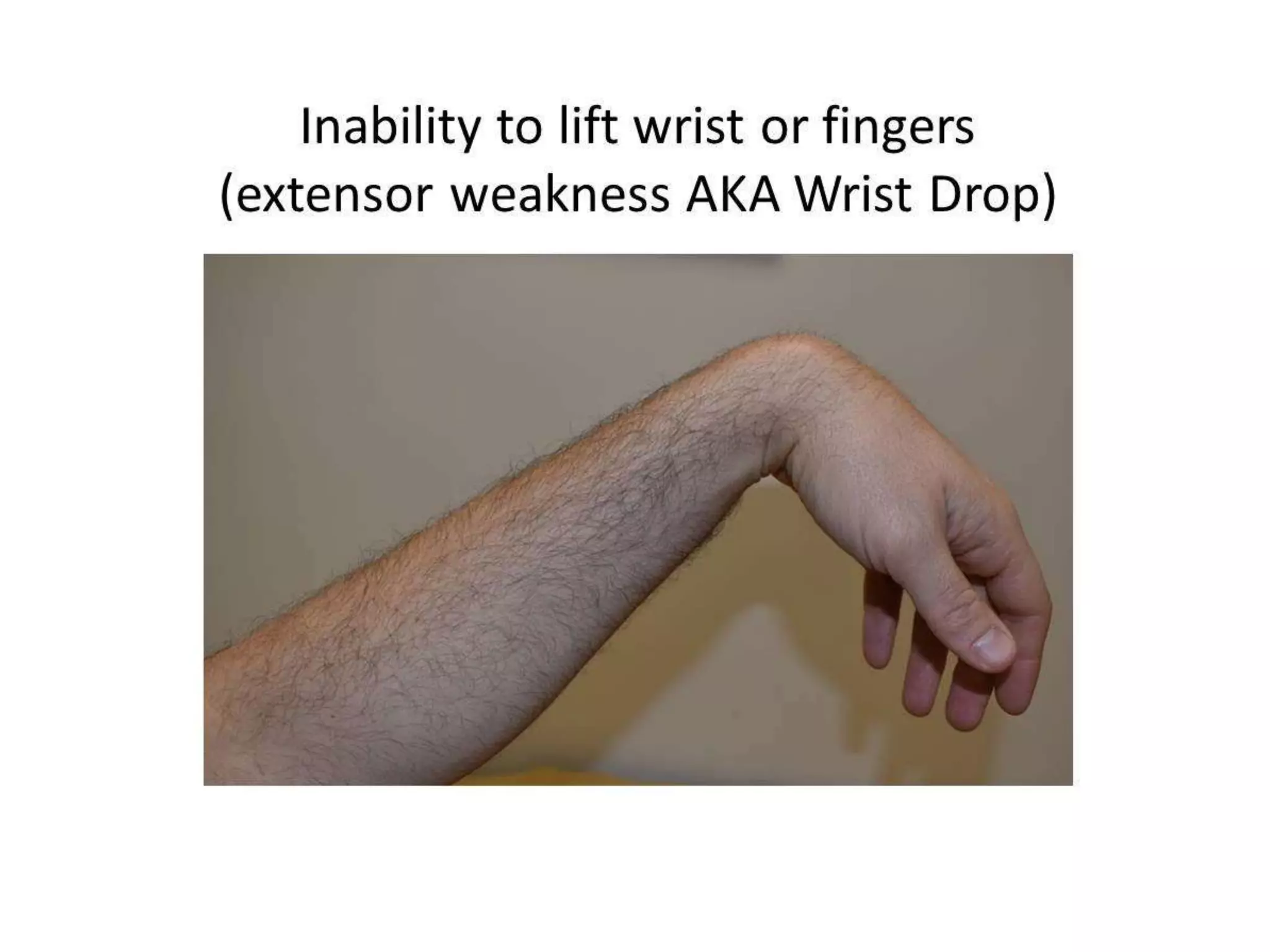
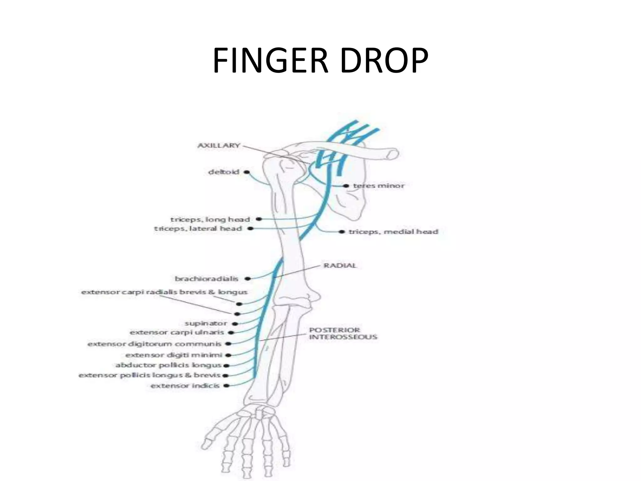
WRIST DROP,FINGER DROP,
FOOT DROP
DR.PARVATHY RAJU.P
JUNIOR RESIDENT
Anatomy of radial nerve
The radial nerve is a continuation of posterior
cord of brachial plexus
It is largest nerve of brachial plexus
Supplies posterior compartment of UL.
Carries fibres from all roots(C5,C6,C7,C8&T1)
of brachial plexus
Course in Axilla
• It lies posterior to third part of axilary artery
• Gives of 3 branches
1.Posterior cutaneous nerve of arm
2.Nerve to long head of triceps
3.Nerve to medial head of triceps
High Radial Nerve Palsy-WRIST DROP
• Injury proximal to elbow- wrist, fingers and
thumb extension are lost result in wrist drop.
• Patient loses the ability to extend the wrist,
fingers and thumb, that are essential for
grasp.
• Loses grip strength because cannot stabilise
wrist during power grip.
FINGER DROP
Low radial nerve palsy-FINGER DROP
• Defined as injury to PIN ,occurs distally to
elbow, wrist extension is preserved because
ECRL remains intact.
• If PIN Injured proximally, ECU function may be
lost, resulting radial deviation .
• If PIN Injured distally, the ECU function is
preserved(no radial deviation) and wrist
extension remains balanced .
ETHIOLOGY
• Crutch palsy
• Compression neuropathies-Saturday night
palsy
• Humeral fractures
• Iatrogenic-during surgery
• Prolonged application of tourniquet
• Intramuscular injection
Treatment of radial palsy
• Non operative-full passive range of motion in
all joint of wrist and hand and prevention of
contractures , including that of the thumb –
index web.
• Splint-cock up splint
OPERATIVE MANAGEMENT
• Nerve repair
• Neurolysis
• Tendon transfer
3 main goals :
 Restoration of finger extension
 Restoration of thumb extension
 Restoration of wrist extension
FOOT DROP
• Foot drop refers to an inability to lift the
forefoot due to weakness of the dorsiflexors.
• This condition may be the result of a
muscular, skeletal, or nervous system
problem.
LOWER EXTREMITY ANATOMY
• Thigh – The thigh contains three muscle
compartments, the anterior, medial, and
posterior compartments, named after their
relationship to the femur .
LEG
• The leg contains 4 compartments
named in relation to the tibia and fibula .
• Anterior compartment – The anterior
compartment contains the tibialis anterior,
extensor hallucis longus, extensor digitorum
longus, and peroneus tertius muscles.
These muscles dorsiflex and invert/evert the
ankle and extend the toes. The arterial supply
is the anterior tibial artery, and the motor
nerve is the deep peroneal nerve .
Lateral compartment – The lateral compartment
contains the peroneus longus and peroneus brevis
muscles .
They evert the foot at the ankle and weakly
plantar flex the foot. The peroneal artery and
superficial peroneal nerve supply these muscles .
• The posterior compartments contains the
muscles that plantarflex and invert the foot at the
ankle and flex the toes.
• The posterior tibial and peroneal arteries supply
both compartments. and innervatinnervated by
tibial nerve.
• The superficial posterior compartment contains
gastronemius, soleus, and plantaris .
• The deep posterior compartment contains the
tibialis posterior, flexor hallucis longus, flexor
digitorum longus, and popliteus muscles.
SENSORY SUPPLY OF LEG
anatomical localisation of foot drop
• Peroneal nerve injury .
• Sciatic nerve .
• L5 radiculopathy .
• Anterior horn cell involvement.
• Stroke .
Etiology
• Compressive .
• Compressive etiologies are the most common
cause of peroneal nerve palsies.
• These occur anywhere along the course of the
nerve.
• Compression of the common peroneal nerve
at the level of the fibular head is the most
common site of entrapment .
• Trauma — Traumatic injuries are the second
most common cause of peroneal nerve palsy.
• Compartment syndrome — Compartment
syndromes affecting the leg, either due to
trauma or related to ischemia-reperfusion
injury, can lead to peroneal nerve ischemia
with subsequent foot drop.
• Iatrogenic — Foot drop secondary to
iatrogenic causes is seen most frequently due
to surgical procedures or protracted
positioning in anesthesia .
• Neurologic disorders — Primary and
secondary neurologic disorders are other
causes of foot drop.
• Charcot-Marie-Tooth, a primary peripheral
nerve disorder is the most commonly
inherited peripheral nerve disorder. It affects
both sensory and motor nerves.
• amyotrophic lateral sclerosis
• multiple sclerosis
Clinical features
• Foot drop, which refers to an inability to lift
the forefoot due to weakness of the
dorsiflexion muscles in the foot, may be
partial or complete, developing acutely or
over a period of days to weeks.
• Depending on the underlying cause of foot
drop, other symptoms may also be present.
Diagnosis
• Extremity imaging (eg, plain radiography or
CT) is useful to evaluate for fractures, masses,
arthritis, or tumor if the history suggests one
of these etiologies.
• Magnetic resonance (MR) imaging of the
lumbar spine, knee, or ankle may be
considered to evaluate for potential soft tissue
lesions .
• Electrodiagnostic studies
• EMG and nerve conduction studies (NCS) can
also be helpful in localizing the lesion.
• Motor nerve conduction studies of the
peroneal nerve and tibial nerve and sensory
nerve conduction studies of the sural and
superficial peroneal nerves are recommended.
Treatment
• Conservative care — The goal of conservative
management is to stabilize the gait and prevent
"foot slap," as well as to prevent contracture of
the denervated muscles .
• Physical therapy — Physical therapy focuses on
stretching and strengthening both the muscles
affected by the palsy, as well as the opposing
musculature. The initial focus of therapy is to
stretch the contralateral muscle groups .
• Splinting — Splints and braces are used in
conjunction with physical therapy to optimize
treatment outcomes. Different types of
footwear and splints are used, depending on
the location of the nerve injury .
Surgical treatment
• Nerve repair
• Neurolysis
• Tendon transfer.
THANK YOU

WRIST DROP,FINGERDROP, by parvathy new.pptx

  • 1.
    WRIST DROP,FINGER DROP, FOOTDROP DR.PARVATHY RAJU.P JUNIOR RESIDENT
  • 2.
    Anatomy of radialnerve The radial nerve is a continuation of posterior cord of brachial plexus It is largest nerve of brachial plexus Supplies posterior compartment of UL. Carries fibres from all roots(C5,C6,C7,C8&T1) of brachial plexus
  • 4.
    Course in Axilla •It lies posterior to third part of axilary artery • Gives of 3 branches 1.Posterior cutaneous nerve of arm 2.Nerve to long head of triceps 3.Nerve to medial head of triceps
  • 9.
    High Radial NervePalsy-WRIST DROP • Injury proximal to elbow- wrist, fingers and thumb extension are lost result in wrist drop. • Patient loses the ability to extend the wrist, fingers and thumb, that are essential for grasp. • Loses grip strength because cannot stabilise wrist during power grip.
  • 11.
  • 12.
    Low radial nervepalsy-FINGER DROP • Defined as injury to PIN ,occurs distally to elbow, wrist extension is preserved because ECRL remains intact. • If PIN Injured proximally, ECU function may be lost, resulting radial deviation . • If PIN Injured distally, the ECU function is preserved(no radial deviation) and wrist extension remains balanced .
  • 13.
    ETHIOLOGY • Crutch palsy •Compression neuropathies-Saturday night palsy • Humeral fractures • Iatrogenic-during surgery • Prolonged application of tourniquet • Intramuscular injection
  • 16.
    Treatment of radialpalsy • Non operative-full passive range of motion in all joint of wrist and hand and prevention of contractures , including that of the thumb – index web. • Splint-cock up splint
  • 18.
    OPERATIVE MANAGEMENT • Nerverepair • Neurolysis • Tendon transfer
  • 19.
    3 main goals:  Restoration of finger extension  Restoration of thumb extension  Restoration of wrist extension
  • 20.
    FOOT DROP • Footdrop refers to an inability to lift the forefoot due to weakness of the dorsiflexors. • This condition may be the result of a muscular, skeletal, or nervous system problem.
  • 21.
    LOWER EXTREMITY ANATOMY •Thigh – The thigh contains three muscle compartments, the anterior, medial, and posterior compartments, named after their relationship to the femur .
  • 23.
    LEG • The legcontains 4 compartments named in relation to the tibia and fibula . • Anterior compartment – The anterior compartment contains the tibialis anterior, extensor hallucis longus, extensor digitorum longus, and peroneus tertius muscles. These muscles dorsiflex and invert/evert the ankle and extend the toes. The arterial supply is the anterior tibial artery, and the motor nerve is the deep peroneal nerve .
  • 24.
    Lateral compartment –The lateral compartment contains the peroneus longus and peroneus brevis muscles . They evert the foot at the ankle and weakly plantar flex the foot. The peroneal artery and superficial peroneal nerve supply these muscles .
  • 25.
    • The posteriorcompartments contains the muscles that plantarflex and invert the foot at the ankle and flex the toes. • The posterior tibial and peroneal arteries supply both compartments. and innervatinnervated by tibial nerve. • The superficial posterior compartment contains gastronemius, soleus, and plantaris . • The deep posterior compartment contains the tibialis posterior, flexor hallucis longus, flexor digitorum longus, and popliteus muscles.
  • 26.
  • 27.
    anatomical localisation offoot drop • Peroneal nerve injury . • Sciatic nerve . • L5 radiculopathy . • Anterior horn cell involvement. • Stroke .
  • 28.
    Etiology • Compressive . •Compressive etiologies are the most common cause of peroneal nerve palsies. • These occur anywhere along the course of the nerve. • Compression of the common peroneal nerve at the level of the fibular head is the most common site of entrapment .
  • 29.
    • Trauma —Traumatic injuries are the second most common cause of peroneal nerve palsy. • Compartment syndrome — Compartment syndromes affecting the leg, either due to trauma or related to ischemia-reperfusion injury, can lead to peroneal nerve ischemia with subsequent foot drop.
  • 30.
    • Iatrogenic —Foot drop secondary to iatrogenic causes is seen most frequently due to surgical procedures or protracted positioning in anesthesia .
  • 31.
    • Neurologic disorders— Primary and secondary neurologic disorders are other causes of foot drop. • Charcot-Marie-Tooth, a primary peripheral nerve disorder is the most commonly inherited peripheral nerve disorder. It affects both sensory and motor nerves. • amyotrophic lateral sclerosis • multiple sclerosis
  • 32.
    Clinical features • Footdrop, which refers to an inability to lift the forefoot due to weakness of the dorsiflexion muscles in the foot, may be partial or complete, developing acutely or over a period of days to weeks. • Depending on the underlying cause of foot drop, other symptoms may also be present.
  • 34.
    Diagnosis • Extremity imaging(eg, plain radiography or CT) is useful to evaluate for fractures, masses, arthritis, or tumor if the history suggests one of these etiologies. • Magnetic resonance (MR) imaging of the lumbar spine, knee, or ankle may be considered to evaluate for potential soft tissue lesions .
  • 35.
    • Electrodiagnostic studies •EMG and nerve conduction studies (NCS) can also be helpful in localizing the lesion. • Motor nerve conduction studies of the peroneal nerve and tibial nerve and sensory nerve conduction studies of the sural and superficial peroneal nerves are recommended.
  • 36.
    Treatment • Conservative care— The goal of conservative management is to stabilize the gait and prevent "foot slap," as well as to prevent contracture of the denervated muscles . • Physical therapy — Physical therapy focuses on stretching and strengthening both the muscles affected by the palsy, as well as the opposing musculature. The initial focus of therapy is to stretch the contralateral muscle groups .
  • 37.
    • Splinting —Splints and braces are used in conjunction with physical therapy to optimize treatment outcomes. Different types of footwear and splints are used, depending on the location of the nerve injury .
  • 38.
    Surgical treatment • Nerverepair • Neurolysis • Tendon transfer.
  • 39.