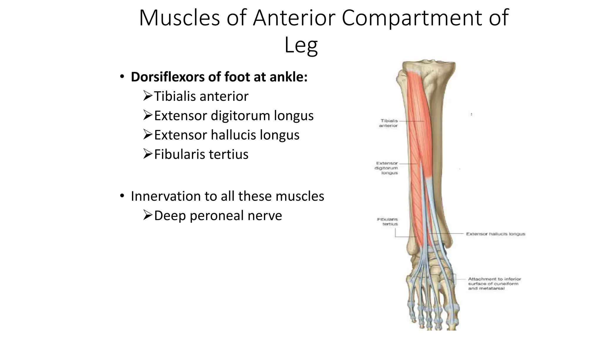
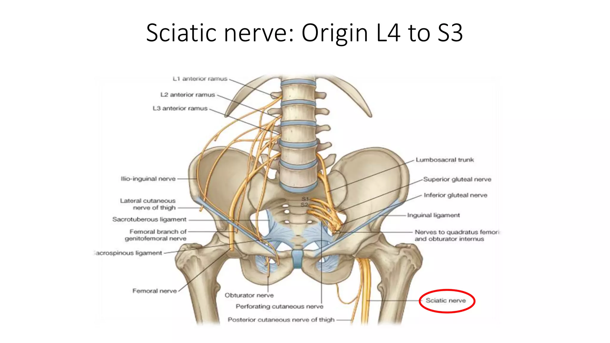
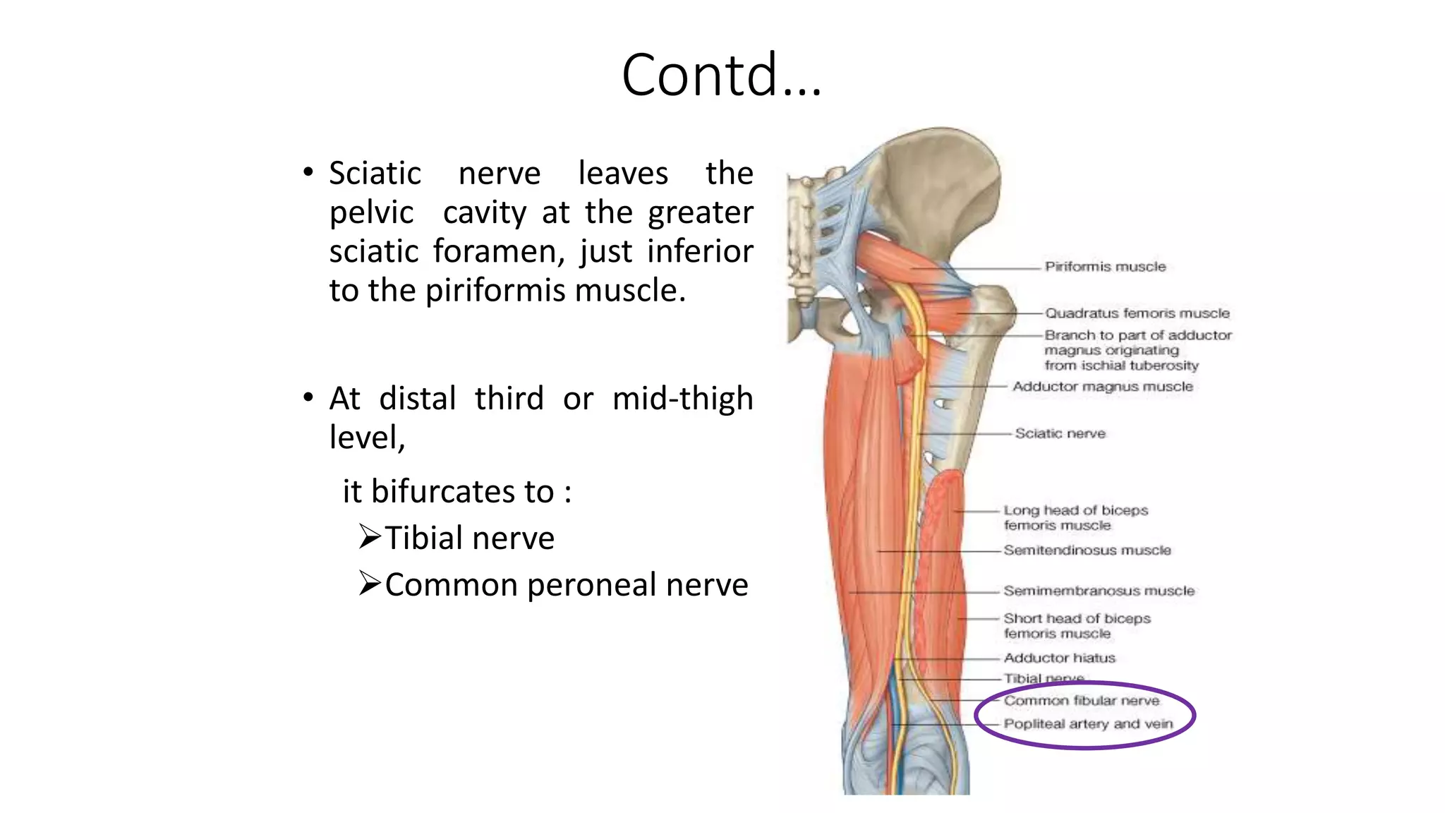
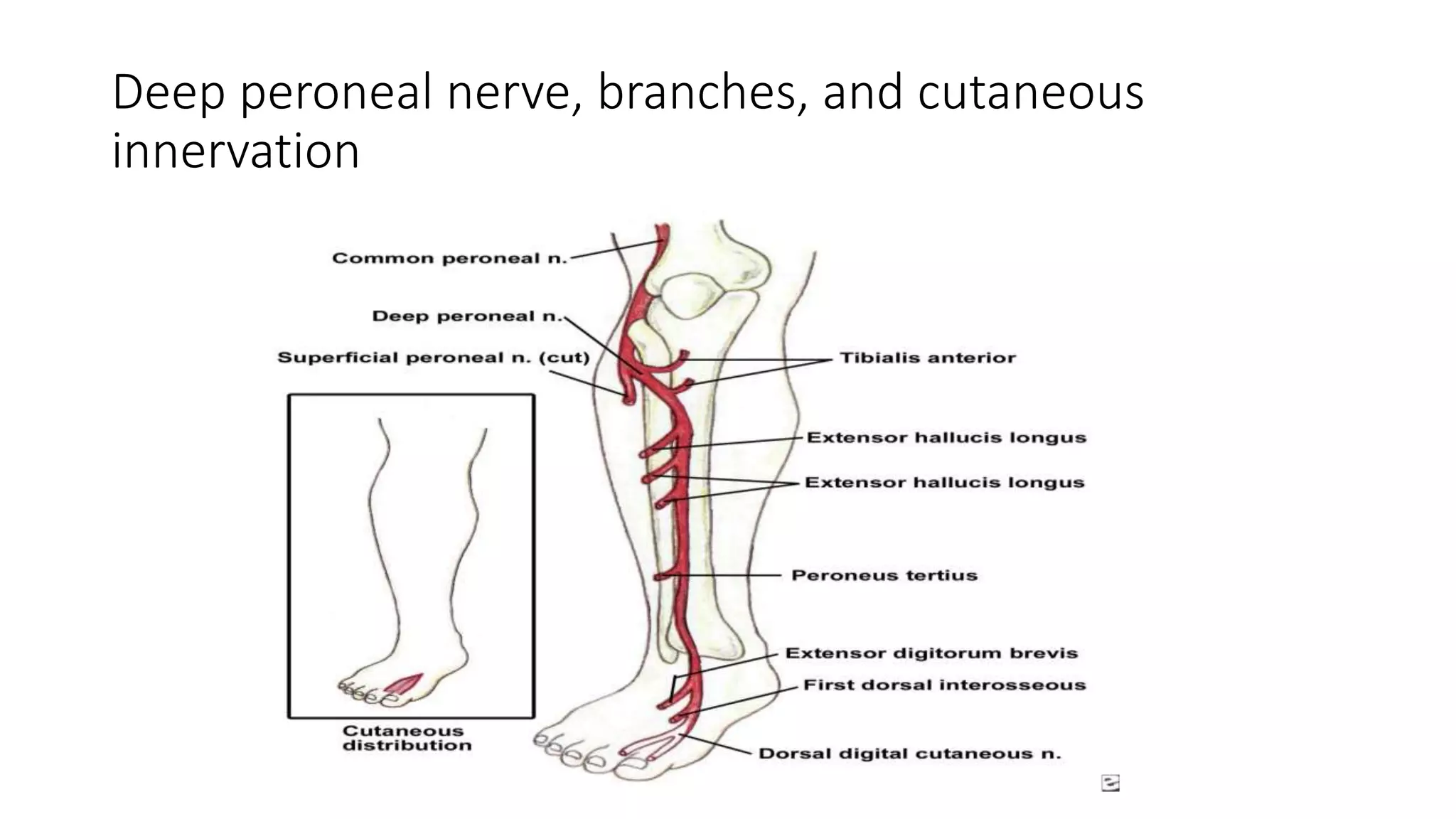
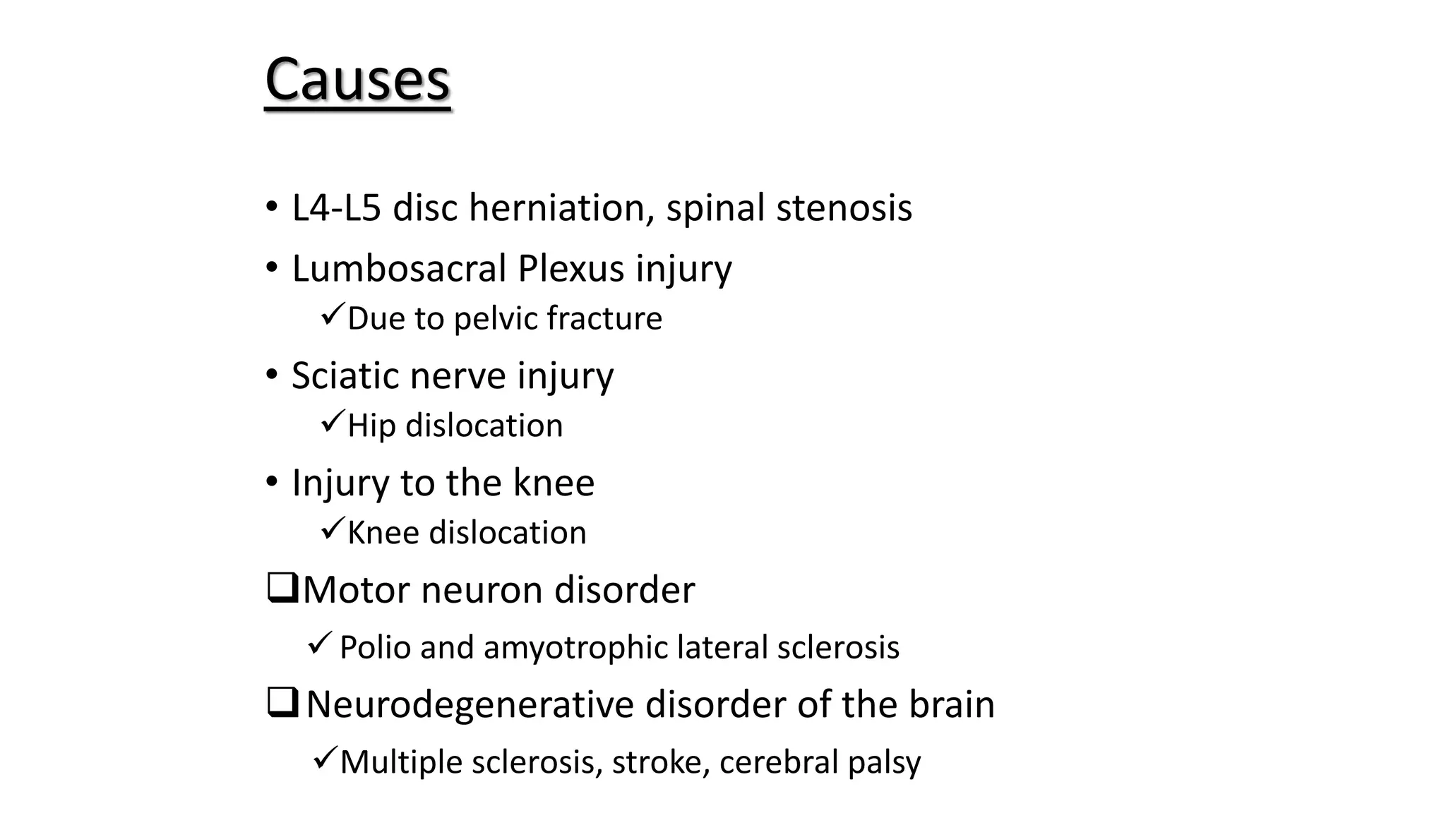
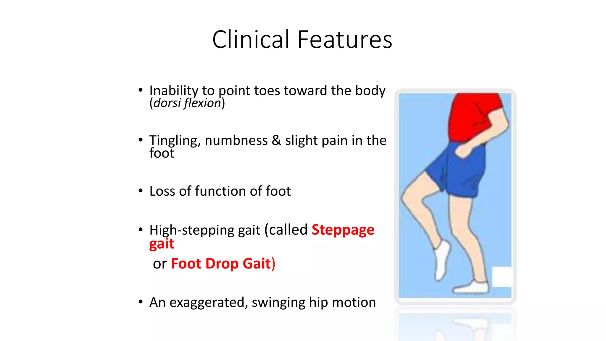
FOOT DROP
PRESENTED BY:
Sunil Kumar Daha
Introduction
• Inability of the dorsiflexion foot  weakness or
paralysis of the muscles that lift the foot
• A symptom
• Can result if there is injury to
• the dorsiflexors or
• any point along the neural pathways that supply them
Muscles of Anterior Compartment of
Leg
• Dorsiflexors of foot at ankle:
Tibialis anterior
Extensor digitorum longus
Extensor hallucis longus
Fibularis tertius
• Innervation to all these muscles
Deep peroneal nerve
Sciatic nerve: Origin L4 to S3
Contd…
• Sciatic nerve leaves the
pelvic cavity at the greater
sciatic foramen, just inferior
to the piriformis muscle.
• At distal third or mid-thigh
level,
it bifurcates to :
Tibial nerve
Common peroneal nerve
Peroneal nerve in Popliteal fossa
• Runs downward through
popliteal fossa following
medial border of biceps
femoris muscle
• Leaves fossa by crossing
superficially, the lateral
head of gastrocnemius
muscle
In the leg
• Passes behind the head
of fibula and winds laterally
around neck of bone
• Pierce peronous longus muscle
and divides into:
Superficial peroneal nerve
Deep peroneal nerve
Common and superficial peroneal nerves, branches, and
cutaneous innervation
Deep peroneal nerve, branches, and cutaneous
innervation
Central
Brain
Ex- Multiple Sclerosis
Spinal Cord
Ex- Disc prolapse
Peripheral
Nerve
Ex- Common
peroneal nerve
injury
Muscle
Ex- Muscle atrophy
Causes
• L4-L5 disc herniation, spinal stenosis
• Lumbosacral Plexus injury
Due to pelvic fracture
• Sciatic nerve injury
Hip dislocation
• Injury to the knee
Knee dislocation
Motor neuron disorder
Polio and amyotrophic lateral sclerosis
Neurodegenerative disorder of the brain
Multiple sclerosis, stroke, cerebral palsy
Causes
• External compression
– During anesthesia, coma, sleep, bed rest
– Plaster cast, braces
– Habitual leg crossing
– Sitting cross legged
– Prolonged squating, kneeling
Direct trauma
– Blunt injuries, lacerations
– Fracture of fibula
– Adduction injuries and dislocations of knee
– Surgery and arthroscopy in popliteal fossa and knee
Causes
• Traction injuries
– Acute ankle injuries
• Masses
– Ganglia, Baker’s cyst, callus, fibular tumors, osteoma, hematomas
Tumors
– Nerve sheath tumors
– Nerve sheath ganglia
– Lipomas
Entrapment
– In the fibular tunnel
– Anterior (tibial) compartment syndrome
Causes
• Vascular
– Vasculitis, local vascular disease
• DM: susceptibility to compression, ischemic damage
• Leprosy
• Idiopathic
Causes
Presentation
• Direct injury to dorsiflexors
• Compartment syndromes
• Anterior compartment syndrome
– Nerve involved: deep peroneal nerve
• Deep posterior compartment syndrome
– Nerve involved: posterior tibial nerve
• Chronic compartment syndrome
• Neurologic defects
Clinical Features
• Inability to point toes toward the body
(dorsi flexion)
• Tingling, numbness & slight pain in the
foot
• Loss of function of foot
• High-stepping gait (called Steppage
gait
or Foot Drop Gait)
• An exaggerated, swinging hip motion
Diagnosis
• History
• Clinical exam, including neurological exams
• Electromyogram
• Nerve conduction test
• Imaging studies, such as X-rays or high-
resolution MRI (magnetic resonance imaging)
Treatment
• Non-surgical
Orthotics, including braces or foot
splint
Physicotherapy including gait training
• Surgical therapy
Decompression surgery
Nerve sutures
Nerve grafting
Nerve transfer
Tendon transfer
The End

2. Foot drop

  • 1.
  • 3.
    Introduction • Inability ofthe dorsiflexion foot  weakness or paralysis of the muscles that lift the foot • A symptom • Can result if there is injury to • the dorsiflexors or • any point along the neural pathways that supply them
  • 4.
    Muscles of AnteriorCompartment of Leg • Dorsiflexors of foot at ankle: Tibialis anterior Extensor digitorum longus Extensor hallucis longus Fibularis tertius • Innervation to all these muscles Deep peroneal nerve
  • 6.
  • 7.
    Contd… • Sciatic nerveleaves the pelvic cavity at the greater sciatic foramen, just inferior to the piriformis muscle. • At distal third or mid-thigh level, it bifurcates to : Tibial nerve Common peroneal nerve
  • 8.
    Peroneal nerve inPopliteal fossa • Runs downward through popliteal fossa following medial border of biceps femoris muscle • Leaves fossa by crossing superficially, the lateral head of gastrocnemius muscle
  • 9.
    In the leg •Passes behind the head of fibula and winds laterally around neck of bone • Pierce peronous longus muscle and divides into: Superficial peroneal nerve Deep peroneal nerve
  • 11.
    Common and superficialperoneal nerves, branches, and cutaneous innervation
  • 12.
    Deep peroneal nerve,branches, and cutaneous innervation
  • 14.
    Central Brain Ex- Multiple Sclerosis SpinalCord Ex- Disc prolapse Peripheral Nerve Ex- Common peroneal nerve injury Muscle Ex- Muscle atrophy Causes
  • 15.
    • L4-L5 discherniation, spinal stenosis • Lumbosacral Plexus injury Due to pelvic fracture • Sciatic nerve injury Hip dislocation • Injury to the knee Knee dislocation Motor neuron disorder Polio and amyotrophic lateral sclerosis Neurodegenerative disorder of the brain Multiple sclerosis, stroke, cerebral palsy Causes
  • 16.
    • External compression –During anesthesia, coma, sleep, bed rest – Plaster cast, braces – Habitual leg crossing – Sitting cross legged – Prolonged squating, kneeling Direct trauma – Blunt injuries, lacerations – Fracture of fibula – Adduction injuries and dislocations of knee – Surgery and arthroscopy in popliteal fossa and knee Causes
  • 17.
    • Traction injuries –Acute ankle injuries • Masses – Ganglia, Baker’s cyst, callus, fibular tumors, osteoma, hematomas Tumors – Nerve sheath tumors – Nerve sheath ganglia – Lipomas Entrapment – In the fibular tunnel – Anterior (tibial) compartment syndrome Causes
  • 18.
    • Vascular – Vasculitis,local vascular disease • DM: susceptibility to compression, ischemic damage • Leprosy • Idiopathic Causes
  • 19.
    Presentation • Direct injuryto dorsiflexors • Compartment syndromes • Anterior compartment syndrome – Nerve involved: deep peroneal nerve • Deep posterior compartment syndrome – Nerve involved: posterior tibial nerve • Chronic compartment syndrome • Neurologic defects
  • 20.
    Clinical Features • Inabilityto point toes toward the body (dorsi flexion) • Tingling, numbness & slight pain in the foot • Loss of function of foot • High-stepping gait (called Steppage gait or Foot Drop Gait) • An exaggerated, swinging hip motion
  • 21.
    Diagnosis • History • Clinicalexam, including neurological exams • Electromyogram • Nerve conduction test • Imaging studies, such as X-rays or high- resolution MRI (magnetic resonance imaging)
  • 22.
    Treatment • Non-surgical Orthotics, includingbraces or foot splint Physicotherapy including gait training • Surgical therapy Decompression surgery Nerve sutures Nerve grafting Nerve transfer Tendon transfer The End

Editor's Notes

  • #2 Peroneal neuropathy caused by compression at the fibular head is the most common compressive neuropathy in the lower extremity. Foot drop is its most notable symptom. All age groups are affected equally, but the condition is more common in males (male-to-female ratio, 2.8:1). About 90% of peroneal lesions are unilateral, and they can affect the right or the left side with equal frequency
  • #3 Inability of the dorsiflexion foot  weakness or paralysis of the muscles that lift the foot
  • #5 The foot and ankle dorsiflexors include the tibialis anterior, the extensor hallucis longus (EHL), and the extensor digitorum longus (EDL). These muscles help the body clear the foot during swing phase and control plantarflexion of the foot on heel strike.
  • #6 The foot and ankle dorsiflexors include the tibialis anterior, the extensor hallucis longus (EHL), and the extensor digitorum longus (EDL). These muscles help the body clear the foot during swing phase and control plantarflexion of the foot on heel strike.
  • #8 Fibers from the dorsal branches of the ventral rami of L4-S1 are found in the peroneal nerve, which is paired with the tibial nerve to constitute the sciatic nerve. The sciatic nerve leaves the pelvic cavity at the greater sciatic foramen, just inferior to the piriformis. It bifurcates to form the peroneal and tibial nerves either in the distal third of the thigh or at the midthigh level. The peroneal nerve crosses laterally to curve over the posterior rim of the fibular neck to the anterior compartment of the lower leg, dividing into superficial and deep branches. The superficial branch travels between the two heads of the peronei and continues down the lower leg to lie between the peroneal tendon and the lateral edge of the gastrocnemius. It then branches to the ankle anterolaterally to supply sensation to the dorsum of the foot (see the image below).
  • #16 The peroneal nerve is susceptible to injury all along its course. In that it is part of the sciatic nerve, its funiculi are relatively isolated from those of the tibial nerve. Therefore, trauma to the sciatic nerve may affect only one of its divisions. The funiculi of the peroneal nerve also are larger and have less protective connective tissue than those of the tibial nerve, making the peroneal nerve more susceptible to trauma. In addition, the peroneal nerve has fewer autonomic fibers; thus, in any injury, motor and sensory fibers bear the brunt of the trauma. The peroneal nerve runs a more superficial course than the tibial nerve does, especially at the fibular neck, and this relatively exposed position makes it vulnerable to direct insult. Its close adherence to the periosteum of the proximal fibula renders it susceptible to injury during surgical procedures in this area.
  • #21 The functional integrity of an axon and its target depends on the continued supply of trophic substances synthesized in the neuronal perikaryon and transported down the axon (axoplasmic flow). A laceration interrupts axoplasmic flow; a crush injury may compromise it as well. A double-crush phenomenon occurs when a proximal insult in a nerve root diminishes axoplasmic flow, making it more susceptible to injury.
  • #23 Weakness in this group of muscles results in an equinovarus deformity. This is sometimes referred to as steppage gait, because the patient tends to walk with an exaggerated flexion of the hip and knee to prevent the toes from catching on the ground during swing phase. During gait, the force of heel strike exceeds body weight, and the direction of the ground reaction vector passes behind the ankle and knee cente This causes the foot to plantarflex and, if uncontrolled, to slap the ground. Ordinarily, eccentric lengthening of the tibialis anterior, which controls plantar flexion, absorbs the shock of heel strike.
  • #24 Prognosis and outcome vary according to the cause of the foot drop. In a peripheral compressive neuropathy, recovery can be expected in up to 3 months, provided that further compression is avoided. A partial peroneal nerve palsy after total knee replacement has a uniformly good prognosis. [5] A variable amount of recovery is seen with a complete postoperative palsy. Follow-up EMG and nerve conduction studies may be useful for assessing recovery. A partial palsy recovers faster because of local sprouting. With complete axonal loss, reinnervation is achieved solely through proximal-to-distal axonal growth, which usually proceeds at a rate of 1 mm/day. Thus, injuries of a nerve close to its target muscle also have a more favorable outcome. In a nerve root compressive neuropathy, one study concluded that severe motor weakness lasting longer than 6 months, a negative straight leg-raising test, and old age were poor prognostic factors for recovery of dorsiflexion. [6] When there is direct injury to the peroneal nerve, the outcome is more favorable for penetrating trauma than for blunt trauma; a traction or stretch injury to the nerve has an intermediate outcome. When nerve grafting is performed, functional recovery depends on the severity of injury and thus on the length of the graft used. With grafts longer than 12 cm, good functional recovery is rare. Wound infection may occur after surgical treatment, as may nerve graft failure. In tendon transfer procedures, recurrent deformity has been reported. In arthrodeses or fusion procedures, pseudoarthrosis, delayed union, or nonunion may be noted
  • #25 Orthotics (Greek: Ορθός, ortho, "to straighten" or "align") is a specialty within the medical field concerned with the design, manufacture and application of orthoses. An orthosis (plural: orthoses) is "an externally applied device used to modify the structural and functional characteristics of the neuromuscular and skeletal system".[1] An orthosis may be used to: Control, guide, limit and/or immobilize an extremities, joint or body segment for a particular reason To restrict movement in a given direction To assist movement generally To reduce weight bearing forces for a particular purpose To aid rehabilitation from fractures after the removal of a cast To otherwise correct the shape and/or function of the body, to provide easier movement capability or reduce pain