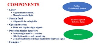
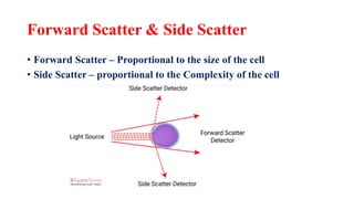
FLOW CYTOMETRY
Dr Tamil Nila
INTRODUCTION
• Laser – based technology
• Identify & quantify cell populations
• Cells are manipulated into a stream of fluid
• Interrogated by an electronic detection system
Flow - Fluid
Cyto - Cell
Metry –
Measurement
Measures properties of the cell as they flow in a fluid
suspension across an illuminated light path
How is it different??
Immunophenotyping
SAMPLE PREPARATION
• Peripheral blood
• Bone marrow
• CSF
• Ascitic fluid
• Pleural fluid
• FNA
• Red cells are lysed
• Appropriate antibody panels
are chosen
Spleen
Lymphnode
Liver
Bone marrow biopsy
After tissue disaggregation
USES
• Identification & quantification of cell populations within a sample
• Normal vs abnormal cells
• Reactive vs neoplastic cells
• Differentiation & maturation stage in a cell population
• Quantification of tumour infiltration
COMPONENTS
• Laser
• Argon (most common)
• Monochromatic light
• Sheath fluid
• Aligns cells in a single file
• Optical systems
• Filter and regulate light signals
• Photomultiplier detectors
• forward light scatter – cell size
• Side light scatter – cell complexity
• Converting fluorescent light signal into electrical signal
• Computer
Fluidics -
Hydrodynamic focusing
Excitation Optics &
Collection Optics
Electronics
How does single filing occur?
• Hydrodynamic focusing
• Sheath fluid exerts pressure onto the sample
• As the nozzle narrows -> a single file is obtained
Forward Scatter & Side Scatter
• Forward Scatter – Proportional to the size of the cell
• Side Scatter – proportional to the Complexity of the cell
Antigen detection
488 nm 510 nm
Light Filter Vs Mirrors
Long pass
Short Pass
Band Pass
Stokes shift
Interpretation
• Intensity of color change is directly proportional to the amount of
antigens present
Processing
• Stain-lyse-wash method
• Stain-lyse-no wash method
• Lyse-stain-wash method
Staining
• STAIN-LYSE-WASH METHOD
• Dilute cell concentration to 1 – 2 x 10⁶ per tube
Pipette 100 μl specimen in round bottom tube +
McAb combination or Multicolor cocktail
Incubate at room temperature in dark room for 15
min
1 ml of NH4Cl based lysing solution &
Incubate for 10 mins
Centrifuge x 5 mins at 300 g
Discard the supernatant & repeat
Resuspend cells in 0.2 – 0.5 ml of sheath fluid
Stain-lyse-no wash method
• No centrifugation
• Rest of the steps are same
• Ideal for samples with few cells
• Cell loss during centrifugation is minimised
Lyse-stain-wash method
• Bulk
specimen
lysis
• Used for
MRD
monitoring
5 – 10 ml of sample
+ same amount of
NH4Cl lysing
solution
Mix gently &
incubate at room
temperature for 10
mins
Centrifuge at 300g
for 5 min
Discard supernatant
Resuspend cell
pellet in 10 ml PBS-
Azide-BSA
Repeat washing &
resuspend
Aliquot a volume of
cell suspension
containing 10 x 106
cells/tube
Add McAb
Incubate in dark
Repeat washing &
resuspend in 0.2 –
0.5 ml of sheath
fluid
Interpret data
Detection of intracellular antigens
• Fix & Perm kit
• Solution A – Fixing agent (paraformaldehyde solution)
• Solution B – lysing agent (lysing solution & detergent)
Pipette 100 μl
of specimen
Add 100 μl of
Solution A &
Incubate at
room temp x
15 mins
Wash twice in
PBS-Azide-
BSA
Add 100 μl of
Solution B +
Ab cocktail
Incubate at
room
temperature
in dark x 15
mins
Wash twice
with PBS-
Azide-BSA
Resuspend in
sheath fluid
Interpret Data
Data Acquisition
• Diagnostic Lymphoproliferative disorder samples – 50000
lymphocytes
• MRD detection – 50000 to 1 lakh lymphocytes needed
MULTICOLOR OR MULTIPARAMETER FLOW
CYTOMETRY
• 8 – 10 color antibody panels
• Provides data of 12 – 14 cellular parameters simultaneously
ADVANTAGES DISADVANTAGES
Increased accuracy Increased complexity of compensation
Smaller sample size Challenges of antibody panel validation
Cost effectiveness Tandem dye conjugate issues
Increased efficiency Increased need for expertise in data
analysis & interpretation
Increased sensitivity for minimal residual
disease
Human error
Scatter plot interpretation
Positive for Y only Positive for both Antigens
Negative for Both Antigens Positive for X only
X
Y
Characteristics of Blasts
B- ALL Blasts
CD 10 +
CD 19+
CD 45 –ve
HLA DR+
CD 34+
TdT+
Either Kappa or
Lambda positive
Case 1
Characteristics of Blasts
T ALL Blasts
CD3+
CD5+
CD7+
CD34+
HLA – DR
TdT +
Case 2
CD 13 -ve
Characteristics of blasts
AML
Pan Myeloid
markers
CD13+
CD 33+
CD 117+
Blast markers
CD 34+
Case 3
References
• Dacie and Lewis Practical hematology
• Flow cytometry of Hematological malignancies, Claudio Ortolani
• Flow cytometry in neoplastic hematology

FLOW CYTOMETRY

  • 1.
  • 2.
    INTRODUCTION • Laser –based technology • Identify & quantify cell populations • Cells are manipulated into a stream of fluid • Interrogated by an electronic detection system Flow - Fluid Cyto - Cell Metry – Measurement Measures properties of the cell as they flow in a fluid suspension across an illuminated light path
  • 3.
    How is itdifferent?? Immunophenotyping
  • 4.
    SAMPLE PREPARATION • Peripheralblood • Bone marrow • CSF • Ascitic fluid • Pleural fluid • FNA • Red cells are lysed • Appropriate antibody panels are chosen Spleen Lymphnode Liver Bone marrow biopsy After tissue disaggregation
  • 5.
    USES • Identification &quantification of cell populations within a sample • Normal vs abnormal cells • Reactive vs neoplastic cells • Differentiation & maturation stage in a cell population • Quantification of tumour infiltration
  • 6.
    COMPONENTS • Laser • Argon(most common) • Monochromatic light • Sheath fluid • Aligns cells in a single file • Optical systems • Filter and regulate light signals • Photomultiplier detectors • forward light scatter – cell size • Side light scatter – cell complexity • Converting fluorescent light signal into electrical signal • Computer Fluidics - Hydrodynamic focusing Excitation Optics & Collection Optics Electronics
  • 8.
    How does singlefiling occur? • Hydrodynamic focusing • Sheath fluid exerts pressure onto the sample • As the nozzle narrows -> a single file is obtained
  • 9.
    Forward Scatter &Side Scatter • Forward Scatter – Proportional to the size of the cell • Side Scatter – proportional to the Complexity of the cell
  • 11.
  • 13.
    Light Filter VsMirrors Long pass Short Pass Band Pass
  • 15.
  • 16.
    Interpretation • Intensity ofcolor change is directly proportional to the amount of antigens present
  • 18.
    Processing • Stain-lyse-wash method •Stain-lyse-no wash method • Lyse-stain-wash method
  • 19.
  • 20.
    • STAIN-LYSE-WASH METHOD •Dilute cell concentration to 1 – 2 x 10⁶ per tube Pipette 100 μl specimen in round bottom tube + McAb combination or Multicolor cocktail Incubate at room temperature in dark room for 15 min 1 ml of NH4Cl based lysing solution & Incubate for 10 mins Centrifuge x 5 mins at 300 g Discard the supernatant & repeat Resuspend cells in 0.2 – 0.5 ml of sheath fluid
  • 21.
    Stain-lyse-no wash method •No centrifugation • Rest of the steps are same • Ideal for samples with few cells • Cell loss during centrifugation is minimised
  • 22.
    Lyse-stain-wash method • Bulk specimen lysis •Used for MRD monitoring 5 – 10 ml of sample + same amount of NH4Cl lysing solution Mix gently & incubate at room temperature for 10 mins Centrifuge at 300g for 5 min Discard supernatant Resuspend cell pellet in 10 ml PBS- Azide-BSA Repeat washing & resuspend Aliquot a volume of cell suspension containing 10 x 106 cells/tube Add McAb Incubate in dark Repeat washing & resuspend in 0.2 – 0.5 ml of sheath fluid Interpret data
  • 23.
    Detection of intracellularantigens • Fix & Perm kit • Solution A – Fixing agent (paraformaldehyde solution) • Solution B – lysing agent (lysing solution & detergent) Pipette 100 μl of specimen Add 100 μl of Solution A & Incubate at room temp x 15 mins Wash twice in PBS-Azide- BSA Add 100 μl of Solution B + Ab cocktail Incubate at room temperature in dark x 15 mins Wash twice with PBS- Azide-BSA Resuspend in sheath fluid Interpret Data
  • 24.
    Data Acquisition • DiagnosticLymphoproliferative disorder samples – 50000 lymphocytes • MRD detection – 50000 to 1 lakh lymphocytes needed
  • 25.
    MULTICOLOR OR MULTIPARAMETERFLOW CYTOMETRY • 8 – 10 color antibody panels • Provides data of 12 – 14 cellular parameters simultaneously ADVANTAGES DISADVANTAGES Increased accuracy Increased complexity of compensation Smaller sample size Challenges of antibody panel validation Cost effectiveness Tandem dye conjugate issues Increased efficiency Increased need for expertise in data analysis & interpretation Increased sensitivity for minimal residual disease Human error
  • 28.
    Scatter plot interpretation Positivefor Y only Positive for both Antigens Negative for Both Antigens Positive for X only X Y
  • 29.
    Characteristics of Blasts B-ALL Blasts CD 10 + CD 19+ CD 45 –ve HLA DR+ CD 34+ TdT+ Either Kappa or Lambda positive
  • 30.
  • 31.
    Characteristics of Blasts TALL Blasts CD3+ CD5+ CD7+ CD34+ HLA – DR TdT +
  • 32.
  • 33.
    Characteristics of blasts AML PanMyeloid markers CD13+ CD 33+ CD 117+ Blast markers CD 34+
  • 34.
  • 35.
    References • Dacie andLewis Practical hematology • Flow cytometry of Hematological malignancies, Claudio Ortolani • Flow cytometry in neoplastic hematology