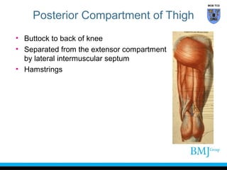
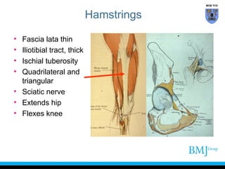
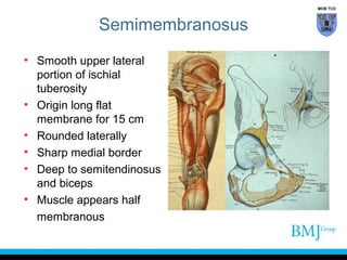
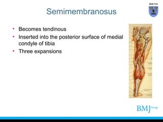
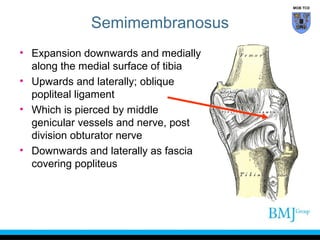
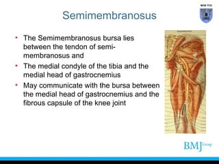
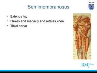
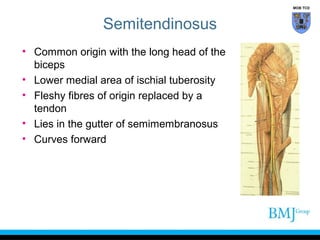
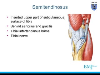
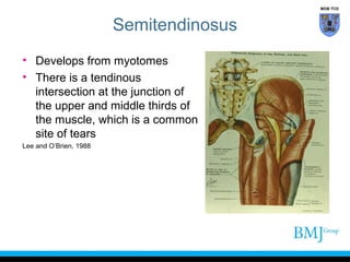
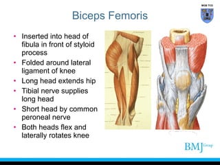
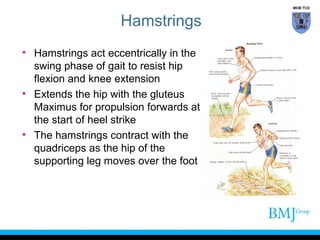
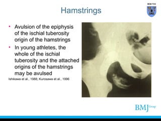
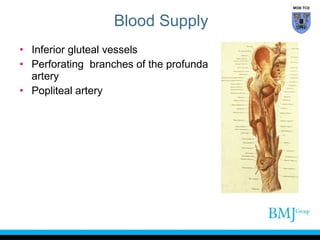
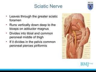
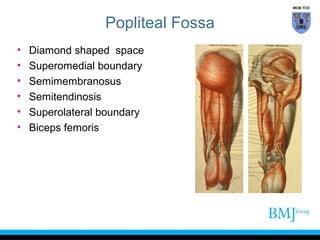
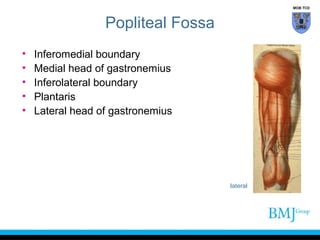
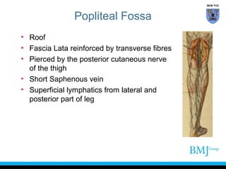
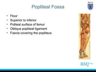
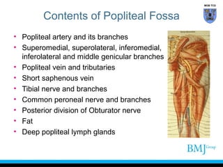
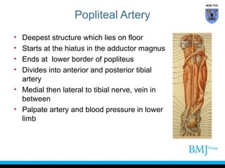
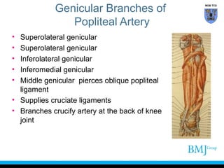
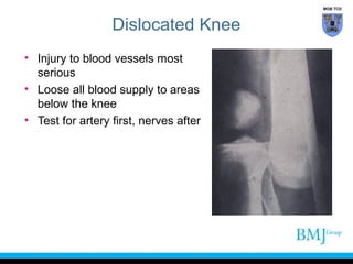
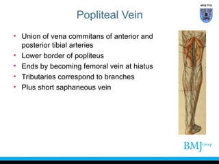
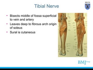
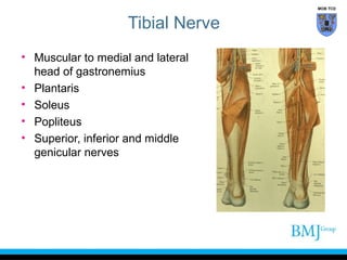
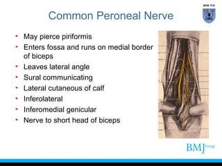
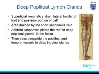
MOB TCD

Posterior Compartment of Thigh
Professor Emeritus Moira O’Brien
FRCPI, FFSEM, FFSEM (UK), FTCD
Trinity College
Dublin
MOB TCD

Posterior Compartment of Thigh
• Buttock to back of knee
• Separated from the extensor compartment
by lateral intermuscular septum
• Hamstrings
MOB TCD

Cutaneous Supply
• Posterior cutaneous nerve of thigh S2
• Posterior branch of lateral cutaneous of thigh
MOB TCD

Hamstrings
•
•
•
•

Fascia lata thin
Iliotibial tract, thick
Ischial tuberosity
Quadrilateral and
triangular
• Sciatic nerve
• Extends hip
• Flexes knee
MOB TCD

Semimembranosus
• Smooth upper lateral
portion of ischial
tuberosity
• Origin long flat
membrane for 15 cm
• Rounded laterally
• Sharp medial border
• Deep to semitendinosus
and biceps
• Muscle appears half
membranous
MOB TCD

Semimembranosus
• Becomes tendinous
• Inserted into the posterior surface of medial
condyle of tibia
• Three expansions
MOB TCD

Semimembranosus
• Expansion downwards and medially
along the medial surface of tibia
• Upwards and laterally; oblique
popliteal ligament
• Which is pierced by middle
genicular vessels and nerve, post
division obturator nerve
• Downwards and laterally as fascia
covering popliteus
MOB TCD

Semimembranosus
• The Semimembranosus bursa lies
between the tendon of semimembranosus and
• The medial condyle of the tibia and the
medial head of gastrocnemius
• May communicate with the bursa between
the medial head of gastrocnemius and the
fibrous capsule of the knee joint
MOB TCD

Semimembranosus
• Extends hip
• Flexes and medially and rotates knee
• Tibial nerve
MOB TCD

Semitendinosus
• Common origin with the long head of the
biceps
• Lower medial area of ischial tuberosity
• Fleshy fibres of origin replaced by a
tendon
• Lies in the gutter of semimembranosus
• Curves forward
MOB TCD

Semitendinosus
• Inserted upper part of subcutaneous
surface of tibia
• Behind sartorius and gracilis
• Tibial intertendinous bursa
• Tibial nerve
MOB TCD

Semitendinosus
• Develops from myotomes
• There is a tendinous
intersection at the junction of
the upper and middle thirds of
the muscle, which is a common
site of tears
Lee and O’Brien, 1988
MOB TCD

Biceps
• Long head has common origin
with the semitendinosus
• Lower medial area of ischial
tuberosity
• Short head from linea aspera
• Upper part of lateral
supracondylar line
MOB TCD

Biceps Femoris
• Inserted into head of
fibula in front of styloid
process
• Folded around lateral
ligament of knee
• Long head extends hip
• Tibial nerve supplies
long head
• Short head by common
peroneal nerve
• Both heads flex and
laterally rotates knee
MOB TCD

Biceps Femoris
• 80% of hamstring strains occur in the occur in the long
head of the biceps femoris muscle
Koulouris & Connell, 2003

• Injuries may occur:
• During the switch between late leg recovery and initial
leg approach in the swing phase of sprinting
Woods et al., 2004

• During the ground contact phase of running
• Poor timing-intermuscular coordination and eccentric
strength in the short head of the biceps femoris muscle
Woods et al., 2004
MOB TCD

Biceps Femoris
• Lack of stiffness and eccentric strength in the short and
long head of the biceps femoris muscle during the
ground contact phase of running
Bosch and Klomp, 2005

• Can be torn at origin from tuberosity
• Middle of thigh
• Prior hamstring injury is a very good indicator of
potential for future injury
Crosier, 2004
MOB TCD

Hamstrings
• Hamstrings act eccentrically in the
swing phase of gait to resist hip
flexion and knee extension
• Extends the hip with the gluteus
Maximus for propulsion forwards at
the start of heel strike
• The hamstrings contract with the
quadriceps as the hip of the
supporting leg moves over the foot
MOB TCD

Hamstrings
• Avulsion of the epiphysis
of the ischial tuberosity
origin of the hamstrings
• In young athletes, the
whole of the ischial
tuberosity and the attached
origins of the hamstrings
may be avulsed
Ishikawa et al., 1988; Kurosawa et al., 1996
MOB TCD

Hamstrings
• Poor posture, stiff lumbar spine and
weak abdominals, will predispose to
tight hamstrings
• Tight hamstrings will shorten the stride
• Resulting in a faster work rate over a
given distance but a slower time
• Hamstrings used in sprinting and
hurdles
MOB TCD

Adductor Magnus
• Triangular area of ischial tuberosity
• Ramus of ischium
• Inserted into medial lip gluteal
tuberosity
• Linea aspera
• Medial supracondylar line
• Inserted into adductor tubercle
MOB TCD

Adductor Magnus
• Adductor portion supplied by
posterior division of obturator
nerve
• Hamstring portion, below hiatus
for femoral vessels
• Supplied by tibial nerve
• Gives origin to the oblique fibres
of the vastus medialis
MOB TCD

Blood Supply
• Inferior gluteal vessels
• Perforating branches of the profunda
artery
• Popliteal artery
MOB TCD

Sciatic Nerve
• Leaves through the greater sciatic
foramen
• Runs vertically down deep to the
biceps on adductor magnus
• Divides into tibial and common
peroneal middle of thigh
• If it divides in the pelvis common
peroneal pierces piriformis
MOB TCD

Popliteal Fossa
•
•
•
•
•
•

Diamond shaped space
Superomedial boundary
Semimembranosus
Semitendinosis
Superolateral boundary
Biceps femoris
MOB TCD

Popliteal Fossa
•
•
•
•
•

Inferomedial boundary
Medial head of gastronemius
Inferolateral boundary
Plantaris
Lateral head of gastronemius

lateral
MOB TCD

Popliteal Fossa
• Roof
• Fascia Lata reinforced by transverse fibres
• Pierced by the posterior cutaneous nerve
of the thigh
• Short Saphenous vein
• Superficial lymphatics from lateral and
posterior part of leg
MOB TCD

Popliteal Fossa
•
•
•
•
•

Floor
Superior to inferior
Politeal surface of femur
Oblique popliteal ligament
Fascia covering the popliteus
MOB TCD

Contents of Popliteal Fossa
• Popliteal artery and its branches
• Superomedial, superolateral, inferomedial,
inferolateral and middle genicular branches
• Popliteal vein and tributaries
• Short saphenous vein
• Tibial nerve and branches
• Common peroneal nerve and branches
• Posterior division of Obturator nerve
• Fat
• Deep popliteal lymph glands
MOB TCD

Popliteal Artery
•
•
•
•

Deepest structure which lies on floor
Starts at the hiatus in the adductor magnus
Ends at lower border of popliteus
Divides into anterior and posterior tibial
artery
• Medial then lateral to tibial nerve, vein in
between
• Palpate artery and blood pressure in lower
limb
Genicular Branches of
Popliteal Artery
•
•
•
•
•

Superolateral genicular
Inferolateral genicular
Inferomedial genicular
Middle genicular pierces oblique popliteal
ligament
• Supplies cruciate ligaments
• Branches crucify artery at the back of knee
joint

MOB TCD
MOB TCD

Dislocated Knee
• Injury to blood vessels most
serious
• Loose all blood supply to areas
below the knee
• Test for artery first, nerves after
MOB TCD

Popliteal Vein
• Union of vena commitans of anterior and
posterior tibial arteries
• Lower border of popliteus
• Ends by becoming femoral vein at hiatus
• Tributaries correspond to branches
• Plus short saphaneous vein
MOB TCD

Tibial Nerve
• Bisects middle of fossa superficial
to vein and artery
• Leaves deep to fibrous arch origin
of soleus
• Sural is cutaneous
MOB TCD

Tibial Nerve
• Muscular to medial and lateral
head of gastronemius
• Plantaris
• Soleus
• Popliteus
• Superior, inferior and middle
genicular nerves
MOB TCD

Common Peroneal Nerve
• May pierce piriformis
• Enters fossa and runs on medial border
of biceps
• Leaves lateral angle
• Sural communicating
• Lateral cutaneous of calf
• Inferolateral
• Inferomedial genicular
• Nerve to short head of biceps
MOB TCD

Deep Popliteal Lymph Glands
• Superficial lymphatics drain lateral border of
foot and posterior portion of calf
• Area drained by the short saphenous vein
• Afferent lymphatics pierce the roof to deep
popliteal glands in the fossa
• Then pass alongside the popliteal and
femoral vessels to deep inguinal glands
“BMJ Publishing Group Limited (“BMJ Group”) 2012. All rights reserved.”

Flexor compartment of thigh

  • 2.
    MOB TCD Posterior Compartmentof Thigh Professor Emeritus Moira O’Brien FRCPI, FFSEM, FFSEM (UK), FTCD Trinity College Dublin
  • 3.
    MOB TCD Posterior Compartmentof Thigh • Buttock to back of knee • Separated from the extensor compartment by lateral intermuscular septum • Hamstrings
  • 4.
    MOB TCD Cutaneous Supply •Posterior cutaneous nerve of thigh S2 • Posterior branch of lateral cutaneous of thigh
  • 5.
    MOB TCD Hamstrings • • • • Fascia latathin Iliotibial tract, thick Ischial tuberosity Quadrilateral and triangular • Sciatic nerve • Extends hip • Flexes knee
  • 6.
    MOB TCD Semimembranosus • Smoothupper lateral portion of ischial tuberosity • Origin long flat membrane for 15 cm • Rounded laterally • Sharp medial border • Deep to semitendinosus and biceps • Muscle appears half membranous
  • 7.
    MOB TCD Semimembranosus • Becomestendinous • Inserted into the posterior surface of medial condyle of tibia • Three expansions
  • 8.
    MOB TCD Semimembranosus • Expansiondownwards and medially along the medial surface of tibia • Upwards and laterally; oblique popliteal ligament • Which is pierced by middle genicular vessels and nerve, post division obturator nerve • Downwards and laterally as fascia covering popliteus
  • 9.
    MOB TCD Semimembranosus • TheSemimembranosus bursa lies between the tendon of semimembranosus and • The medial condyle of the tibia and the medial head of gastrocnemius • May communicate with the bursa between the medial head of gastrocnemius and the fibrous capsule of the knee joint
  • 10.
    MOB TCD Semimembranosus • Extendship • Flexes and medially and rotates knee • Tibial nerve
  • 11.
    MOB TCD Semitendinosus • Commonorigin with the long head of the biceps • Lower medial area of ischial tuberosity • Fleshy fibres of origin replaced by a tendon • Lies in the gutter of semimembranosus • Curves forward
  • 12.
    MOB TCD Semitendinosus • Insertedupper part of subcutaneous surface of tibia • Behind sartorius and gracilis • Tibial intertendinous bursa • Tibial nerve
  • 13.
    MOB TCD Semitendinosus • Developsfrom myotomes • There is a tendinous intersection at the junction of the upper and middle thirds of the muscle, which is a common site of tears Lee and O’Brien, 1988
  • 14.
    MOB TCD Biceps • Longhead has common origin with the semitendinosus • Lower medial area of ischial tuberosity • Short head from linea aspera • Upper part of lateral supracondylar line
  • 15.
    MOB TCD Biceps Femoris •Inserted into head of fibula in front of styloid process • Folded around lateral ligament of knee • Long head extends hip • Tibial nerve supplies long head • Short head by common peroneal nerve • Both heads flex and laterally rotates knee
  • 16.
    MOB TCD Biceps Femoris •80% of hamstring strains occur in the occur in the long head of the biceps femoris muscle Koulouris & Connell, 2003 • Injuries may occur: • During the switch between late leg recovery and initial leg approach in the swing phase of sprinting Woods et al., 2004 • During the ground contact phase of running • Poor timing-intermuscular coordination and eccentric strength in the short head of the biceps femoris muscle Woods et al., 2004
  • 17.
    MOB TCD Biceps Femoris •Lack of stiffness and eccentric strength in the short and long head of the biceps femoris muscle during the ground contact phase of running Bosch and Klomp, 2005 • Can be torn at origin from tuberosity • Middle of thigh • Prior hamstring injury is a very good indicator of potential for future injury Crosier, 2004
  • 18.
    MOB TCD Hamstrings • Hamstringsact eccentrically in the swing phase of gait to resist hip flexion and knee extension • Extends the hip with the gluteus Maximus for propulsion forwards at the start of heel strike • The hamstrings contract with the quadriceps as the hip of the supporting leg moves over the foot
  • 19.
    MOB TCD Hamstrings • Avulsionof the epiphysis of the ischial tuberosity origin of the hamstrings • In young athletes, the whole of the ischial tuberosity and the attached origins of the hamstrings may be avulsed Ishikawa et al., 1988; Kurosawa et al., 1996
  • 20.
    MOB TCD Hamstrings • Poorposture, stiff lumbar spine and weak abdominals, will predispose to tight hamstrings • Tight hamstrings will shorten the stride • Resulting in a faster work rate over a given distance but a slower time • Hamstrings used in sprinting and hurdles
  • 21.
    MOB TCD Adductor Magnus •Triangular area of ischial tuberosity • Ramus of ischium • Inserted into medial lip gluteal tuberosity • Linea aspera • Medial supracondylar line • Inserted into adductor tubercle
  • 22.
    MOB TCD Adductor Magnus •Adductor portion supplied by posterior division of obturator nerve • Hamstring portion, below hiatus for femoral vessels • Supplied by tibial nerve • Gives origin to the oblique fibres of the vastus medialis
  • 23.
    MOB TCD Blood Supply •Inferior gluteal vessels • Perforating branches of the profunda artery • Popliteal artery
  • 24.
    MOB TCD Sciatic Nerve •Leaves through the greater sciatic foramen • Runs vertically down deep to the biceps on adductor magnus • Divides into tibial and common peroneal middle of thigh • If it divides in the pelvis common peroneal pierces piriformis
  • 25.
    MOB TCD Popliteal Fossa • • • • • • Diamondshaped space Superomedial boundary Semimembranosus Semitendinosis Superolateral boundary Biceps femoris
  • 26.
    MOB TCD Popliteal Fossa • • • • • Inferomedialboundary Medial head of gastronemius Inferolateral boundary Plantaris Lateral head of gastronemius lateral
  • 27.
    MOB TCD Popliteal Fossa •Roof • Fascia Lata reinforced by transverse fibres • Pierced by the posterior cutaneous nerve of the thigh • Short Saphenous vein • Superficial lymphatics from lateral and posterior part of leg
  • 28.
    MOB TCD Popliteal Fossa • • • • • Floor Superiorto inferior Politeal surface of femur Oblique popliteal ligament Fascia covering the popliteus
  • 29.
    MOB TCD Contents ofPopliteal Fossa • Popliteal artery and its branches • Superomedial, superolateral, inferomedial, inferolateral and middle genicular branches • Popliteal vein and tributaries • Short saphenous vein • Tibial nerve and branches • Common peroneal nerve and branches • Posterior division of Obturator nerve • Fat • Deep popliteal lymph glands
  • 30.
    MOB TCD Popliteal Artery • • • • Deepeststructure which lies on floor Starts at the hiatus in the adductor magnus Ends at lower border of popliteus Divides into anterior and posterior tibial artery • Medial then lateral to tibial nerve, vein in between • Palpate artery and blood pressure in lower limb
  • 31.
    Genicular Branches of PoplitealArtery • • • • • Superolateral genicular Superolateral genicular Inferolateral genicular Inferomedial genicular Middle genicular pierces oblique popliteal ligament • Supplies cruciate ligaments • Branches crucify artery at the back of knee joint MOB TCD
  • 32.
    MOB TCD Dislocated Knee •Injury to blood vessels most serious • Loose all blood supply to areas below the knee • Test for artery first, nerves after
  • 33.
    MOB TCD Popliteal Vein •Union of vena commitans of anterior and posterior tibial arteries • Lower border of popliteus • Ends by becoming femoral vein at hiatus • Tributaries correspond to branches • Plus short saphaneous vein
  • 34.
    MOB TCD Tibial Nerve •Bisects middle of fossa superficial to vein and artery • Leaves deep to fibrous arch origin of soleus • Sural is cutaneous
  • 35.
    MOB TCD Tibial Nerve •Muscular to medial and lateral head of gastronemius • Plantaris • Soleus • Popliteus • Superior, inferior and middle genicular nerves
  • 36.
    MOB TCD Common PeronealNerve • May pierce piriformis • Enters fossa and runs on medial border of biceps • Leaves lateral angle • Sural communicating • Lateral cutaneous of calf • Inferolateral • Inferomedial genicular • Nerve to short head of biceps
  • 37.
    MOB TCD Deep PoplitealLymph Glands • Superficial lymphatics drain lateral border of foot and posterior portion of calf • Area drained by the short saphenous vein • Afferent lymphatics pierce the roof to deep popliteal glands in the fossa • Then pass alongside the popliteal and femoral vessels to deep inguinal glands
  • 38.
    “BMJ Publishing GroupLimited (“BMJ Group”) 2012. All rights reserved.”

Editor's Notes