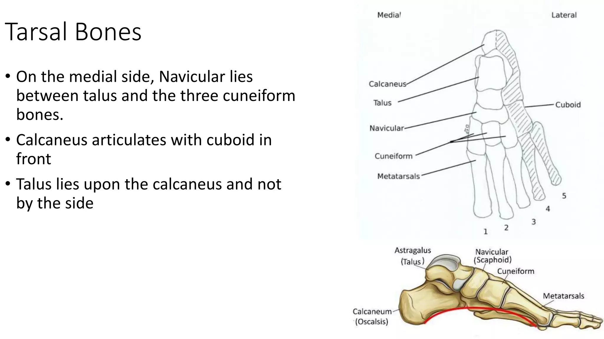
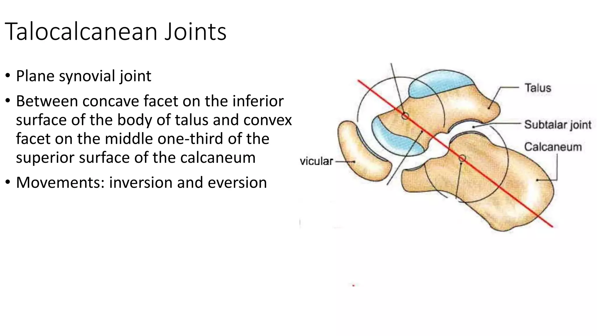
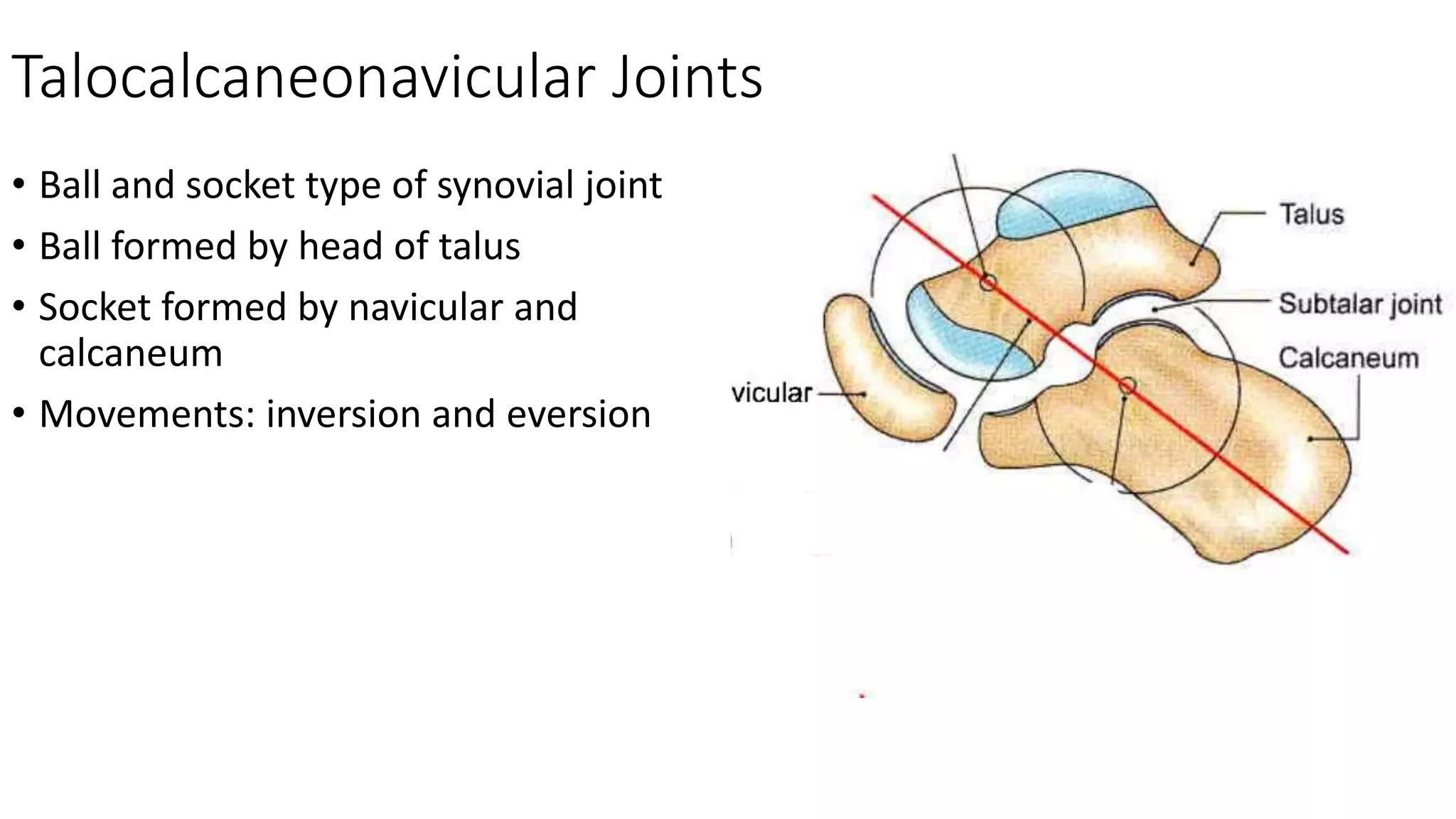
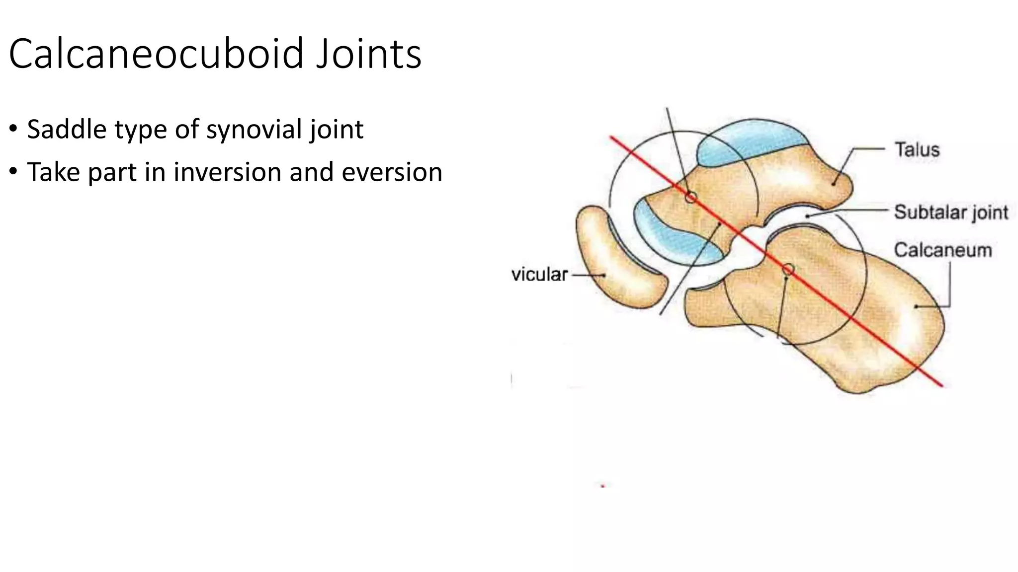
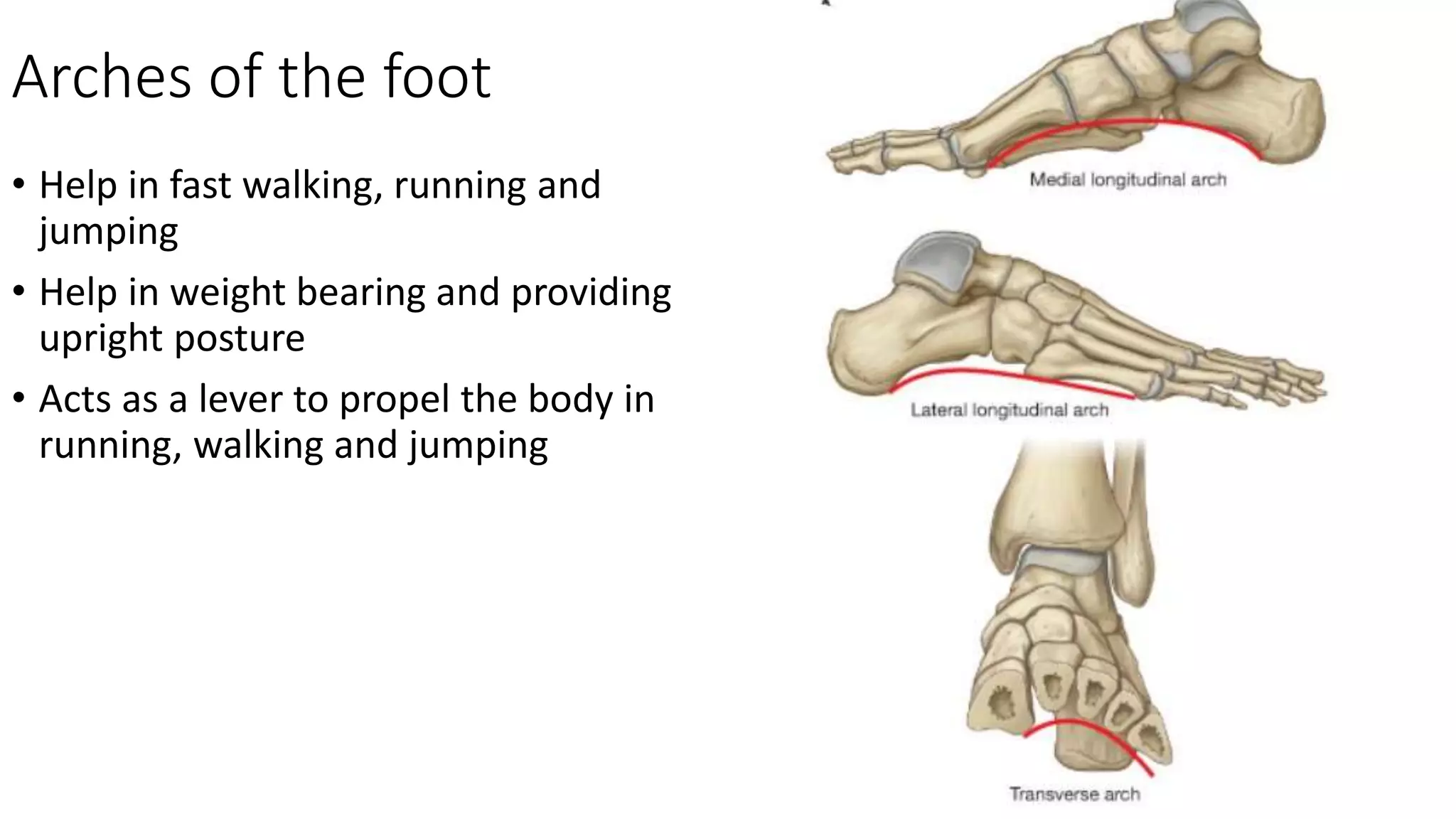
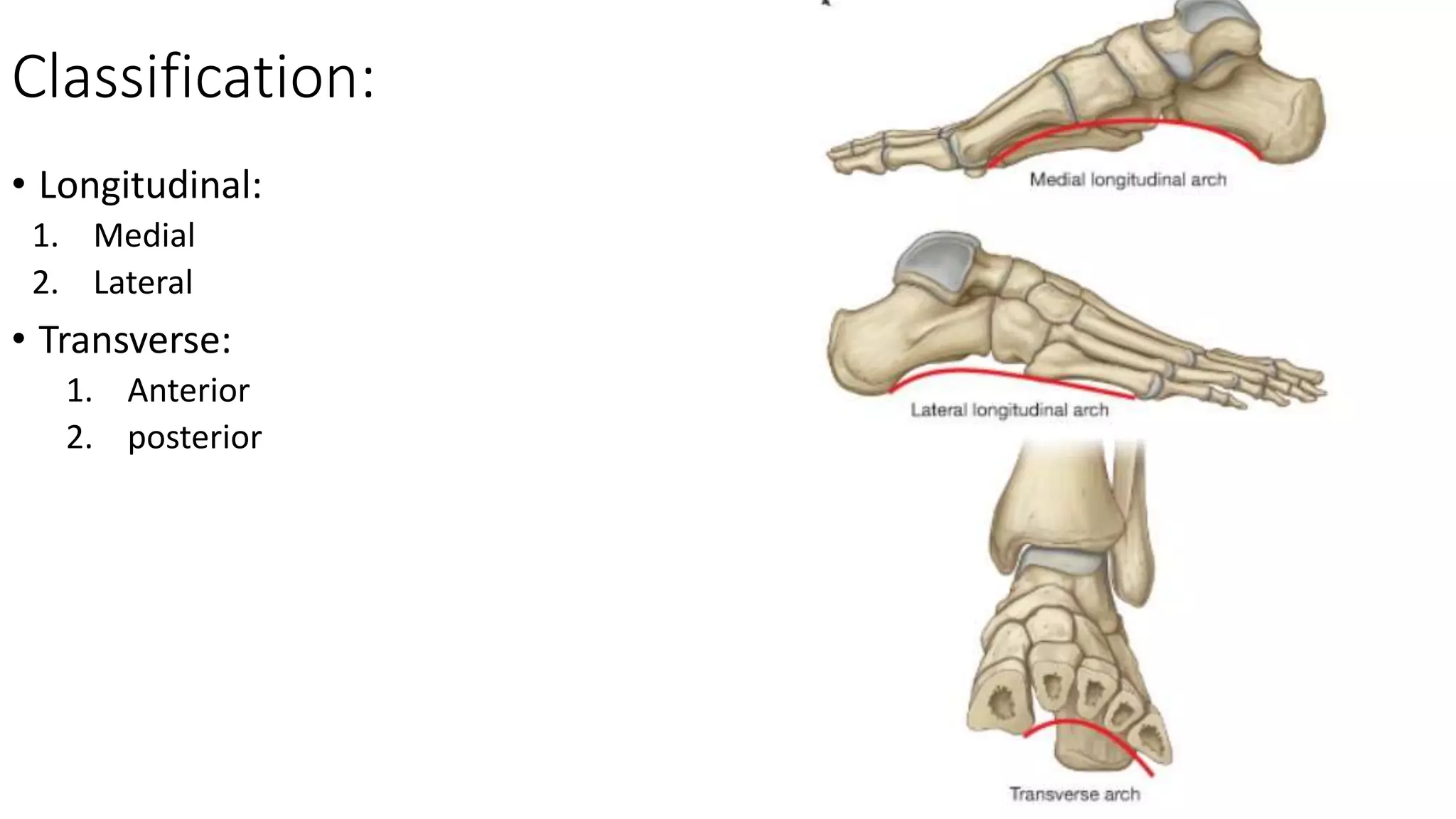
Bones and Joints of Foot
Dr. Mushfiqul Hoque
Lecturer
Dhaka National Medical College
Tarsal Bones
Tarsal Bones
• 7 in number
• Arranged in two rows, proximal and
distal
• On the medial side between the two
rows there is one bone, navicular
• Proximal row bones: Talus and
calcaneus
• Distal row bones: (from medial to
lateral side) medial cuneiform,
intermediate cuneiform, lateral
cuneiform and cuboid
Tarsal Bones
• On the medial side, Navicular lies
between talus and the three cuneiform
bones.
• Calcaneus articulates with cuboid in
front
• Talus lies upon the calcaneus and not
by the side
Metatarsal Bones
• 5 in number
• First metatarsal is the on the medial
side which is the shortest and thickest.
• Second metatarsal is the longest
Joints of the foot
Classification
• Joints of the foot:
1. Intertarsal
2. Tarsometatarsal
3. Intermetatarsal
4. Metatarsophalangeal
5. interphalangeal
Intertarsal Joints
• Subtalar or talocalcanean joint
• Talocalcaneonavicular joint
• Calcanocuboid joint
Talocalcanean Joints
• Plane synovial joint
• Between concave facet on the inferior
surface of the body of talus and convex
facet on the middle one-third of the
superior surface of the calcaneum
• Movements: inversion and eversion
Talocalcaneonavicular Joints
• Ball and socket type of synovial joint
• Ball formed by head of talus
• Socket formed by navicular and
calcaneum
• Movements: inversion and eversion
Calcaneocuboid Joints
• Saddle type of synovial joint
• Take part in inversion and eversion
Inversion and eversion of the foot
• Joints taking part:
1. Subtalar (talocalcanean)
2. Talocancaneonavicular
• Accessory joints:
1. Cancaneocuboid
2. Talonavicular
Smaller joints of the foot:
• Tarsometatarsal, intertarsal,
intermetatarsal joints are plane
type of synovial joints
• Metatarsophalangeal joints are
ellipsoid type of synovial joints
• Interphalangeal joints are hinge
type of synovial joints
Arches of the Foot
Arches of the foot
• Help in fast walking, running and
jumping
• Help in weight bearing and providing
upright posture
• Acts as a lever to propel the body in
running, walking and jumping
Classification:
• Longitudinal:
1. Medial
2. Lateral
• Transverse:
1. Anterior
2. posterior
Medial Longitudinal Arch
• Considerably higher
• More mobile and resilient than lateral
• Ends: anterior end is formed by heads
of first and second metatarsal
• Posterior end forms by medial tubercle
of the calcaneum
• Summit of the arch is superior articular
surface of the body of talus
• Pillars are talus, navicular, 3 cuneiform
and the first 3 metatarsal
• main joint is talocalcaneonavicular
Lateral Longitudinal Arch
• Characteristically low
• Limited mobility
• Built to transmit weight and thrust to
the ground
• Anterior end is formed by the heads of
the 4th and 5th metatarsals
• Posterior end is formed by lateral
tubercle of the calcaneum
• Summit is at the level of articular
facets of the superior surface of
calcaneum
• Pillars are cuboid and 4th and 5th
metatarsal
Anterior Transverse Arch
• Formed by the heads of the 5
metatarsal bones
• Complete because both heads of 1st
and 5th metatarsal come in contact
with the ground
Posterior Transverse Arch
• Formed by greater part of tarsus and
metatarsus
• Incomplete because only lateral end
comes in contact with the ground
• Forms a "half dome"
Factors responsible for maintenance of the arches
• Shape of the bones
• Intersegmental ties holding
the different segments
together
• Tie beams or bowstrings
that connect the two ends
of an arch
• Slings that keep the summit
pulled up
• Suspension
Intersegmental Ties:
• The Spring ligament for medial
longitudinal arch
• The long and short plantar ligaments
for the lateral longitudinal arch
• Interosseous muscles for transverse
arch.
Tie beams:
• Plantar aponeurosis and muscles of the
first layer of foot for longitudinal
arches
• Adductor hellucis for transverse arches
Slings:
• Tendons of posterior compartment
into the sole I.e. tibialis posterior,
flexor hellucis longus, flexor digitorum
longus for medial arch
• Peroneus longus and brevis for lateral
arch
• Tendons of tibialis anterior and
peroneus longus together form a sling
to support both longitudinal arches
• Tendon of peroneus longus for
transverse arches along with tibialis
posterior.

Bones and joint of foot

  • 1.
    Bones and Jointsof Foot Dr. Mushfiqul Hoque Lecturer Dhaka National Medical College
  • 2.
  • 3.
    Tarsal Bones • 7in number • Arranged in two rows, proximal and distal • On the medial side between the two rows there is one bone, navicular • Proximal row bones: Talus and calcaneus • Distal row bones: (from medial to lateral side) medial cuneiform, intermediate cuneiform, lateral cuneiform and cuboid
  • 4.
    Tarsal Bones • Onthe medial side, Navicular lies between talus and the three cuneiform bones. • Calcaneus articulates with cuboid in front • Talus lies upon the calcaneus and not by the side
  • 5.
    Metatarsal Bones • 5in number • First metatarsal is the on the medial side which is the shortest and thickest. • Second metatarsal is the longest
  • 6.
  • 7.
    Classification • Joints ofthe foot: 1. Intertarsal 2. Tarsometatarsal 3. Intermetatarsal 4. Metatarsophalangeal 5. interphalangeal
  • 8.
    Intertarsal Joints • Subtalaror talocalcanean joint • Talocalcaneonavicular joint • Calcanocuboid joint
  • 9.
    Talocalcanean Joints • Planesynovial joint • Between concave facet on the inferior surface of the body of talus and convex facet on the middle one-third of the superior surface of the calcaneum • Movements: inversion and eversion
  • 10.
    Talocalcaneonavicular Joints • Balland socket type of synovial joint • Ball formed by head of talus • Socket formed by navicular and calcaneum • Movements: inversion and eversion
  • 11.
    Calcaneocuboid Joints • Saddletype of synovial joint • Take part in inversion and eversion
  • 12.
    Inversion and eversionof the foot • Joints taking part: 1. Subtalar (talocalcanean) 2. Talocancaneonavicular • Accessory joints: 1. Cancaneocuboid 2. Talonavicular
  • 14.
    Smaller joints ofthe foot: • Tarsometatarsal, intertarsal, intermetatarsal joints are plane type of synovial joints • Metatarsophalangeal joints are ellipsoid type of synovial joints • Interphalangeal joints are hinge type of synovial joints
  • 15.
  • 16.
    Arches of thefoot • Help in fast walking, running and jumping • Help in weight bearing and providing upright posture • Acts as a lever to propel the body in running, walking and jumping
  • 17.
    Classification: • Longitudinal: 1. Medial 2.Lateral • Transverse: 1. Anterior 2. posterior
  • 18.
    Medial Longitudinal Arch •Considerably higher • More mobile and resilient than lateral • Ends: anterior end is formed by heads of first and second metatarsal • Posterior end forms by medial tubercle of the calcaneum • Summit of the arch is superior articular surface of the body of talus • Pillars are talus, navicular, 3 cuneiform and the first 3 metatarsal • main joint is talocalcaneonavicular
  • 19.
    Lateral Longitudinal Arch •Characteristically low • Limited mobility • Built to transmit weight and thrust to the ground • Anterior end is formed by the heads of the 4th and 5th metatarsals • Posterior end is formed by lateral tubercle of the calcaneum • Summit is at the level of articular facets of the superior surface of calcaneum • Pillars are cuboid and 4th and 5th metatarsal
  • 20.
    Anterior Transverse Arch •Formed by the heads of the 5 metatarsal bones • Complete because both heads of 1st and 5th metatarsal come in contact with the ground
  • 21.
    Posterior Transverse Arch •Formed by greater part of tarsus and metatarsus • Incomplete because only lateral end comes in contact with the ground • Forms a "half dome"
  • 22.
    Factors responsible formaintenance of the arches • Shape of the bones • Intersegmental ties holding the different segments together • Tie beams or bowstrings that connect the two ends of an arch • Slings that keep the summit pulled up • Suspension
  • 23.
    Intersegmental Ties: • TheSpring ligament for medial longitudinal arch • The long and short plantar ligaments for the lateral longitudinal arch • Interosseous muscles for transverse arch.
  • 24.
    Tie beams: • Plantaraponeurosis and muscles of the first layer of foot for longitudinal arches • Adductor hellucis for transverse arches
  • 25.
    Slings: • Tendons ofposterior compartment into the sole I.e. tibialis posterior, flexor hellucis longus, flexor digitorum longus for medial arch • Peroneus longus and brevis for lateral arch • Tendons of tibialis anterior and peroneus longus together form a sling to support both longitudinal arches • Tendon of peroneus longus for transverse arches along with tibialis posterior.