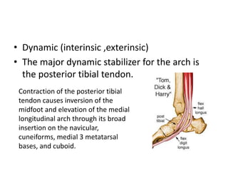
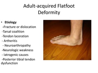
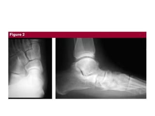
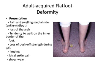
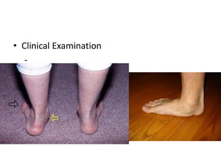
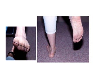
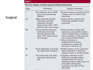
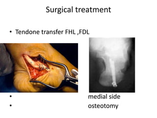
Adult-acquired Flatfoot
Deformity
• adult-acquired flatfoot deformity
encompasses a wide range of deformities.
• These deformities vary in location, severity,
and rate of progression.
• Anatomy
• Two arches held by
tendons & ligaments
• Allow foot to support
weight of the body: ball
of foot – 40% weight.
Heel – 60% weight
• Leverage for walking
• Fully developed by age
13
• Longitudinal arch:
medial and lateral parts
• Transverse arch
Two longitudinal
arches
Medial longitudinal
arch - extends from
calcaneus bone to
talus, navicular, 3
cuneiforms, and
proximal ends of 3
medial metatarsals
Lateral longitudinal
arch - extends from
calcaneus to cuboid
and proximal ends of
4th & 5th metatarsals
Transverse arch
extends across foot
from 1st metatarsal to
the 5th metatarsal
• The 3 most important static contributors to
arch stability:
• plantar fascia, the long and short plantar
ligaments, and the spring ligament
(calcaneonavicular ligament)
• Dynamic (interinsic ,exterinsic)
• The major dynamic stabilizer for the arch is
the posterior tibial tendon.
Contraction of the posterior tibial
tendon causes inversion of the
midfoot and elevation of the medial
longitudinal arch through its broad
insertion on the navicular,
cuneiforms, medial 3 metatarsal
bases, and cuboid.
Adult-acquired Flatfoot
Deformity
• Etiology
-Fracture or dislocation
-Tarsal coalition
-Tendon laceration
- Artheritis
- Neuroarthropathy
-Neurologic weakness
- Iatrogenic causes
-Posterior tibial tendon
dysfunction
Adult-acquired Flatfoot
Deformity
• Originally known as posterior tibial tendon
dysfunction or insufficiency
• first described as tendon failure.
• However, failure of the ligaments that support
the arch also occurs,
Adult-acquired Flatfoot
Deformity
• Etiology
• The etiology of the condition is multifactorial
attributed to degenerative, inflammatory, and
traumatic causes
obesity , preexisting flatfoot
• Pathology
- PT tendon deg.
- tendon failure most common (1-1.5 cm)
distal to MM (hypovascular)
- ligaments failure (spring )
- talonavi. Subluxation  medioplantar
migration talar head  further deformity
- hind foot valgus
• Diagnosis
Patient History
Clinical examination
Standing Radiograph of foot and ankle
Adult-acquired Flatfoot
Deformity
• Presentation
- Pain and swelling medial side
(ankle-midfoot)
- loss of the arch
- Tendency to walk on the inner
border of the
foot.
- Loss of push-off strength during
gait
- limping
- latral ankle pain
- shoes wear.
• Clinical Examination
-
• Radiological:
Stages:
- Described by
Johnson and
Strom, 4 stages
Stage 3
Treatment
• Nonsurgical
- Recommended first because it may be helpful in
alleviating symptoms.
- NSAD
- Removable boot or cast is most often helpful.
- Support with customized brace (articulated
ankle-foot orthosis)
- Foot orthosis e (medial arch+ medial heel wedg)
- Physiotherapy
• Nonsurgical
• No study has been done to document whether
these devices slow or prevent the progression
of deformity. ( Jonathan T. AAOS 2008 ,)
Treatment
• Surgical
- Failed nonsurgical Rx in alleviate symptoms.
- Increasing deformity.
-When the deformities become more severe
and fixed, the results of treatment are more
limited.
- Controversies persist regarding how to treat
• Surgical
Surgical
Surgical treatment
• Tendone transfer FHL ,FDL
• medial side
• osteotomy
Surgical treatment
• In all stages, there are benefits to achieve
proper alignment and maintaining as much
flexibility as possible.

Adult flatfoot

  • 1.
  • 2.
    • adult-acquired flatfootdeformity encompasses a wide range of deformities. • These deformities vary in location, severity, and rate of progression.
  • 3.
  • 4.
    • Two archesheld by tendons & ligaments • Allow foot to support weight of the body: ball of foot – 40% weight. Heel – 60% weight • Leverage for walking • Fully developed by age 13 • Longitudinal arch: medial and lateral parts • Transverse arch
  • 5.
    Two longitudinal arches Medial longitudinal arch- extends from calcaneus bone to talus, navicular, 3 cuneiforms, and proximal ends of 3 medial metatarsals Lateral longitudinal arch - extends from calcaneus to cuboid and proximal ends of 4th & 5th metatarsals Transverse arch extends across foot from 1st metatarsal to the 5th metatarsal
  • 6.
    • The 3most important static contributors to arch stability: • plantar fascia, the long and short plantar ligaments, and the spring ligament (calcaneonavicular ligament)
  • 8.
    • Dynamic (interinsic,exterinsic) • The major dynamic stabilizer for the arch is the posterior tibial tendon. Contraction of the posterior tibial tendon causes inversion of the midfoot and elevation of the medial longitudinal arch through its broad insertion on the navicular, cuneiforms, medial 3 metatarsal bases, and cuboid.
  • 9.
    Adult-acquired Flatfoot Deformity • Etiology -Fractureor dislocation -Tarsal coalition -Tendon laceration - Artheritis - Neuroarthropathy -Neurologic weakness - Iatrogenic causes -Posterior tibial tendon dysfunction
  • 10.
    Adult-acquired Flatfoot Deformity • Originallyknown as posterior tibial tendon dysfunction or insufficiency • first described as tendon failure. • However, failure of the ligaments that support the arch also occurs,
  • 11.
    Adult-acquired Flatfoot Deformity • Etiology •The etiology of the condition is multifactorial attributed to degenerative, inflammatory, and traumatic causes obesity , preexisting flatfoot
  • 12.
    • Pathology - PTtendon deg. - tendon failure most common (1-1.5 cm) distal to MM (hypovascular) - ligaments failure (spring ) - talonavi. Subluxation  medioplantar migration talar head  further deformity - hind foot valgus
  • 14.
    • Diagnosis Patient History Clinicalexamination Standing Radiograph of foot and ankle
  • 15.
    Adult-acquired Flatfoot Deformity • Presentation -Pain and swelling medial side (ankle-midfoot) - loss of the arch - Tendency to walk on the inner border of the foot. - Loss of push-off strength during gait - limping - latral ankle pain - shoes wear.
  • 16.
  • 18.
  • 20.
    Stages: - Described by Johnsonand Strom, 4 stages
  • 21.
  • 22.
    Treatment • Nonsurgical - Recommendedfirst because it may be helpful in alleviating symptoms. - NSAD - Removable boot or cast is most often helpful. - Support with customized brace (articulated ankle-foot orthosis) - Foot orthosis e (medial arch+ medial heel wedg) - Physiotherapy
  • 23.
    • Nonsurgical • Nostudy has been done to document whether these devices slow or prevent the progression of deformity. ( Jonathan T. AAOS 2008 ,)
  • 24.
    Treatment • Surgical - Failednonsurgical Rx in alleviate symptoms. - Increasing deformity. -When the deformities become more severe and fixed, the results of treatment are more limited. - Controversies persist regarding how to treat
  • 25.
  • 26.
    Surgical treatment • Tendonetransfer FHL ,FDL • medial side • osteotomy
  • 27.
    Surgical treatment • Inall stages, there are benefits to achieve proper alignment and maintaining as much flexibility as possible.