SlideShare a Scribd company logo
FLAT FOOT- POSTERIOR
TIBIALIS TENDON
DYSFUNCTION- ACCESSORY
NAVICULARIS
RIZQI D ROSANDI
FK UNIVERSITAS BRAWIJAYA
APRIL 2020
Pengampu : dr. Ananto Satyo Pradana,
Sp. OT
ANATOMY OF FOOT
• The foot is able to sustain large weight-bearing
stresses while accommodating to a variety of
surfaces and activities.
• The foot must be stable to provide an adequate
base of support and function as a rigid lever for
pushing-off when walking, running, or jumping.
• The foot must also be mobile to adapt to uneven
terrain, absorb shock as the foot hits the ground,
ARCHES OF THE FOOT
The arches of the foot, formed by the tarsal
and metatarsal bones, strengthened by
ligaments and tendons, allow the foot to
support the weight of the body in the erect
posture with the least weight.
USE OF THE ARCHED FOOT
 Supports body weight in upright posture
 Acts as a lever to propel the body forwards in walking,
running and jumping
 Acts as a shock absorber
 Concavity of the arches protects the soft tissues of the sole
against pressure
Medial longitudinal arch
• Higher than lateral
• Composed of – Calcaneous
- Talus
- Navicular
- 3 cuneiform
- 3 metatarsals
• Talar head is key stone of this arch
• Tibialis anterior attached to – 1st metatarsal,medial cuneiform –
strength for this arch.
• Peroneus longus tendon – pass laterally to this arch providing
support
Lateral longitudinal Arch
• Flatter than medial
longitudinal arch.
• Rests on the ground during
standing.
• It is made up of – calcaneous,
cuboid, 2 lateral metatarsals.
Transverse arch
• Runs from side to side
• It is formed by – cuboid,
cuneiforms, bases of
metatarsals
• Medial and lateral parts
of longitudinal arch act as
pillars
• Tendons of fibularis
longus and tibialis
posterior
BIOMECHANIC OF FOOT
Integrity of bony arches
• Maintained by passive factors and dynamic
supports
Passive factors• Shape of the united bones (bony
congruency)
• Four successive layers of fibrous
tissue – bowstring the longitudinal
arch
– Plantar aponeurosis
– Long plantar ligament
– Plantar calcaneocuboid (short
plantar) ligament
– Plantar calcaneonavicular
(spring) ligament
Dynamic supports
• Active bracing action of intrinsic muscles of foot
• Active and tonic contraction of muscles with long
tendons extending in to foot
– Flexor hallusis and digitorum longus – longitudinal arch
– Fibularis longus and tibialis posterior – transverse arch
• Plantar ligaments and plantar aponeurosis bear
greatest stress and important in maintaining arches
MECHANISM OF ARCH SUPPORT
SHAPE OF BONES
• Bones are wedge-shaped with the thin edge lying inferiorly
• This applies particularly to the bone occupying the center of
the arch“keystone”
MECHANISM OF ARCH SUPPORT
Inferior edges of bone tied together
MECHANISM OF ARCH SUPPORT
Tying the ends of the arch together
MECHANISM OF ARCH SUPPORT
SUSPENDING THE ARCH FROM ABOVE
• Medial longitudinal arch: Tibialis anterior, Tibialis
posterior, medial ligament of ankle joint
• Lateral longitudinal arch: Peroneus longus, Peroneus
brevis
• Transverse arch: Peroneus longus
MECHANISM OF ARCH SUPPORT
SUSPENDING THE ARCH FROM ABOVE
SO…
FLAT FOOT
‘OUR FEET ARE NO MORE ALIKE THAN OUR FACES.’
Synonyms
• Pes planovalgus
• Fallen arches
• Pronation of feet
Definition
• Absence of normal medial longitudinal arch
• Instep of the foot collapses and comes in
contact with the ground.
• In some individuals, this arch never develops
• Flat feet are a common condition.
• In infants and toddlers, the longitudinal arch is not developed
and flat feet are normal.
• The arch develops in childhood
• By adulthood (12-13yrs), most people have developed
normal arches
Types
 Flexible
 Rigid
Can be
painless
Painful
Types
• Flexible –on weight bearing it disappears and
on non weight bearing it reappears
• Rigid – acceptable medial longitudinal arch
does not seen even on non weight bearing
• Flexible, painless is most common
disappears Appears
Etiology
Flexible
Developmental – the most common
Hypermobile (ligamentous hyperlaxity; Ehlers-Donlos, Marfans)
Neurogenic (rare and usually cause the reverse-Pes Cavus)
Rigid
 Congenital (Tarsal coalition,Vertical talus)
Aquired (inflammatory)
SYMPTOMS
• Deformity
• Foot pain ,ankle pain, leg pain
• Heel tilts away from the midline of the body more than
usual
• Abnormal shoe wear
FLAT FEET CAN produce
• Tendonitis. posterior tibial tendon and it can either fail,
rupture, stretch or just hurt. This condition is called
POSTERIOR TIBIAL DYSFUNCTION (PTD OR TPD) .
• Arthritis.
• Plantar fasciitis
• Bunions & Hammertoes
• Corns and callosities
Radiography
• Asymptomatic flatfoot radiological evaluation unnecessary
• First Anteroposterior and lateral views of the foot should
be taken to evaluate severity of deformity
• Antero-posterior ankle to rule out valgus at the distal end of
tibia
• Special view - 45 degree eversion oblique for accessory
navicular bone
Radiography
• AP standing view is to asses heel valgus
, talocalcaneal (Kite’s) angle more than
35 degree is associated with incresed
heel valgus
• CT scan accurately defines anatomy
of subtalar joint , allows surgical
planning if it is involved.
Meary’s Angle
• Most common angle to indicate
flat foot
• Intersects at apex of the
deformity
• Meary’s angle - between long
axis of talus and long axis of
first metatarsal on a standing
lateral X ray
 Normal Meary's angle:long axis
of the talus should bisect the
navicular and first metatarsal
0 degrees – normal
0 – 15 degrees – mild
15 – 40 degrees – moderate
> 40 degrees – severe
The long axis of the talus is angled plantarward in relation to
the first metatarsal, consistent with pes planus
Other radiologic signs
Calcaneal Pitch
CYMA Line
Pedobarography
 A record of pressure can be obtained
by making the patient to stand and
walk on a force plate.
 Mainly used to compare pre & post
operative function
Treatment
0-3 years old:
 No treatment unless very
strong family hx of
persistent flatfeet
 Orthotic shoes with thomas
heels , medial heel wedges
and navicular pads
 Convince the parents.
3-9 years
• Conservative management
• No surgery
• Custom orthosis inserted
with leather ,cork, propylene
Exercise
 Toe-walking and multiple toe-ups
 If tendo-achilles is contracted, stretching it actively and passively is an
important form of management
 Grasping marbles with toes Heel to toe walking
 Playing in sand
 Ballet dancing
 Walking on a supination board
10-14 yrs
• No symptom- No treatment
• Symptomatic – conservative management
initially
• Surgical
Surgical treatment
Indications
1. Pain
2. Failure to respond to orthotic control
3. Ulceration or callus under the head of the plantiflexed
talus
4. Excessive shoe wear
Surgical treatment
• The surgeon , patient, and parents must be willing to
exchange loss of eversion and inversion of the foot
for relief of pain and disability .
Surgical treatment
• Arthrodesis for relieving painful flat foot have been
most successful when the subtalar joint is involved .
• Although midtarsal arthtrodesis without inclusion of
the subtalar joint has gained popularity
Surgeries
• Durham flatfoot plasty
• Posterior calcaneal displacement osteotomy
• Anterior calcaneal lengthening – distraction
wedge osteotomy
• Triple atrhrodesis (triplane)
Durham Plasty for Pes Planus
A, Incision.
B, Elevation of posterior tibial
tendon.
C, Elevation of osteo-periosteal
flap from proximal to distal.
D, Arthrodesis of navicular–first
cuneiform joint.
E, Extent of arthrodesis resection
through midfoot.
F, Internal fixation of navicular–
first cuneiform joint.
•
pull the posterior tibial tendon taut
into its prepared bed on the plantar
surface of the waist of the
navicular, and tie the suture
dorsally
• Lengthening of lateral
column of the foot by
inserting a tibial bone graft
and calcaneocuboidal
fusion
Calcaneal osteotomy (Dilwyn- Evana, Mosca)
• Symptomatic patients with excessive heel valgus , a calcaneal osteotomy
is intended to displace the posterior part of the calcaneum medially , to
restore normal weight bearing alignment
Posterior calcaneal displacement osteotomy (koutsgiannis)
Triple Arthrodesis
Joints fused are:
• Subtalar joint
• Calcaneo cuboid joint
• Talo navicular joint
AGE
• Usually done after the age of 12
• Triple arthrodesis tend to have a high (50%) failure rate in
children under 10 years of age;
• contra-indicated in young children (less than 10-12 yrs)
because the procedure limits foot growth
Complications
• Nonunion
• Degenerative joint disease
• Avascular necrosis
• Lateral instability
• Stiff foot
POSTERIOR TIBIALIS TENDON
DYSFUNCTION
THE MOST COMMON CAUSE OF ADULT-ACQUIRED FLATFOOT DEFORMITY
Epidemiology
Demographics
 > women
 > present in the 6th decade
Risk Factor
 obesity
 hypertension
 diabetes
 increased age
 corticosteroid use
 seronegative inflammatory disorders
Mechanism
 Exact etiology is unknown
 Acute injury vs long standing tendon degeneration
Pathoanatomy
Foot deformity
 pes planus
 hindfoot valgus
 forefoot varus
 forefoot abduction
Early Disease Late Disease
• Early tenosynovitis  PPTD
• leads to loss of medial longitudinal
arch dynamic stabilization
• PTTD  attritional failure of static
hindfoot stabilizers & collapse of the
medial longitudinal arch
• Fixed degenerative joint changes occur
at late stages
Associated condition
 Inflammatory arthropathy
 tarsal coalition
Classification of Tibialis Posterior
Tendon Dysfunction
Clinical Presentation
Symptoms
Physical Exam
• Medial ankle/ foot pain &
weakness
• Progressive loss of arch
• Lateral ankle pain
Inspection & Palpation:
• Pes planus
• Hindfoot valgus deformity
• Forefoot abduction
• Tenderness in tip of medial
Range of Motion:
• Single-limb heel rise
• PTT power
• Deformity – flexible or fixed
Imaging
 Radiographs :
 Ankle AP/ Lateral
 Ankle Mortise
 MRI :
 Tendon degeneration and arthritic changes in the talonavicular, subtalar,
and tibiotalar joints
 Ultrasound:
• increasing role in the evaluation of pathology within the PTT
Differential Diagnostic
 Pes planus, secondary to – midfoor pathology or incompetence of the
spring ligament
Treatment
 Nonoperative :
 Ankle foot orthosis
 Immobilization in walking cast (3-4 months)
 Custom molded in shoe orthosis
 Operative :
 Tenosynevectomy
 FDL transfer, calcaneal osteotomy, TAL, +/- forefoot correction osteotomy
+/- spring ligament repair +/- lateral column lengthening +/- medial
column arthrodesis +/- PTT debridement
Gatzoulis, 2014: Tibialis posterior dysfunction: a common & treatable cause
of adult acquired flatfoot. BMJ Journal
Management of Tibialis Posterior
Tendon Dysfunction
Abousayed et al.,
2017: JBJS Journal
Treatment
 First TMT joint arthrodesis, calcaneal osteotomy, TAL +/- lateral column
lengthening +/- PTT debridement
 Isolated subtalar arthrodesis
 Hindfoot arthrodesis
 Triple arthrodesis & TAL+ deltoid ligament reconstruction
 Tibiotalocalcaneal arthrodesis
ACCESORY NAVICULAR
FIRST DESCRIBED BY BAUHIN IN 1605
OTHER NAMES : PREHALLUX, ACCESSORY SCAPHOID, NAVICULAR SECUNDUM
Accessory navicular bone
• It is a most common accessory bone in the foot
• Listed as a cause of flat foot
Pathoanatomy
• Abnormal insertion of Tibialis Posterior into
accessory navicular bone believe to cause the flat
foot
Clinical presentation
• Often incidental, many patients are asymptomatic
• Pain
• Prominence of medial aspect of foot
• On attempted inversion of the foot against resistance
, Tibialis posterior tendon is inserted into the bump
and this maneuver produces pain
Radiography
• Special view - 45 degree eversion oblique for
accessory navicular bone
• Antero-Posterior view and Lateral weight bearing
views of the foot should be taken to evaluate other
deformities
Radiological types
• TypeI– Small ossicle in the substance of Tibialis
Posterior tendon (os tibiale externum or naviculam
secondorium )
• Type II –Triangular frangment larger than type I connected
to navicular bone by a cartilaginous synchondrosis
• Type III – Cornuate navicular resulting from fusion of the
accessory navicular with main body of navicular
Treatment
INITIAL TREATMENT:
Conservative- stretcing shoes, avoiding activity
that irritates foot
SURGICAL:
Kidners procedure
Kidners procedure• Excision of accessory
navicular bone and rerouting
of tibialis Posterior tendon
into a more plantar position
(navicular)
• Parents should be informed
before surgery that pain
may not be alleviated
completely
THANK YOU
MALANG, 16 APRIL 2020

More Related Content

What's hot

Ortho Journal Club 11 by Dr Saumya Agarwal
Ortho Journal Club 11 by Dr Saumya AgarwalOrtho Journal Club 11 by Dr Saumya Agarwal
Uncemented femoral stem
Uncemented  femoral stemUncemented  femoral stem
Uncemented femoral stem
Sameer Ashar
 
Acl graft fixation options
Acl graft fixation optionsAcl graft fixation options
Acl graft fixation options
orthoprinciples
 
talus fracture
talus fracturetalus fracture
talus fracture
Harsimran Sidhu
 
Templating of total hip replacement (THR)
Templating of total hip replacement (THR)Templating of total hip replacement (THR)
Templating of total hip replacement (THR)
Govt service, Osmania Medical College, Hyderabad.
 
Poller screw
Poller screwPoller screw
Poller screw
drsiddharthdubey
 
Cavus foot
Cavus footCavus foot
Cavus foot
Dr. Anurag Mittal
 
Posterolateral corner injuries of knee joint
Posterolateral corner injuries of knee joint Posterolateral corner injuries of knee joint
Posterolateral corner injuries of knee joint
Samir Dwidmuthe
 
Recurrent patellar dislocation
Recurrent patellar dislocationRecurrent patellar dislocation
Recurrent patellar dislocation
boneheallerortho
 
Posterior malleolus fracture
Posterior malleolus fracturePosterior malleolus fracture
Posterior malleolus fracture
AnuragSai7
 
Ankle & Foot Xray & Surgical Approaches
Ankle & Foot Xray & Surgical ApproachesAnkle & Foot Xray & Surgical Approaches
Ankle & Foot Xray & Surgical Approaches
Mirant Dave
 
Calcaneus fracture
Calcaneus fractureCalcaneus fracture
Calcaneus fracture
akhilmk123
 
Terrible triad - elbow
Terrible triad - elbow Terrible triad - elbow
Terrible triad - elbow jatinder12345
 
Medial Opening Wedge High Tibial Osteotomy
Medial Opening Wedge High Tibial Osteotomy Medial Opening Wedge High Tibial Osteotomy
Medial Opening Wedge High Tibial Osteotomy
washingtonortho
 
Protrusio acetabuli
Protrusio acetabuliProtrusio acetabuli
Protrusio acetabuli
Ghazwan Bayaty
 
Recurrent Dislocation of patella
Recurrent Dislocation of patellaRecurrent Dislocation of patella
Recurrent Dislocation of patella
sabir khadka
 
Ankle fractures
Ankle fracturesAnkle fractures
Ankle fractures
Dr.Anshu Sharma
 
osteotomies around hip
osteotomies around hiposteotomies around hip
osteotomies around hip
Prashanth Kumar
 
Surgical Approaches to Hip Joint
Surgical Approaches to Hip JointSurgical Approaches to Hip Joint
Surgical Approaches to Hip Joint
Apoorv Jain
 
Limb length discrepancy
Limb length discrepancyLimb length discrepancy
Limb length discrepancy
sudarshan731
 

What's hot (20)

Ortho Journal Club 11 by Dr Saumya Agarwal
Ortho Journal Club 11 by Dr Saumya AgarwalOrtho Journal Club 11 by Dr Saumya Agarwal
Ortho Journal Club 11 by Dr Saumya Agarwal
 
Uncemented femoral stem
Uncemented  femoral stemUncemented  femoral stem
Uncemented femoral stem
 
Acl graft fixation options
Acl graft fixation optionsAcl graft fixation options
Acl graft fixation options
 
talus fracture
talus fracturetalus fracture
talus fracture
 
Templating of total hip replacement (THR)
Templating of total hip replacement (THR)Templating of total hip replacement (THR)
Templating of total hip replacement (THR)
 
Poller screw
Poller screwPoller screw
Poller screw
 
Cavus foot
Cavus footCavus foot
Cavus foot
 
Posterolateral corner injuries of knee joint
Posterolateral corner injuries of knee joint Posterolateral corner injuries of knee joint
Posterolateral corner injuries of knee joint
 
Recurrent patellar dislocation
Recurrent patellar dislocationRecurrent patellar dislocation
Recurrent patellar dislocation
 
Posterior malleolus fracture
Posterior malleolus fracturePosterior malleolus fracture
Posterior malleolus fracture
 
Ankle & Foot Xray & Surgical Approaches
Ankle & Foot Xray & Surgical ApproachesAnkle & Foot Xray & Surgical Approaches
Ankle & Foot Xray & Surgical Approaches
 
Calcaneus fracture
Calcaneus fractureCalcaneus fracture
Calcaneus fracture
 
Terrible triad - elbow
Terrible triad - elbow Terrible triad - elbow
Terrible triad - elbow
 
Medial Opening Wedge High Tibial Osteotomy
Medial Opening Wedge High Tibial Osteotomy Medial Opening Wedge High Tibial Osteotomy
Medial Opening Wedge High Tibial Osteotomy
 
Protrusio acetabuli
Protrusio acetabuliProtrusio acetabuli
Protrusio acetabuli
 
Recurrent Dislocation of patella
Recurrent Dislocation of patellaRecurrent Dislocation of patella
Recurrent Dislocation of patella
 
Ankle fractures
Ankle fracturesAnkle fractures
Ankle fractures
 
osteotomies around hip
osteotomies around hiposteotomies around hip
osteotomies around hip
 
Surgical Approaches to Hip Joint
Surgical Approaches to Hip JointSurgical Approaches to Hip Joint
Surgical Approaches to Hip Joint
 
Limb length discrepancy
Limb length discrepancyLimb length discrepancy
Limb length discrepancy
 

Similar to Flat Foot, Tibialis Posterior Tendon Dysfunction & Accessory Navicularis

ARCHES OF FOOT.pptx
ARCHES OF FOOT.pptxARCHES OF FOOT.pptx
ARCHES OF FOOT.pptx
VasanthakohilaMuthuk
 
flat foot.ppt [pes planus ] #physio.# rehabilitation
flat foot.ppt [pes planus ] #physio.# rehabilitationflat foot.ppt [pes planus ] #physio.# rehabilitation
flat foot.ppt [pes planus ] #physio.# rehabilitation
Priyankaranawat4
 
Foot arch deformities 2
Foot arch deformities 2Foot arch deformities 2
Foot arch deformities 2
Jamia Millia Islamia
 
Flat foot and Cavus foot
 Flat foot and Cavus foot Flat foot and Cavus foot
Flat foot and Cavus foot
Dr Thouseef Abdul Majeed
 
Heel Pain and Plantar Fasciitis
Heel Pain and Plantar FasciitisHeel Pain and Plantar Fasciitis
Heel Pain and Plantar Fasciitis
Kevin Ambadan
 
Flat foot By Dr.Mahbub
Flat foot By Dr.MahbubFlat foot By Dr.Mahbub
Flat foot By Dr.Mahbub
dr_mhb21
 
Adult Flatfoot.ppt
Adult Flatfoot.pptAdult Flatfoot.ppt
Adult Flatfoot.ppt
Ahmed Ashour dr.
 
Adult flatfoot
Adult flatfootAdult flatfoot
Adult flatfoot
Amr Mansour Hassan
 
Foot Drop
Foot DropFoot Drop
Foot Drop
Kevin Ambadan
 
Ctev
CtevCtev
Foot pain problems
Foot pain problems Foot pain problems
Foot pain problems
mohamed aouini
 
CONGENITAL TALIPES EQUINO VARUS (CTEV)
CONGENITAL TALIPES EQUINO VARUS (CTEV)CONGENITAL TALIPES EQUINO VARUS (CTEV)
CONGENITAL TALIPES EQUINO VARUS (CTEV)
Ashish kumar Sharma
 
Arches of the foot
Arches of the footArches of the foot
Arches of the foot
peebujaanu
 
امراض القدم عند الاطفال Pediatric foot 1, البروفيسور فريح ابوحسان - استشاري ...
امراض القدم عند الاطفال Pediatric foot  1, البروفيسور فريح ابوحسان - استشاري ...امراض القدم عند الاطفال Pediatric foot  1, البروفيسور فريح ابوحسان - استشاري ...
امراض القدم عند الاطفال Pediatric foot 1, البروفيسور فريح ابوحسان - استشاري ...
Prof Freih Abu Hassan البروفيسور فريح ابوحسان
 
ankleassessment-180926221008.pdf
ankleassessment-180926221008.pdfankleassessment-180926221008.pdf
ankleassessment-180926221008.pdf
a7med7amdy2
 
ankleassessment-180926221008 (1).pdf
ankleassessment-180926221008 (1).pdfankleassessment-180926221008 (1).pdf
ankleassessment-180926221008 (1).pdf
a7med7amdy2
 
Examination evaluation & Assessment of Ankle & Foot
Examination evaluation & Assessment of Ankle & Foot Examination evaluation & Assessment of Ankle & Foot
Examination evaluation & Assessment of Ankle & Foot
Lady Reading Hospital-Medical Teaching Institution, Peshawar
 
Orthotics aruncb
Orthotics  aruncbOrthotics  aruncb
Orthotics aruncb
Arun Cb
 
An approach to limping child
An approach to limping childAn approach to limping child
An approach to limping child
manoj das
 

Similar to Flat Foot, Tibialis Posterior Tendon Dysfunction & Accessory Navicularis (20)

ARCHES OF FOOT.pptx
ARCHES OF FOOT.pptxARCHES OF FOOT.pptx
ARCHES OF FOOT.pptx
 
flat foot.ppt [pes planus ] #physio.# rehabilitation
flat foot.ppt [pes planus ] #physio.# rehabilitationflat foot.ppt [pes planus ] #physio.# rehabilitation
flat foot.ppt [pes planus ] #physio.# rehabilitation
 
Foot arch deformities 2
Foot arch deformities 2Foot arch deformities 2
Foot arch deformities 2
 
Flat foot
Flat footFlat foot
Flat foot
 
Flat foot and Cavus foot
 Flat foot and Cavus foot Flat foot and Cavus foot
Flat foot and Cavus foot
 
Heel Pain and Plantar Fasciitis
Heel Pain and Plantar FasciitisHeel Pain and Plantar Fasciitis
Heel Pain and Plantar Fasciitis
 
Flat foot By Dr.Mahbub
Flat foot By Dr.MahbubFlat foot By Dr.Mahbub
Flat foot By Dr.Mahbub
 
Adult Flatfoot.ppt
Adult Flatfoot.pptAdult Flatfoot.ppt
Adult Flatfoot.ppt
 
Adult flatfoot
Adult flatfootAdult flatfoot
Adult flatfoot
 
Foot Drop
Foot DropFoot Drop
Foot Drop
 
Ctev
CtevCtev
Ctev
 
Foot pain problems
Foot pain problems Foot pain problems
Foot pain problems
 
CONGENITAL TALIPES EQUINO VARUS (CTEV)
CONGENITAL TALIPES EQUINO VARUS (CTEV)CONGENITAL TALIPES EQUINO VARUS (CTEV)
CONGENITAL TALIPES EQUINO VARUS (CTEV)
 
Arches of the foot
Arches of the footArches of the foot
Arches of the foot
 
امراض القدم عند الاطفال Pediatric foot 1, البروفيسور فريح ابوحسان - استشاري ...
امراض القدم عند الاطفال Pediatric foot  1, البروفيسور فريح ابوحسان - استشاري ...امراض القدم عند الاطفال Pediatric foot  1, البروفيسور فريح ابوحسان - استشاري ...
امراض القدم عند الاطفال Pediatric foot 1, البروفيسور فريح ابوحسان - استشاري ...
 
ankleassessment-180926221008.pdf
ankleassessment-180926221008.pdfankleassessment-180926221008.pdf
ankleassessment-180926221008.pdf
 
ankleassessment-180926221008 (1).pdf
ankleassessment-180926221008 (1).pdfankleassessment-180926221008 (1).pdf
ankleassessment-180926221008 (1).pdf
 
Examination evaluation & Assessment of Ankle & Foot
Examination evaluation & Assessment of Ankle & Foot Examination evaluation & Assessment of Ankle & Foot
Examination evaluation & Assessment of Ankle & Foot
 
Orthotics aruncb
Orthotics  aruncbOrthotics  aruncb
Orthotics aruncb
 
An approach to limping child
An approach to limping childAn approach to limping child
An approach to limping child
 

More from Rizqi D Rosandi MD

Spine_ Management of Si Joint Dysfunction
Spine_ Management of Si Joint DysfunctionSpine_ Management of Si Joint Dysfunction
Spine_ Management of Si Joint Dysfunction
Rizqi D Rosandi MD
 
Lower Patellofemoral Instability
Lower Patellofemoral InstabilityLower Patellofemoral Instability
Lower Patellofemoral Instability
Rizqi D Rosandi MD
 
Lower avn of talus
Lower avn of talusLower avn of talus
Lower avn of talus
Rizqi D Rosandi MD
 
Lower avn hip
Lower avn hipLower avn hip
Lower avn hip
Rizqi D Rosandi MD
 
Management of knee cartilage defect & meniscus tear
Management of knee cartilage defect & meniscus tearManagement of knee cartilage defect & meniscus tear
Management of knee cartilage defect & meniscus tear
Rizqi D Rosandi MD
 
Update of management polytrauma patient
Update of management polytrauma patientUpdate of management polytrauma patient
Update of management polytrauma patient
Rizqi D Rosandi MD
 
Management of tibial plateau fracture
Management of tibial plateau fractureManagement of tibial plateau fracture
Management of tibial plateau fracture
Rizqi D Rosandi MD
 

More from Rizqi D Rosandi MD (7)

Spine_ Management of Si Joint Dysfunction
Spine_ Management of Si Joint DysfunctionSpine_ Management of Si Joint Dysfunction
Spine_ Management of Si Joint Dysfunction
 
Lower Patellofemoral Instability
Lower Patellofemoral InstabilityLower Patellofemoral Instability
Lower Patellofemoral Instability
 
Lower avn of talus
Lower avn of talusLower avn of talus
Lower avn of talus
 
Lower avn hip
Lower avn hipLower avn hip
Lower avn hip
 
Management of knee cartilage defect & meniscus tear
Management of knee cartilage defect & meniscus tearManagement of knee cartilage defect & meniscus tear
Management of knee cartilage defect & meniscus tear
 
Update of management polytrauma patient
Update of management polytrauma patientUpdate of management polytrauma patient
Update of management polytrauma patient
 
Management of tibial plateau fracture
Management of tibial plateau fractureManagement of tibial plateau fracture
Management of tibial plateau fracture
 

Recently uploaded

Report Back from SGO 2024: What’s the Latest in Cervical Cancer?
Report Back from SGO 2024: What’s the Latest in Cervical Cancer?Report Back from SGO 2024: What’s the Latest in Cervical Cancer?
Report Back from SGO 2024: What’s the Latest in Cervical Cancer?
bkling
 
ANATOMY AND PHYSIOLOGY OF URINARY SYSTEM.pptx
ANATOMY AND PHYSIOLOGY OF URINARY SYSTEM.pptxANATOMY AND PHYSIOLOGY OF URINARY SYSTEM.pptx
ANATOMY AND PHYSIOLOGY OF URINARY SYSTEM.pptx
Swetaba Besh
 
Cervical & Brachial Plexus By Dr. RIG.pptx
Cervical & Brachial Plexus By Dr. RIG.pptxCervical & Brachial Plexus By Dr. RIG.pptx
Cervical & Brachial Plexus By Dr. RIG.pptx
Dr. Rabia Inam Gandapore
 
Pulmonary Thromboembolism - etilogy, types, medical- Surgical and nursing man...
Pulmonary Thromboembolism - etilogy, types, medical- Surgical and nursing man...Pulmonary Thromboembolism - etilogy, types, medical- Surgical and nursing man...
Pulmonary Thromboembolism - etilogy, types, medical- Surgical and nursing man...
VarunMahajani
 
Alcohol_Dr. Jeenal Mistry MD Pharmacology.pdf
Alcohol_Dr. Jeenal Mistry MD Pharmacology.pdfAlcohol_Dr. Jeenal Mistry MD Pharmacology.pdf
Alcohol_Dr. Jeenal Mistry MD Pharmacology.pdf
Dr Jeenal Mistry
 
Are There Any Natural Remedies To Treat Syphilis.pdf
Are There Any Natural Remedies To Treat Syphilis.pdfAre There Any Natural Remedies To Treat Syphilis.pdf
Are There Any Natural Remedies To Treat Syphilis.pdf
Little Cross Family Clinic
 
For Better Surat #ℂall #Girl Service ❤85270-49040❤ Surat #ℂall #Girls
For Better Surat #ℂall #Girl Service ❤85270-49040❤ Surat #ℂall #GirlsFor Better Surat #ℂall #Girl Service ❤85270-49040❤ Surat #ℂall #Girls
For Better Surat #ℂall #Girl Service ❤85270-49040❤ Surat #ℂall #Girls
Savita Shen $i11
 
New Directions in Targeted Therapeutic Approaches for Older Adults With Mantl...
New Directions in Targeted Therapeutic Approaches for Older Adults With Mantl...New Directions in Targeted Therapeutic Approaches for Older Adults With Mantl...
New Directions in Targeted Therapeutic Approaches for Older Adults With Mantl...
i3 Health
 
POST OPERATIVE OLIGURIA and its management
POST OPERATIVE OLIGURIA and its managementPOST OPERATIVE OLIGURIA and its management
POST OPERATIVE OLIGURIA and its management
touseefaziz1
 
24 Upakrama.pptx class ppt useful in all
24 Upakrama.pptx class ppt useful in all24 Upakrama.pptx class ppt useful in all
24 Upakrama.pptx class ppt useful in all
DrSathishMS1
 
Physiology of Chemical Sensation of smell.pdf
Physiology of Chemical Sensation of smell.pdfPhysiology of Chemical Sensation of smell.pdf
Physiology of Chemical Sensation of smell.pdf
MedicoseAcademics
 
Ophthalmology Clinical Tests for OSCE exam
Ophthalmology Clinical Tests for OSCE examOphthalmology Clinical Tests for OSCE exam
Ophthalmology Clinical Tests for OSCE exam
KafrELShiekh University
 
MANAGEMENT OF ATRIOVENTRICULAR CONDUCTION BLOCK.pdf
MANAGEMENT OF ATRIOVENTRICULAR CONDUCTION BLOCK.pdfMANAGEMENT OF ATRIOVENTRICULAR CONDUCTION BLOCK.pdf
MANAGEMENT OF ATRIOVENTRICULAR CONDUCTION BLOCK.pdf
Jim Jacob Roy
 
Triangles of Neck and Clinical Correlation by Dr. RIG.pptx
Triangles of Neck and Clinical Correlation by Dr. RIG.pptxTriangles of Neck and Clinical Correlation by Dr. RIG.pptx
Triangles of Neck and Clinical Correlation by Dr. RIG.pptx
Dr. Rabia Inam Gandapore
 
ARTHROLOGY PPT NCISM SYLLABUS AYURVEDA STUDENTS
ARTHROLOGY PPT NCISM SYLLABUS AYURVEDA STUDENTSARTHROLOGY PPT NCISM SYLLABUS AYURVEDA STUDENTS
ARTHROLOGY PPT NCISM SYLLABUS AYURVEDA STUDENTS
Dr. Vinay Pareek
 
Superficial & Deep Fascia of the NECK.pptx
Superficial & Deep Fascia of the NECK.pptxSuperficial & Deep Fascia of the NECK.pptx
Superficial & Deep Fascia of the NECK.pptx
Dr. Rabia Inam Gandapore
 
The hemodynamic and autonomic determinants of elevated blood pressure in obes...
The hemodynamic and autonomic determinants of elevated blood pressure in obes...The hemodynamic and autonomic determinants of elevated blood pressure in obes...
The hemodynamic and autonomic determinants of elevated blood pressure in obes...
Catherine Liao
 
Charaka Samhita Sutra Sthana 9 Chapter khuddakachatuspadadhyaya
Charaka Samhita Sutra Sthana 9 Chapter khuddakachatuspadadhyayaCharaka Samhita Sutra Sthana 9 Chapter khuddakachatuspadadhyaya
Charaka Samhita Sutra Sthana 9 Chapter khuddakachatuspadadhyaya
Dr KHALID B.M
 
Phone Us ❤85270-49040❤ #ℂall #gIRLS In Surat By Surat @ℂall @Girls Hotel With...
Phone Us ❤85270-49040❤ #ℂall #gIRLS In Surat By Surat @ℂall @Girls Hotel With...Phone Us ❤85270-49040❤ #ℂall #gIRLS In Surat By Surat @ℂall @Girls Hotel With...
Phone Us ❤85270-49040❤ #ℂall #gIRLS In Surat By Surat @ℂall @Girls Hotel With...
Savita Shen $i11
 
Lung Cancer: Artificial Intelligence, Synergetics, Complex System Analysis, S...
Lung Cancer: Artificial Intelligence, Synergetics, Complex System Analysis, S...Lung Cancer: Artificial Intelligence, Synergetics, Complex System Analysis, S...
Lung Cancer: Artificial Intelligence, Synergetics, Complex System Analysis, S...
Oleg Kshivets
 

Recently uploaded (20)

Report Back from SGO 2024: What’s the Latest in Cervical Cancer?
Report Back from SGO 2024: What’s the Latest in Cervical Cancer?Report Back from SGO 2024: What’s the Latest in Cervical Cancer?
Report Back from SGO 2024: What’s the Latest in Cervical Cancer?
 
ANATOMY AND PHYSIOLOGY OF URINARY SYSTEM.pptx
ANATOMY AND PHYSIOLOGY OF URINARY SYSTEM.pptxANATOMY AND PHYSIOLOGY OF URINARY SYSTEM.pptx
ANATOMY AND PHYSIOLOGY OF URINARY SYSTEM.pptx
 
Cervical & Brachial Plexus By Dr. RIG.pptx
Cervical & Brachial Plexus By Dr. RIG.pptxCervical & Brachial Plexus By Dr. RIG.pptx
Cervical & Brachial Plexus By Dr. RIG.pptx
 
Pulmonary Thromboembolism - etilogy, types, medical- Surgical and nursing man...
Pulmonary Thromboembolism - etilogy, types, medical- Surgical and nursing man...Pulmonary Thromboembolism - etilogy, types, medical- Surgical and nursing man...
Pulmonary Thromboembolism - etilogy, types, medical- Surgical and nursing man...
 
Alcohol_Dr. Jeenal Mistry MD Pharmacology.pdf
Alcohol_Dr. Jeenal Mistry MD Pharmacology.pdfAlcohol_Dr. Jeenal Mistry MD Pharmacology.pdf
Alcohol_Dr. Jeenal Mistry MD Pharmacology.pdf
 
Are There Any Natural Remedies To Treat Syphilis.pdf
Are There Any Natural Remedies To Treat Syphilis.pdfAre There Any Natural Remedies To Treat Syphilis.pdf
Are There Any Natural Remedies To Treat Syphilis.pdf
 
For Better Surat #ℂall #Girl Service ❤85270-49040❤ Surat #ℂall #Girls
For Better Surat #ℂall #Girl Service ❤85270-49040❤ Surat #ℂall #GirlsFor Better Surat #ℂall #Girl Service ❤85270-49040❤ Surat #ℂall #Girls
For Better Surat #ℂall #Girl Service ❤85270-49040❤ Surat #ℂall #Girls
 
New Directions in Targeted Therapeutic Approaches for Older Adults With Mantl...
New Directions in Targeted Therapeutic Approaches for Older Adults With Mantl...New Directions in Targeted Therapeutic Approaches for Older Adults With Mantl...
New Directions in Targeted Therapeutic Approaches for Older Adults With Mantl...
 
POST OPERATIVE OLIGURIA and its management
POST OPERATIVE OLIGURIA and its managementPOST OPERATIVE OLIGURIA and its management
POST OPERATIVE OLIGURIA and its management
 
24 Upakrama.pptx class ppt useful in all
24 Upakrama.pptx class ppt useful in all24 Upakrama.pptx class ppt useful in all
24 Upakrama.pptx class ppt useful in all
 
Physiology of Chemical Sensation of smell.pdf
Physiology of Chemical Sensation of smell.pdfPhysiology of Chemical Sensation of smell.pdf
Physiology of Chemical Sensation of smell.pdf
 
Ophthalmology Clinical Tests for OSCE exam
Ophthalmology Clinical Tests for OSCE examOphthalmology Clinical Tests for OSCE exam
Ophthalmology Clinical Tests for OSCE exam
 
MANAGEMENT OF ATRIOVENTRICULAR CONDUCTION BLOCK.pdf
MANAGEMENT OF ATRIOVENTRICULAR CONDUCTION BLOCK.pdfMANAGEMENT OF ATRIOVENTRICULAR CONDUCTION BLOCK.pdf
MANAGEMENT OF ATRIOVENTRICULAR CONDUCTION BLOCK.pdf
 
Triangles of Neck and Clinical Correlation by Dr. RIG.pptx
Triangles of Neck and Clinical Correlation by Dr. RIG.pptxTriangles of Neck and Clinical Correlation by Dr. RIG.pptx
Triangles of Neck and Clinical Correlation by Dr. RIG.pptx
 
ARTHROLOGY PPT NCISM SYLLABUS AYURVEDA STUDENTS
ARTHROLOGY PPT NCISM SYLLABUS AYURVEDA STUDENTSARTHROLOGY PPT NCISM SYLLABUS AYURVEDA STUDENTS
ARTHROLOGY PPT NCISM SYLLABUS AYURVEDA STUDENTS
 
Superficial & Deep Fascia of the NECK.pptx
Superficial & Deep Fascia of the NECK.pptxSuperficial & Deep Fascia of the NECK.pptx
Superficial & Deep Fascia of the NECK.pptx
 
The hemodynamic and autonomic determinants of elevated blood pressure in obes...
The hemodynamic and autonomic determinants of elevated blood pressure in obes...The hemodynamic and autonomic determinants of elevated blood pressure in obes...
The hemodynamic and autonomic determinants of elevated blood pressure in obes...
 
Charaka Samhita Sutra Sthana 9 Chapter khuddakachatuspadadhyaya
Charaka Samhita Sutra Sthana 9 Chapter khuddakachatuspadadhyayaCharaka Samhita Sutra Sthana 9 Chapter khuddakachatuspadadhyaya
Charaka Samhita Sutra Sthana 9 Chapter khuddakachatuspadadhyaya
 
Phone Us ❤85270-49040❤ #ℂall #gIRLS In Surat By Surat @ℂall @Girls Hotel With...
Phone Us ❤85270-49040❤ #ℂall #gIRLS In Surat By Surat @ℂall @Girls Hotel With...Phone Us ❤85270-49040❤ #ℂall #gIRLS In Surat By Surat @ℂall @Girls Hotel With...
Phone Us ❤85270-49040❤ #ℂall #gIRLS In Surat By Surat @ℂall @Girls Hotel With...
 
Lung Cancer: Artificial Intelligence, Synergetics, Complex System Analysis, S...
Lung Cancer: Artificial Intelligence, Synergetics, Complex System Analysis, S...Lung Cancer: Artificial Intelligence, Synergetics, Complex System Analysis, S...
Lung Cancer: Artificial Intelligence, Synergetics, Complex System Analysis, S...
 

Flat Foot, Tibialis Posterior Tendon Dysfunction & Accessory Navicularis

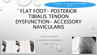
  • 1. FLAT FOOT- POSTERIOR TIBIALIS TENDON DYSFUNCTION- ACCESSORY NAVICULARIS RIZQI D ROSANDI FK UNIVERSITAS BRAWIJAYA APRIL 2020 Pengampu : dr. Ananto Satyo Pradana, Sp. OT
  • 3. • The foot is able to sustain large weight-bearing stresses while accommodating to a variety of surfaces and activities. • The foot must be stable to provide an adequate base of support and function as a rigid lever for pushing-off when walking, running, or jumping. • The foot must also be mobile to adapt to uneven terrain, absorb shock as the foot hits the ground,
  • 4.
  • 5.
  • 6. ARCHES OF THE FOOT The arches of the foot, formed by the tarsal and metatarsal bones, strengthened by ligaments and tendons, allow the foot to support the weight of the body in the erect posture with the least weight.
  • 7. USE OF THE ARCHED FOOT  Supports body weight in upright posture  Acts as a lever to propel the body forwards in walking, running and jumping  Acts as a shock absorber  Concavity of the arches protects the soft tissues of the sole against pressure
  • 8. Medial longitudinal arch • Higher than lateral • Composed of – Calcaneous - Talus - Navicular - 3 cuneiform - 3 metatarsals • Talar head is key stone of this arch
  • 9. • Tibialis anterior attached to – 1st metatarsal,medial cuneiform – strength for this arch. • Peroneus longus tendon – pass laterally to this arch providing support
  • 10. Lateral longitudinal Arch • Flatter than medial longitudinal arch. • Rests on the ground during standing. • It is made up of – calcaneous, cuboid, 2 lateral metatarsals.
  • 11. Transverse arch • Runs from side to side • It is formed by – cuboid, cuneiforms, bases of metatarsals • Medial and lateral parts of longitudinal arch act as pillars • Tendons of fibularis longus and tibialis posterior
  • 13. Integrity of bony arches • Maintained by passive factors and dynamic supports
  • 14. Passive factors• Shape of the united bones (bony congruency) • Four successive layers of fibrous tissue – bowstring the longitudinal arch – Plantar aponeurosis – Long plantar ligament – Plantar calcaneocuboid (short plantar) ligament – Plantar calcaneonavicular (spring) ligament
  • 15. Dynamic supports • Active bracing action of intrinsic muscles of foot • Active and tonic contraction of muscles with long tendons extending in to foot – Flexor hallusis and digitorum longus – longitudinal arch – Fibularis longus and tibialis posterior – transverse arch • Plantar ligaments and plantar aponeurosis bear greatest stress and important in maintaining arches
  • 16. MECHANISM OF ARCH SUPPORT SHAPE OF BONES • Bones are wedge-shaped with the thin edge lying inferiorly • This applies particularly to the bone occupying the center of the arch“keystone”
  • 17. MECHANISM OF ARCH SUPPORT Inferior edges of bone tied together
  • 18. MECHANISM OF ARCH SUPPORT Tying the ends of the arch together
  • 19. MECHANISM OF ARCH SUPPORT SUSPENDING THE ARCH FROM ABOVE • Medial longitudinal arch: Tibialis anterior, Tibialis posterior, medial ligament of ankle joint • Lateral longitudinal arch: Peroneus longus, Peroneus brevis • Transverse arch: Peroneus longus
  • 20. MECHANISM OF ARCH SUPPORT SUSPENDING THE ARCH FROM ABOVE
  • 21. SO…
  • 22. FLAT FOOT ‘OUR FEET ARE NO MORE ALIKE THAN OUR FACES.’
  • 23. Synonyms • Pes planovalgus • Fallen arches • Pronation of feet
  • 24. Definition • Absence of normal medial longitudinal arch • Instep of the foot collapses and comes in contact with the ground. • In some individuals, this arch never develops
  • 25. • Flat feet are a common condition. • In infants and toddlers, the longitudinal arch is not developed and flat feet are normal. • The arch develops in childhood • By adulthood (12-13yrs), most people have developed normal arches
  • 26.
  • 27. Types  Flexible  Rigid Can be painless Painful
  • 28. Types • Flexible –on weight bearing it disappears and on non weight bearing it reappears • Rigid – acceptable medial longitudinal arch does not seen even on non weight bearing • Flexible, painless is most common disappears Appears
  • 29. Etiology Flexible Developmental – the most common Hypermobile (ligamentous hyperlaxity; Ehlers-Donlos, Marfans) Neurogenic (rare and usually cause the reverse-Pes Cavus) Rigid  Congenital (Tarsal coalition,Vertical talus) Aquired (inflammatory)
  • 30. SYMPTOMS • Deformity • Foot pain ,ankle pain, leg pain • Heel tilts away from the midline of the body more than usual • Abnormal shoe wear
  • 31.
  • 32. FLAT FEET CAN produce • Tendonitis. posterior tibial tendon and it can either fail, rupture, stretch or just hurt. This condition is called POSTERIOR TIBIAL DYSFUNCTION (PTD OR TPD) . • Arthritis. • Plantar fasciitis • Bunions & Hammertoes • Corns and callosities
  • 33. Radiography • Asymptomatic flatfoot radiological evaluation unnecessary • First Anteroposterior and lateral views of the foot should be taken to evaluate severity of deformity • Antero-posterior ankle to rule out valgus at the distal end of tibia • Special view - 45 degree eversion oblique for accessory navicular bone
  • 34. Radiography • AP standing view is to asses heel valgus , talocalcaneal (Kite’s) angle more than 35 degree is associated with incresed heel valgus • CT scan accurately defines anatomy of subtalar joint , allows surgical planning if it is involved.
  • 35. Meary’s Angle • Most common angle to indicate flat foot • Intersects at apex of the deformity • Meary’s angle - between long axis of talus and long axis of first metatarsal on a standing lateral X ray
  • 36.  Normal Meary's angle:long axis of the talus should bisect the navicular and first metatarsal 0 degrees – normal 0 – 15 degrees – mild 15 – 40 degrees – moderate > 40 degrees – severe The long axis of the talus is angled plantarward in relation to the first metatarsal, consistent with pes planus
  • 38. Pedobarography  A record of pressure can be obtained by making the patient to stand and walk on a force plate.  Mainly used to compare pre & post operative function
  • 39. Treatment 0-3 years old:  No treatment unless very strong family hx of persistent flatfeet  Orthotic shoes with thomas heels , medial heel wedges and navicular pads  Convince the parents.
  • 40. 3-9 years • Conservative management • No surgery • Custom orthosis inserted with leather ,cork, propylene
  • 41. Exercise  Toe-walking and multiple toe-ups  If tendo-achilles is contracted, stretching it actively and passively is an important form of management  Grasping marbles with toes Heel to toe walking  Playing in sand  Ballet dancing  Walking on a supination board
  • 42.
  • 43. 10-14 yrs • No symptom- No treatment • Symptomatic – conservative management initially • Surgical
  • 44. Surgical treatment Indications 1. Pain 2. Failure to respond to orthotic control 3. Ulceration or callus under the head of the plantiflexed talus 4. Excessive shoe wear
  • 45. Surgical treatment • The surgeon , patient, and parents must be willing to exchange loss of eversion and inversion of the foot for relief of pain and disability .
  • 46. Surgical treatment • Arthrodesis for relieving painful flat foot have been most successful when the subtalar joint is involved . • Although midtarsal arthtrodesis without inclusion of the subtalar joint has gained popularity
  • 47. Surgeries • Durham flatfoot plasty • Posterior calcaneal displacement osteotomy • Anterior calcaneal lengthening – distraction wedge osteotomy • Triple atrhrodesis (triplane)
  • 48. Durham Plasty for Pes Planus A, Incision. B, Elevation of posterior tibial tendon. C, Elevation of osteo-periosteal flap from proximal to distal. D, Arthrodesis of navicular–first cuneiform joint. E, Extent of arthrodesis resection through midfoot. F, Internal fixation of navicular– first cuneiform joint.
  • 49. • pull the posterior tibial tendon taut into its prepared bed on the plantar surface of the waist of the navicular, and tie the suture dorsally
  • 50. • Lengthening of lateral column of the foot by inserting a tibial bone graft and calcaneocuboidal fusion Calcaneal osteotomy (Dilwyn- Evana, Mosca)
  • 51. • Symptomatic patients with excessive heel valgus , a calcaneal osteotomy is intended to displace the posterior part of the calcaneum medially , to restore normal weight bearing alignment Posterior calcaneal displacement osteotomy (koutsgiannis)
  • 52. Triple Arthrodesis Joints fused are: • Subtalar joint • Calcaneo cuboid joint • Talo navicular joint
  • 53. AGE • Usually done after the age of 12 • Triple arthrodesis tend to have a high (50%) failure rate in children under 10 years of age; • contra-indicated in young children (less than 10-12 yrs) because the procedure limits foot growth
  • 54. Complications • Nonunion • Degenerative joint disease • Avascular necrosis • Lateral instability • Stiff foot
  • 55. POSTERIOR TIBIALIS TENDON DYSFUNCTION THE MOST COMMON CAUSE OF ADULT-ACQUIRED FLATFOOT DEFORMITY
  • 56. Epidemiology Demographics  > women  > present in the 6th decade Risk Factor  obesity  hypertension  diabetes  increased age  corticosteroid use  seronegative inflammatory disorders
  • 57. Mechanism  Exact etiology is unknown  Acute injury vs long standing tendon degeneration
  • 58. Pathoanatomy Foot deformity  pes planus  hindfoot valgus  forefoot varus  forefoot abduction Early Disease Late Disease • Early tenosynovitis  PPTD • leads to loss of medial longitudinal arch dynamic stabilization • PTTD  attritional failure of static hindfoot stabilizers & collapse of the medial longitudinal arch • Fixed degenerative joint changes occur at late stages
  • 59. Associated condition  Inflammatory arthropathy  tarsal coalition
  • 60. Classification of Tibialis Posterior Tendon Dysfunction
  • 61. Clinical Presentation Symptoms Physical Exam • Medial ankle/ foot pain & weakness • Progressive loss of arch • Lateral ankle pain Inspection & Palpation: • Pes planus • Hindfoot valgus deformity • Forefoot abduction • Tenderness in tip of medial Range of Motion: • Single-limb heel rise • PTT power • Deformity – flexible or fixed
  • 62. Imaging  Radiographs :  Ankle AP/ Lateral  Ankle Mortise  MRI :  Tendon degeneration and arthritic changes in the talonavicular, subtalar, and tibiotalar joints  Ultrasound: • increasing role in the evaluation of pathology within the PTT
  • 63. Differential Diagnostic  Pes planus, secondary to – midfoor pathology or incompetence of the spring ligament
  • 64. Treatment  Nonoperative :  Ankle foot orthosis  Immobilization in walking cast (3-4 months)  Custom molded in shoe orthosis  Operative :  Tenosynevectomy  FDL transfer, calcaneal osteotomy, TAL, +/- forefoot correction osteotomy +/- spring ligament repair +/- lateral column lengthening +/- medial column arthrodesis +/- PTT debridement
  • 65. Gatzoulis, 2014: Tibialis posterior dysfunction: a common & treatable cause of adult acquired flatfoot. BMJ Journal
  • 66. Management of Tibialis Posterior Tendon Dysfunction Abousayed et al., 2017: JBJS Journal
  • 67. Treatment  First TMT joint arthrodesis, calcaneal osteotomy, TAL +/- lateral column lengthening +/- PTT debridement  Isolated subtalar arthrodesis  Hindfoot arthrodesis  Triple arthrodesis & TAL+ deltoid ligament reconstruction  Tibiotalocalcaneal arthrodesis
  • 68. ACCESORY NAVICULAR FIRST DESCRIBED BY BAUHIN IN 1605 OTHER NAMES : PREHALLUX, ACCESSORY SCAPHOID, NAVICULAR SECUNDUM
  • 69. Accessory navicular bone • It is a most common accessory bone in the foot • Listed as a cause of flat foot
  • 70. Pathoanatomy • Abnormal insertion of Tibialis Posterior into accessory navicular bone believe to cause the flat foot
  • 71. Clinical presentation • Often incidental, many patients are asymptomatic • Pain • Prominence of medial aspect of foot • On attempted inversion of the foot against resistance , Tibialis posterior tendon is inserted into the bump and this maneuver produces pain
  • 72. Radiography • Special view - 45 degree eversion oblique for accessory navicular bone • Antero-Posterior view and Lateral weight bearing views of the foot should be taken to evaluate other deformities
  • 73.
  • 74. Radiological types • TypeI– Small ossicle in the substance of Tibialis Posterior tendon (os tibiale externum or naviculam secondorium ) • Type II –Triangular frangment larger than type I connected to navicular bone by a cartilaginous synchondrosis • Type III – Cornuate navicular resulting from fusion of the accessory navicular with main body of navicular
  • 75.
  • 76. Treatment INITIAL TREATMENT: Conservative- stretcing shoes, avoiding activity that irritates foot SURGICAL: Kidners procedure
  • 77. Kidners procedure• Excision of accessory navicular bone and rerouting of tibialis Posterior tendon into a more plantar position (navicular) • Parents should be informed before surgery that pain may not be alleviated completely
  • 78. THANK YOU MALANG, 16 APRIL 2020