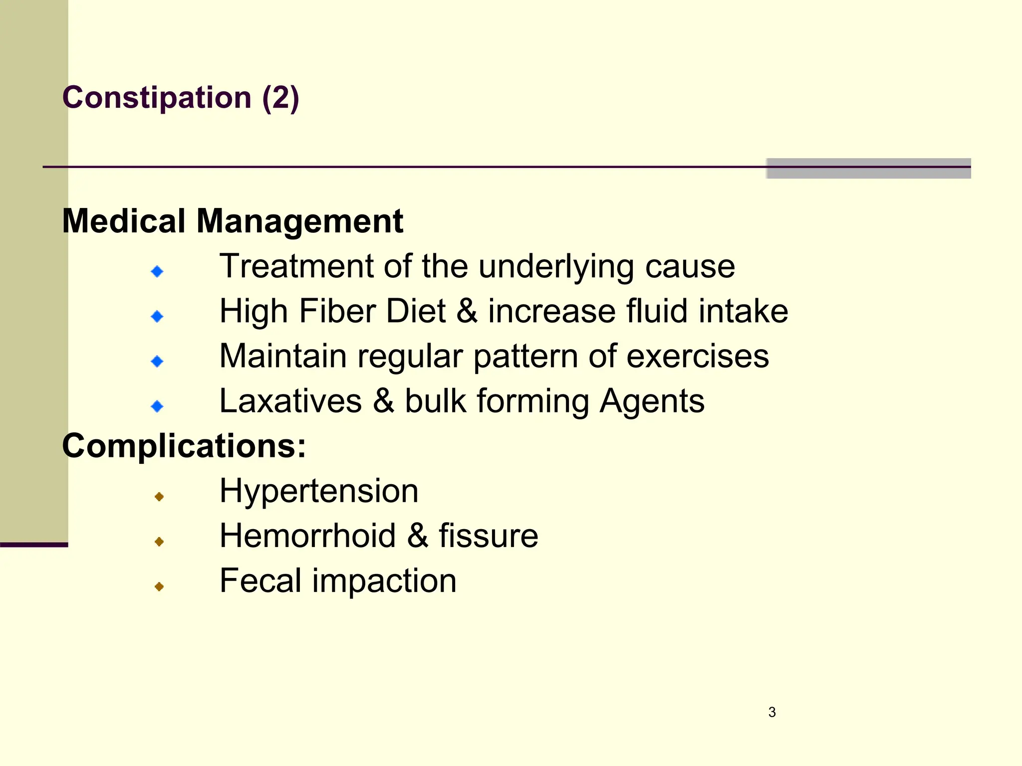
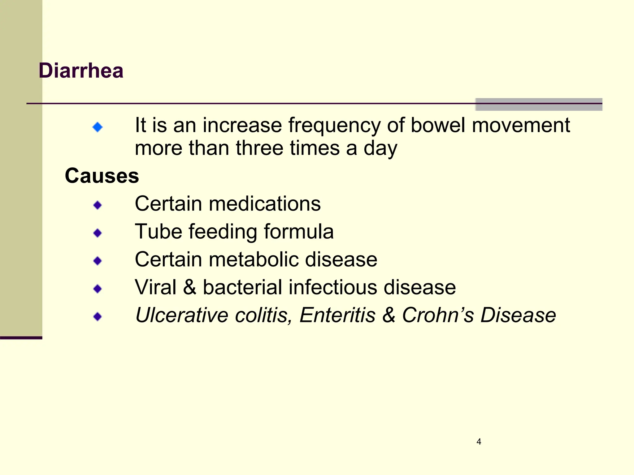
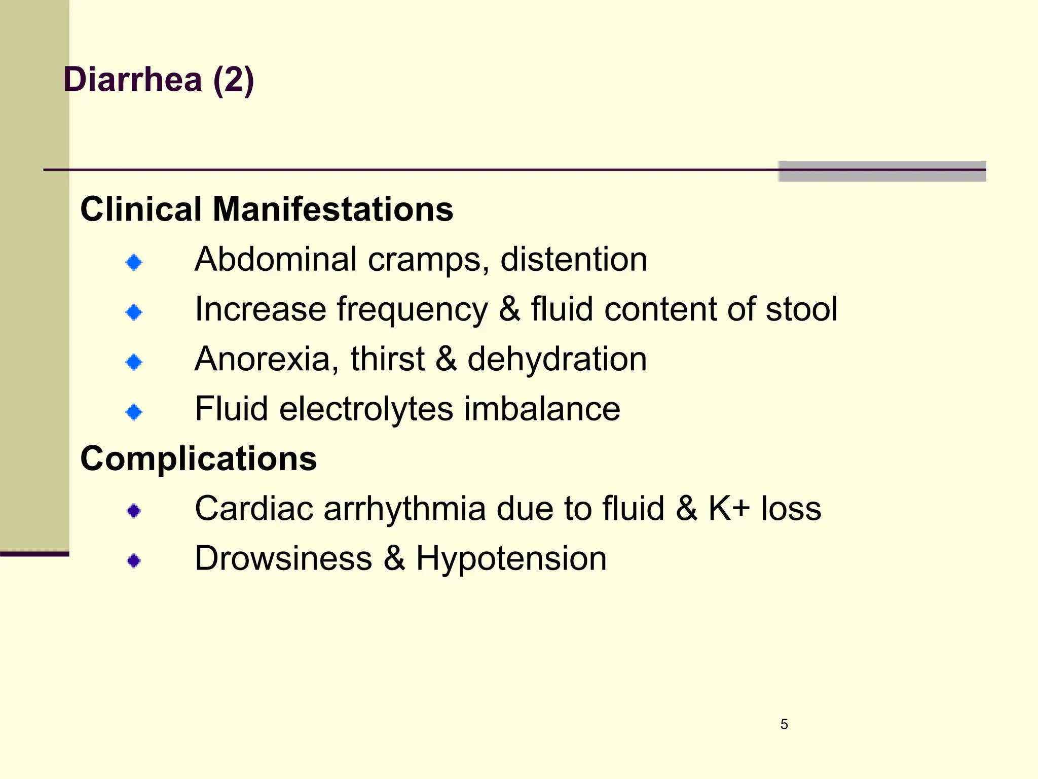
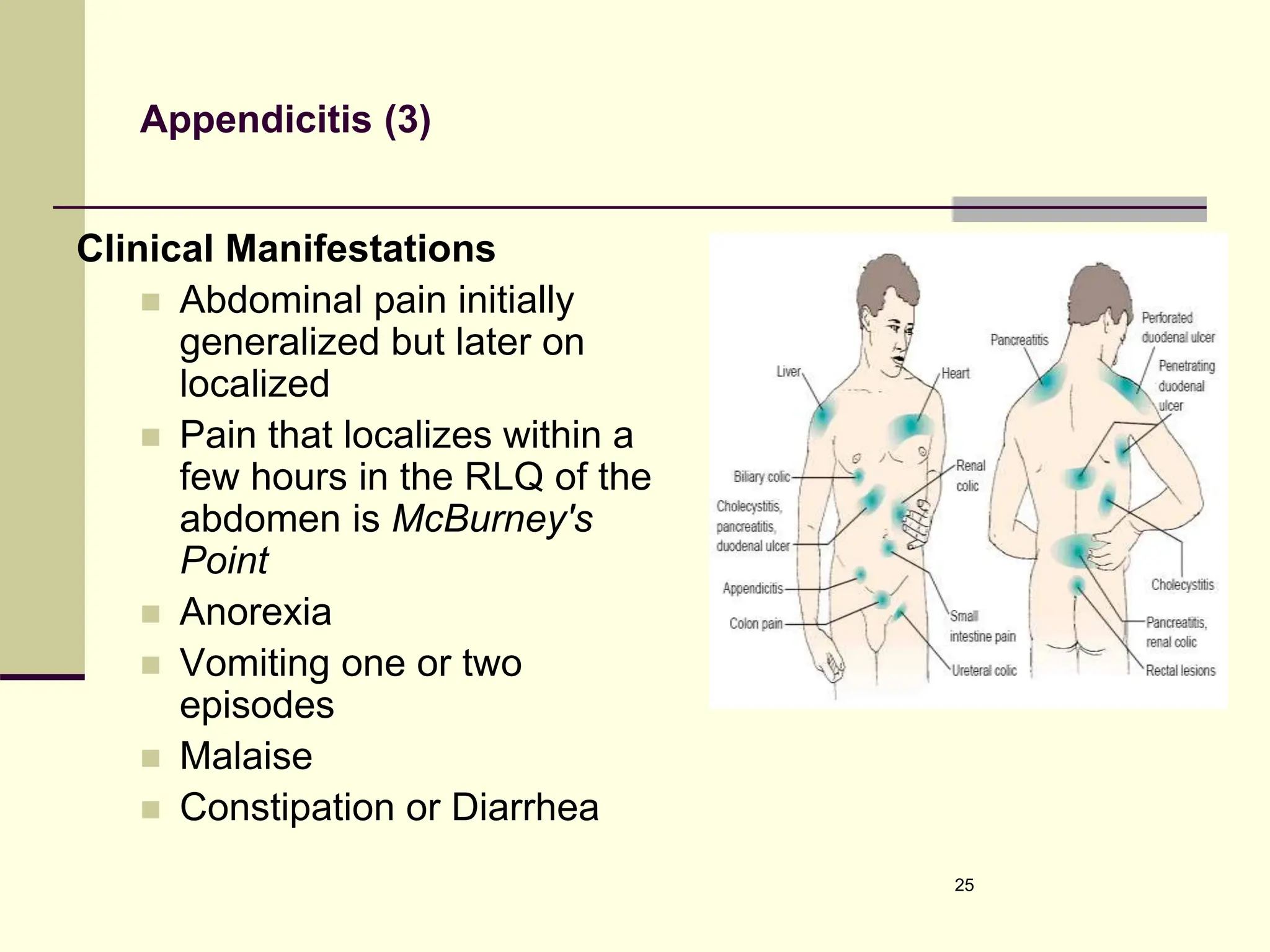
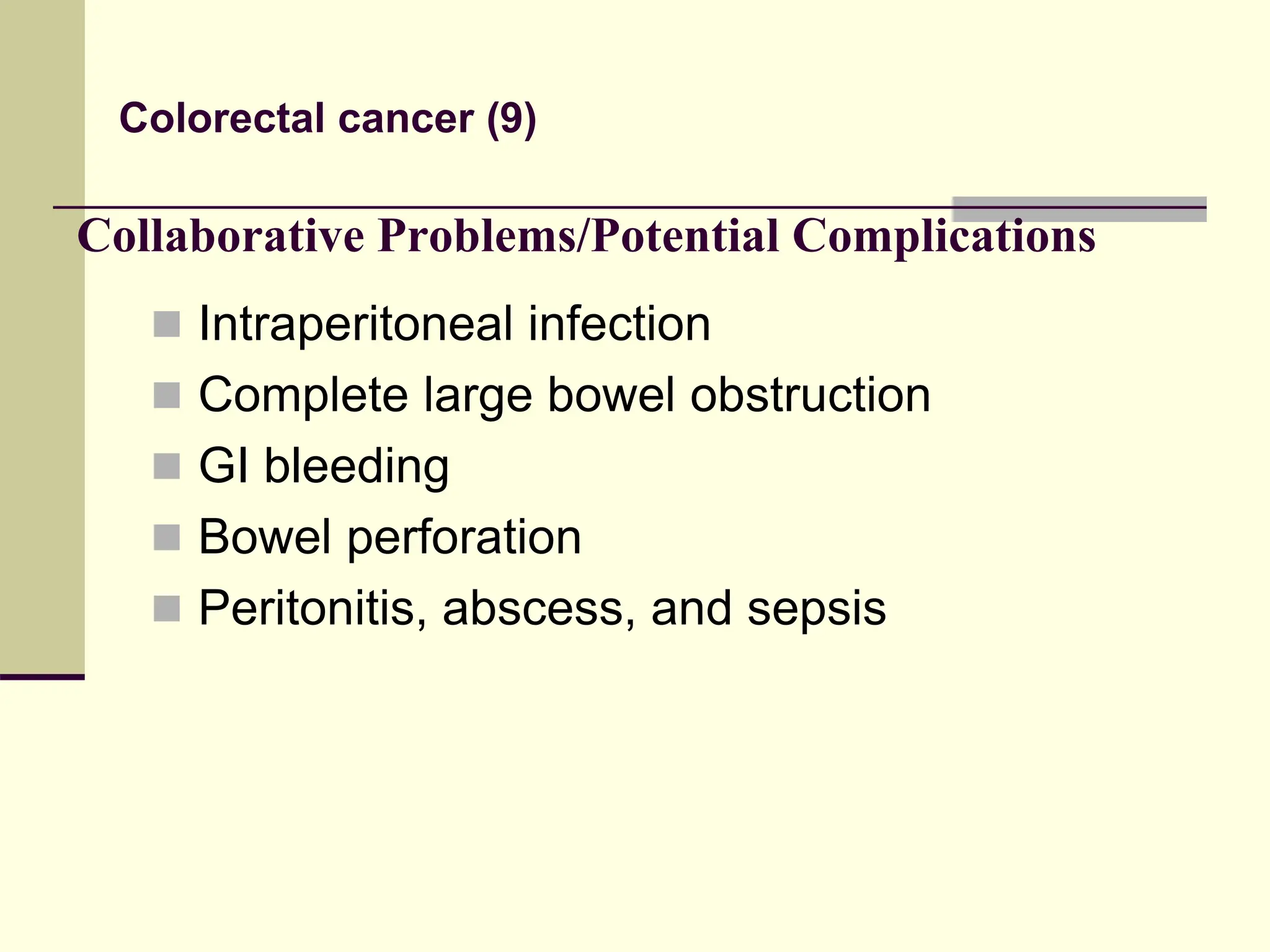
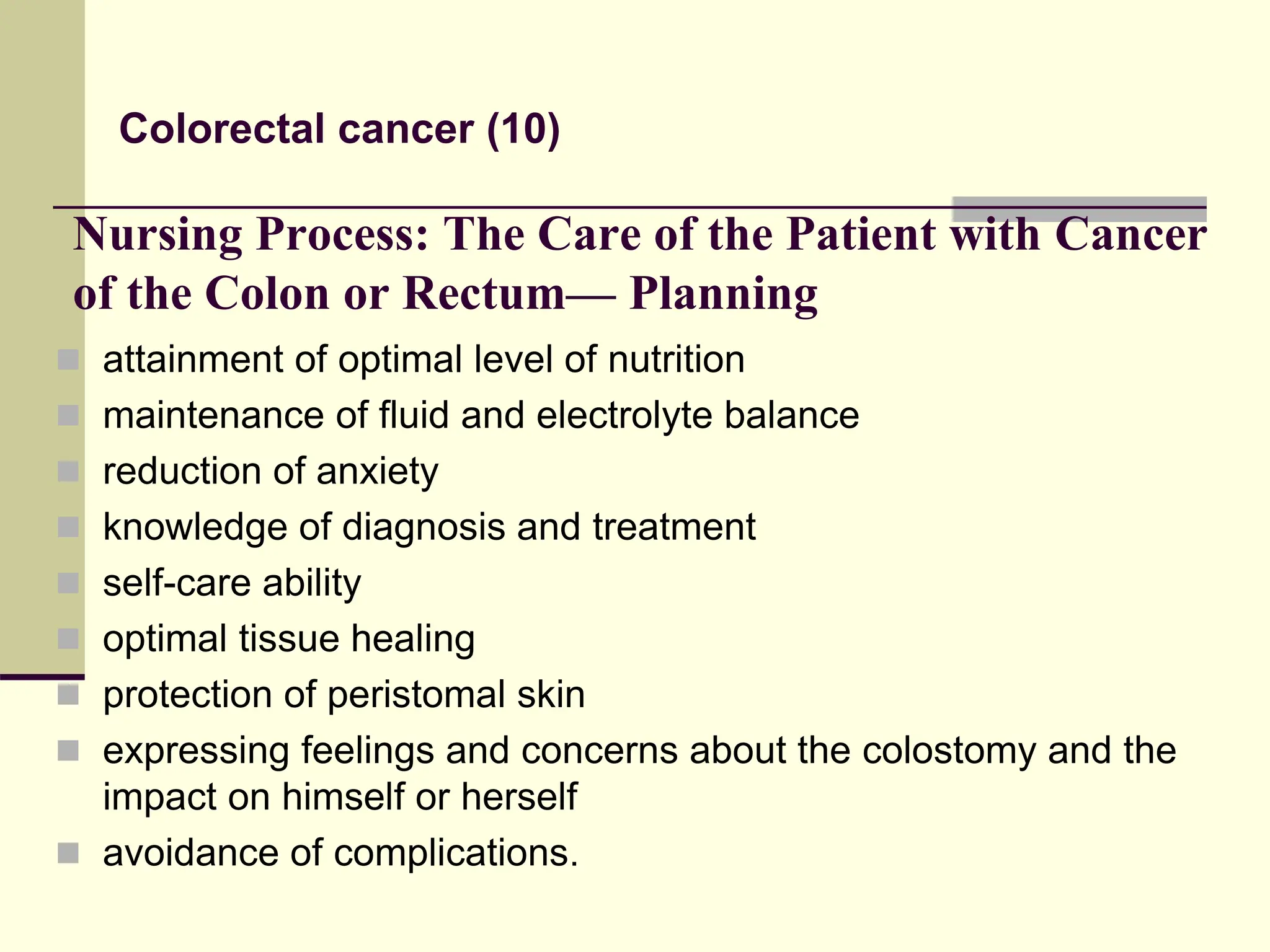
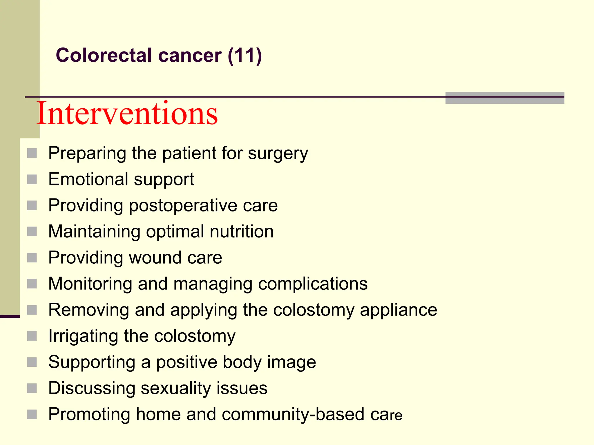
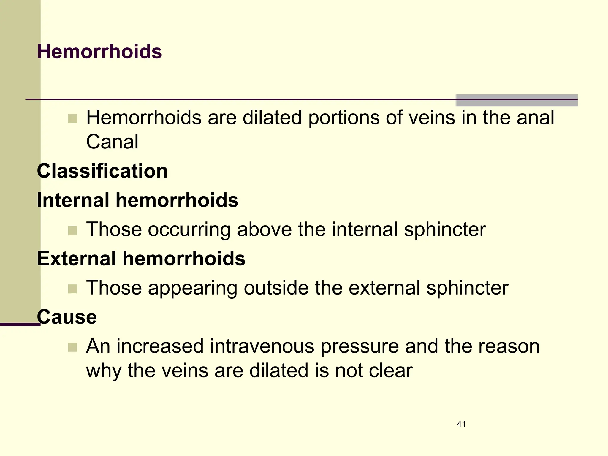
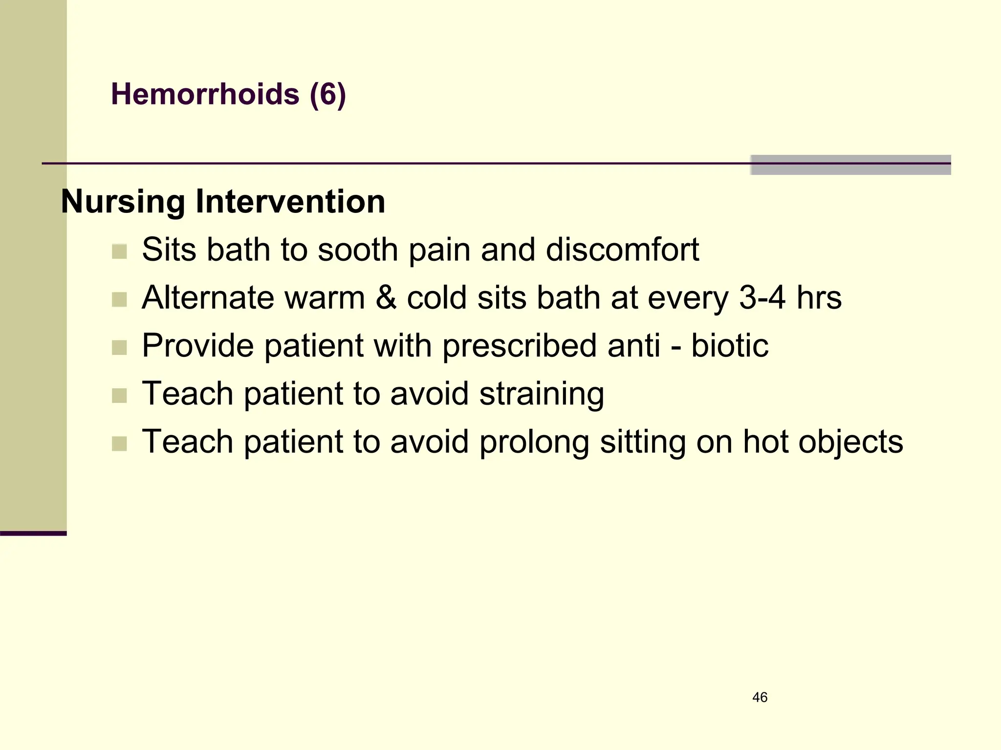
2. Intestinal and Rectal Disorders
Constipation
Abnormal hardening of stool
Retention of stool on rectum for prolonged period
Clinical Manifestations
Abdominal distention
Pain & pressure
Anorexia fatigue & headache
Incomplete emptying & strain defecation
2
3. Constipation (2)
Medical Management
Treatment of the underlying cause
High Fiber Diet & increase fluid intake
Maintain regular pattern of exercises
Laxatives & bulk forming Agents
Complications:
Hypertension
Hemorrhoid & fissure
Fecal impaction
3
4. Diarrhea
It is an increase frequency of bowel movement
more than three times a day
Causes
Certain medications
Tube feeding formula
Certain metabolic disease
Viral & bacterial infectious disease
Ulcerative colitis, Enteritis & Crohn’s Disease
4
5. Diarrhea (2)
Clinical Manifestations
Abdominal cramps, distention
Increase frequency & fluid content of stool
Anorexia, thirst & dehydration
Fluid electrolytes imbalance
Complications
Cardiac arrhythmia due to fluid & K+ loss
Drowsiness & Hypotension
5
6. Diarrhea (3)
Medical Management
1. Treatment of the underlying cause
2. Controlling symptoms & preventing
complications
3. Antibiotics & anti-inflammatory agents
4. Anti-diarrheal & antispasmoic agents
Opiate related drugs, act on opoid receptors in GI
tract decreased motility.
Loperamide (Lomotril)
Defenoxin with atropine (Motofen)
Diphenoxylate with Atropine (Lomotil)
6
7. Diarrhea (4)
Nursing Managements
1. Assessment of the nature & pattern of the
diarrhea
2. Bed rest & monitoring of fluid status
3. Serum electrolytes (K)
4. Perineal care
7
8. Intestinal Obstruction
Intestinal obstruction exists when blockage prevents
the normal flow of intestinal contents through the
intestinal tract.
Obstruction can be Partial or Complete
Severity of intestinal obstruction depends on
The region of bowel that is affected
The degree to which the lumen is occluded
The degree to which the blood circulation in the
bowel wall is disturbed
8
9. Intestinal Obstruction (2)
Processes of Intestinal Obstruction
Mechanical
This is due to the obstruction of the lumen of the intestine
The problem is directly to the lumen
No problem with the muscles.
Cause
Strictures
Adhesion (SBO)
Hernia
Tumors
Volvulus
Intussusceptions
9
10. Intestinal Obstruction (3)
Functional /Non mechanical/
The intestinal musculature is unable to propel the
contents along the bowel.
The problem is due to dysfunction of intestinal
musculature.
Cause
Muscular dystrophy
Amyloidosis
Neurologic disorders
10
11. Types of Intestinal Obstruction
Small Intestine Obstruction
Acute in onset
Sever lower abdominal cramp
Abdominal distention
Nausea & early profuse
vomiting
Sever fluid & electrolyte
imbalance
Metabolic alkalosis
Accounts for about 85% of an
obstruction
Mortality rate is about 10%
If obstruction is due to
strangulation mortality rate is
about 20 - 75%
Large Intestine Obstruction
Gradual in onset
Abdominal discomfort
Abdominal distention is more
pronounced
Minimal or no vomiting (may
contain fecal)
No major fluid & electrolyte
imbalance
Metabolic acidosis
Accounts for about 15% of all
obstruction
Mortality rate is about 30%
If obstruction is due to
strangulation mortality rate is
about 60%.
11
12. Intestinal Obstruction (5)
The most common site for large bowel obstruction is the
Sigmoid Colon
Cause
Carcinoma
Diverticulitis
Benign Tumors
Inflammatory Bowel Disorders
Volvulus of Sigmoid colon
12
14. Intestinal Obstruction (8)
Treatment
Medical Management
Abdominal decompression by using NGT
IV fluid (replacement of fluid and electrolytes)
NPO the patient
Surgical Mx
Indication of surgical Mx
If obstruction is due to Strangulation, Adhesion ,Hernia
If pt has increased pulse, increased Temperature,
Decreased B.P, and increased WBC
Surgery depends on the cause of the obstruction and
severity and size of the obstruction
14
16. Intestinal Obstruction (10)
Nursing intervention
Teach the patient to eat high fiber foods
To eat raw fruits & vegetables & whole grain products
Encourage patient to take plenty of fluids
To exercise regularly
Close follow up of the NGT
Monitor input and output
Preoperative preparation of the patient
Monitor the patient for passing of feces & flatus
Observe the color of the stool
16
17. Hernia
It is a weakness in the abdominal muscle wall through
which a segment of bowel or other abdominal structure
protrudes.
The most common types of abdominal hernias
Indirect - sac formed from the peritoneum that contains a
portion of the intestine
Direct /through a weak point in the ab. wall
Femoral (protrude through the femoral region
Umbilical congenital (in infant) /acquired increased intra abd.
pressure
Incisional (ventral) occur at the site of previous surgical incision
17
19. Hernia (3)
Types of Hernias
Reducible:- Content of the hernia sac can be replaced in to
the abdominal cavity by gentle pressure
Irreducible :-(Incarcerated) can't be reduced or placed back
in to the abdominal cavity
Strangulated:-when the blood supply to the herniated
segment of the bowel is cut off by pressure from the
hernia ring (the band of muscle around the hernia) If a
hernia is strangulated.
Strangulated hernia is characterized by adhesion and constricted
neck of hernia
19
20. Hernia (4)
Cause
Weakness of abdominal muscles
Congenital
Aging
Injury
Surgery
Increased intra abdominal pressure
Pregnancy
Obesity
Straining
Coughing
Lifting of heavy objects
20
21. Hernia (5)
Clinical Manifestations of Strangulates
Abdominal distention
Vomiting
Nausea
Pain
Fever
Tachycardia
N.B. any hernia that is not reducible requires immediate
and careful surgical evaluation
Diagnostic evaluation
C/M
Abdominal X-ray
WBC count
21
22. Hernia (6)
Treatment
Medical (non surgical MX)
Truss - for elderly or debilitated clients who are poor
surgical risks.
Surgical Management
Repair of the hernia is generally Rx of choice
Herniorrhaphy - is surgical correction of the defected
structure
Hernioplasty (less frequently used)
22
23. Appendicitis
Is an acute inflammation of the vermiform appendix.
Most common cause of acute inflammation in the RLQ
of the abdominal cavity.
It is more common between the age of 5 and 30 years
Perforation is relatively more common in the elderly
because of difficulty in diagnosis.
23
25. Appendicitis (3)
Clinical Manifestations
Abdominal pain initially
generalized but later on
localized
Pain that localizes within a
few hours in the RLQ of the
abdomen is McBurney's
Point
Anorexia
Vomiting one or two
episodes
Malaise
Constipation or Diarrhea
25
26. Appendicitis (4)
Low grade fever (37.2- 380C)
Later palpation may show tenderness in the RLQ of
the abdomen that worsen when the patient is asked
to cough or during gentle percussion
Sudden cessation of abdominal pain signals
perforation or infarction
Diagnostic evaluations
C/M
Slight Leukocytes
X-Ray
26
27. Appendicitis (5)
RX
Appendectomy is the treatment of choice
Nursing management
IV fluids
Anti-pain while patient is ready for surgery
Reducing anxiety
Elimination of infection
Maintain skin integrity
Attaining optimum nutrition
27
28. Appendicitis (6)
Preoperative Nursing Management
Physical & Psychological support
Secure IV line
NG tube
Enema is CI
Postoperative Nursing Management
Position patient in Semi Fowler's Position
Administering Antibiotics (if ordered)
28
29. Appendicitis (7)
Give oral fluids and food on the day of surgical if
tolerated.
Discharge patient on the day of surgery (if it is un
complicated)
Provided that the temperature is in a normal limits &
there is no discomfort in the operative area
Complication
Rupture or Perforation
Peritonitis
Appendical abscess
Pyelonephritis
29
30. Colorectal cancer
Cancer colon is the abnormal growth of cancer cells
in the lower digestive tract.
Risk factors
Increasing age
Family history of colon cancer or polyps
Previous colon cancer or adenomatous polyps
High consumption of alcohol
Cigarette smoking
Obesity
History of gastrectomy
History of inflammatory bowel disease
High-fat, high-protein (with high intake of beef), low-fiber diet
Genital cancer (eg, endometrial cancer, ovarian cancer) or
breast cancer (in women)
30
31. Colorectal cancer (2)
Clinical manifestations :determined by the location of the tumor, the
stage of the disease, and the function of the affected intestinal segment:
a change in bowel habits (most common) .
The passage of blood in or on the stools (second most common)
unexplained anemia, anorexia, weight loss, and fatigue.
Symptoms of Right sided lesions are dull abdominal pain and melena.
Symptoms of left-sided lesions are abdominal pain and cramping,
narrowing stools, constipation, distention), as well as bright-red blood
in the stool
rectal lesions symptoms are tenesmus, rectal pain, the feeling of
incomplete evacuation after a bowel movement, alternating
constipation and diarrhea, and bloody stool.
31
32. Colorectal cancer (3)
Assessment and diagnostic findings
abdominal and rectal examination,
fecal occult blood testing,
barium enema,
proctosigmoidoscopy
Colonoscopy with biopsy.
Carcinoembryonic antigen (CEA): With complete excision
of the tumor, the elevated levels of CEA should return to
normal within 48 hours. Elevations of CEA at a later date
suggest recurrence.
32
33. Colorectal cancer (4)
Stages of colorectal cancer (Duke's classification)
Class A: Tumor limited to muscular mucosa and
submucosa
Class B1: Tumor extends into mucosa
Class B2: Tumor extends through entire bowel wall into
serosa or pericolic fat, no nodal involvement
Class C1: Positive nodes, tumor is limited to bowel wall
Class C2: Positive nodes, tumor extends through entire
bowel wall
Class D: Advanced and metastasis to liver, lung, or
bone
33
34. Colorectal cancer (5)
Medical management
IV fluids and nasogastric suction
blood component therapy
Surgical management to remove the tumor(curative or palliative)
Segmental resection with anastomosis
Abdominoperineal resection with permanent sigmoid colostomy
Temporary colostomy
Permanent colostomy or ileostomy for palliation of unresectable
obstructing lesions
Construction of a coloanal reservoir called a colonic J pouch
Adjuvant therapy
Chemotherapy
Radiotherapy
Immunotherapy 34
35. Colorectal cancer (6)
A colostomy is the surgical creation of
an opening (ie, stoma) into the colon. It
can be created as a temporary or
permanent fecal diversion. It allows the
drainage or evacuation of colon
contents to the outside of the body. The
consistency of the drainage is related
to the placement of the colostomy,
which is dictated by the location of the
tumor and the extent of invasion into
surrounding tissues 35
Placement of Colostomies
36. Nursing Process: The Care of the Patient with
Cancer of the Colon or Rectum— Assessment
Health history
Fatigue and weakness
Abdominal or rectal pain
Nutritional status and dietary habits
Elimination patterns
Abdominal assessment
Characteristics of stool
Colorectal cancer (7)
37. Colorectal cancer (8)
Nursing diagnosis
o Imbalanced nutrition, less than body requirements, related to nausea and
anorexia
o Risk for deficient fluid volume related to vomiting and decrease fluid intake
o Anxiety related to impending surgery and the diagnosis of cancer
o Risk for ineffective therapeutic regimen management related to knowledge
deficit concerning the diagnosis, the surgical procedure, and self-care after
discharge
o Impaired skin integrity related to the surgical incisions, the formation of a
stoma, and frequent fecal contamination of peristomal skin
o Disturbed body image related to colostomy
o Ineffective sexuality patterns related to presence of ostomy and changes in
body image and self-concept
37
38. Collaborative Problems/Potential Complications
Intraperitoneal infection
Complete large bowel obstruction
GI bleeding
Bowel perforation
Peritonitis, abscess, and sepsis
Colorectal cancer (9)
39. Nursing Process: The Care of the Patient with Cancer
of the Colon or Rectum— Planning
attainment of optimal level of nutrition
maintenance of fluid and electrolyte balance
reduction of anxiety
knowledge of diagnosis and treatment
self-care ability
optimal tissue healing
protection of peristomal skin
expressing feelings and concerns about the colostomy and the
impact on himself or herself
avoidance of complications.
Colorectal cancer (10)
40. Interventions
Preparing the patient for surgery
Emotional support
Providing postoperative care
Maintaining optimal nutrition
Providing wound care
Monitoring and managing complications
Removing and applying the colostomy appliance
Irrigating the colostomy
Supporting a positive body image
Discussing sexuality issues
Promoting home and community-based care
Colorectal cancer (11)
41. Hemorrhoids
Hemorrhoids are dilated portions of veins in the anal
Canal
Classification
Internal hemorrhoids
Those occurring above the internal sphincter
External hemorrhoids
Those appearing outside the external sphincter
Cause
An increased intravenous pressure and the reason
why the veins are dilated is not clear
41
42. Hemorrhoids (2)
Risk Factor
Chronic Constipation
Intestinal disorders
Prolonged sitting on hot objects
Presence of varicose vein in the lower leg.
Hereditary
Aggravating Factors
Pregnancy
Straining
42
43. Hemorrhoids (3)
Clinical manifestation
Anal pain and discomfort
Itching in the anus
Inflammation of the anal canal
Visible blood in stool
Visible dilated vein
Thrombosis/Clotting of blood within the hemorrhoid
43
44. Hemorrhoids (4)
Treatment
Palliative
Cold & warm sits bath alternatively
High fluid intake to make stool soft
High roughage foods
Laxatives in severe constipation
Avoidance of
Straining during defecation
Lifting heavy objects
Prolonged sitting
44
45. Hemorrhoids (5)
Radical treatment
Surgical treatment
Elastic Band /Rubber Band/ the dilated blood vessels
will be tighten up.
Cryotherapy: The dilated blood vessels will be applied
excessive cold.
Sclerotherapy: injection of a solution (salt) to veins
make them swell and stick together. Overtime the scar
tissue will heal and make the veins normal.
Haemorrhoidectomy : Surgical removal of the dilated
blood vessels
45
46. Hemorrhoids (6)
Nursing Intervention
Sits bath to sooth pain and discomfort
Alternate warm & cold sits bath at every 3-4 hrs
Provide patient with prescribed anti - biotic
Teach patient to avoid straining
Teach patient to avoid prolong sitting on hot objects
46
It increases the bulk in your stool, an effect that helps to cause movement of the intestines. It also works by increasing the amount of water in the stool, making the stool softer and easier to pass.
e.g. Psyllium
Bisacodyl, Pedia-Lax, Dulcolax (laxatives)
Atropine belongs to a class of drugs known as anticholinergics, which help to dry up body fluids and also slow gut movement.
Diphenoxylate is similar to narcotic pain relievers, but it acts mainly to slow the gut.
Small Bowel Obstruction
Adhesion: The union of two opposing tissue surfaces.
A stricture is a narrowing of a section of intestine
Volvulus - an obstruction caused by twisting of the stomach or intestine
Intussusception - an instance of the inversion of one portion of the intestine within another.
Amyloid is an abnormal protein that is usually produced in your bone marrow and can be deposited in any tissue or organ.
outpocketings of the colonic mucosa and submucosa through weaknesses of muscle layers in the colon wall
Omentum - membrane lining the abdominal wall
Hernia Truss – supportive undergarment designed to keep the protruding organ/hernia in place.
Mesh is used in Hernioplasty
Mcburney’s Point – point in the abdomen that is one third in distance between anterior superior iliac spine and the umbilicus.
Enema increases the risk of RUPTURED APPENDIX
PN- infection of the kidneys, as in ascending UTI.