SlideShare a Scribd company logo
RESEARCH POSTER PRESENTATION DESIGN © 2012
www.PosterPresentation
s.com
During the investigation, it was shown that gold nanoparticles of a
larger size (around 0.02 M HAuCl4) and at a certain quantity are
more efficient, when being used as substrates of analytes on lab
filter paper strips, to enhance the detection of Adenine in SERS.
In contrast, particles of a smaller size will have little to no effect
on the reading, as show in Figure 6.
As expected, the process of analyzing the strips on SERS took
only a few seconds using commonly available and portable
materials.
Further work on this investigation should include testing the strips
with other DNA components: Guanine, Cytosine, Thymine, as
well as RNA component Uracil. In addition, the use of an Ink-Jet
printer to quickly reproduce the sensor strips.
• Pedro M. Fierro-Mercado and Samuel P. Hérnandez Rivera (2012) “Highly
Sensitive Filter Paper Substrate for SERS Trace Explosives Detection”
International Journal of Spectroscopy, Vol. 2012, 1-7.
• Huixiang Li and Lewis Rothberg (2004) “DNA Sequence Detection Using
Selective Fluorescence Quenching of Tagged Oligonucleotide Probes by Gold
Nanoparticles” Analytical Chemistry, Vol. 76 (18), pp. 5414-5417.
• http://www.semrock.com/Data/Sites/1/semrockimages/technote_images/fig-9-
sers.gif
• http://sbir.gsfc.nasa.gov/SBIR/successes/images/9-028pic.jpg
• http://2.bp.blogspot.com/_WMdGdPSdn0U/TVEhvTNFl2I/AAAAAAAABIM/
UujDNgsBxWg/s400/gold_noparticle
• http://upload.wikimedia.org/wikipedia/commons/thumb/e/e4/DNA_chemical_st
ructure.svg/300px-DNA_chemical_structure.svg.png
• AGMUS Institute of Mathematics
• National Science Foundation
• Universidad Metropolitana
• Dr. Juan Arratia
• Saturday Academy
• Dr. Oliva M. Primera-Pedrozo
• Dr. Samuel P. Hernandez-Rivera
• Pedro Fierro
• Marcos Rodríguez
• Ricardo Lorenzana
• Gold Nanoparticles have a wide range of applications in
science, particularly for their absorption of light.
• As sensors, they are particularly useful as substrates in the
Surface Enhanced Raman Spectroscopy (SERS) to detect the
energy in chemical bonds.
• This technique allows for detection of specific molecules in
analytes.
• Raman Spectroscopy is a technique that uses photons at
different wavelengths in order to determine the properties of
compounds.
• The atoms in the analytes scatter light from the Raman
emission. This will result in a change in the energy of the
photons, which can be used to characterize the chemical bonds
of the atoms.
• Surface Enhanced Raman Spectroscopy (SERS) is a technique
that produces silver plasmons on the analyte by using a metal
substrate so that it resonates with a metal tip.
• This process greatly enhances the Raman emission, which
makes it ideal to detect low concentrations of compounds.
1Escuela Secundaria de la Universidad de Puerto Rico, San Juan, Puerto Rico
2Colegio Luterano Resurrección, Inc., Carolina, Puerto Rico
3Nanomaterials Science Lab. Universidad Metropolitana, Recinto de Cupey, San Juan, Puerto Rico
Ricardo Rodriguez-Garcia1, Stephanie Chan-Yau2, Marcos Rodriguez3, Ricardo Lorenzana3 ,
Oliva M. Primera-Pedrozo3
Gold nanoparticle printer-strip sensors for detection of DNA
components using SERS: Model of adsorption at 785nm
• Synthesize gold nanoparticles at two sizes and analyze their
effect in the detection of DNA components using SERS.
• Prepare lab-filter paper sensors using gold nanoparticles as
substrates, which will efficiently detect DNA components in a
short amount of time.
• Determine what amount and size of gold nanoparticles is best for
detecting DNA components.
Figure 6: Gold Nanoparticles (HAuCl4 0.01 M) are tested in SERS in liquid using
different concentrations of Adenine and 0.01 M NaCl. (A) First Vial: 1000µL AuNPs.
(B) 1000 µL AuNPs with Adenine 1x10⁻² M and NaCl 0.01 M. (C) 1000µL AuNPs
with Adenine 1x10⁻² M. (D) Second Vial: 1000µL AuNPs. (E) 1000µL AuNPs with
Adenine 1x10⁻⁵ M. (F) 1000µL AuNPs with Adenine 1x10⁻⁵ M and NaCl 0.01 M.
Figure 8: SERS test of strips with different amounts of AuNPs (.02 M HAuCl4) and
50µL of adenine. (A) no AuNPs. (B) 50µL AuNPs. (C) 100µL AuNPs. (D) 150µL
AuNPs. (E) 200µL AuNPs. (F) 250µL AuNPs.
Results
Methodology
Acknowledgements
References
Conclusions
Objectives
Introduction
Abstract
Gold Nanoparticles have a wide range of applications in biology,
chemistry, and other related fields. These particles can strongly
absorb light and dissipate it in their surroundings, thus making them
great for manipulating heat. In addition, they are used to deliver
therapeutic agents. Due to their density, they can be utilized as
probes for transmission electron microscopy. As sensors, they are
particularly useful as substrates in the Surface Enhanced Raman
Spectroscopy (SERS) to detect the energy in chemical bonds. This
technique allows for detection of specific molecules in analytes.
Currently, gold nanoparticles are utilized as substrates in sensors to
detect low concentrations of hazardous materials, especially those
relating to explosives, through the use of Surface Enhanced Raman
Spectroscopy (SERS). This process enhances the Raman emission
through the use of metal (Gold) which will resonate with a metal
tip. These properties allowed the development of lab filter paper
strip-based sensors in order to detect DNA components in analytes.
Gold nanoparticles were synthesized using 0.01M and 0.02M
hydrogen tetrachloroaurate (III) trihydrate solution with sodium
citrate tribasic dihydrate (TSC, 1%) as both a reducing agent and
capping agent. For this investigation, the strips were tested with the
DNA component adenine. After obtaining results from SERS, it can
be concluded that the use of larger gold nanoparticles (0.02M of
gold salt) is best to accurately detect adenine. Also, the strip
sensors proved to be a fast and simple method to detect DNA
components, which, if perfected, can be useful for medical
examinations and the detection of biological agents. Further work
on this investigation should include testing on other DNA
components guanine, cytosine, and thymine, as well as RNA
component Uracil.
Figure 2: SERS test
using vial or liquid
Figure 3: SERS test using a
flat surface.
Synthesis:
UHP H2O
Add Sodium
Citrate 1 .0 %
(dropwise)
Purification/Characterization:
Add HAuCl4 trihydrate
0.01M/0.02 M
Gold
Nanoparticles
1 mL of AuNPs in
each centrifuge tube
Centrifuge at
10,000 rpm for
1.0 H
Remove supernatant
and re-disperse in
2-propanol
UV-Visible
Spectroscopy
SERS
Strips-substrate preparationLiquid
30
50
70
90
110
130
150
400 900 1400 1900
Counts
Raman Shift /cm^-1
A
B
C
D
E
F
I
519nm
I 523nm
-0.02
0.18
0.38
0.58
0.78
0.98
1.18
1.38
1.58
1.78
1.98
400 500 600 700 800
Absorbance
Wavelength (nm)
A
B
Figure 5: UV-Visible Spectroscopy of AuNPs at different concentrations. (A) AuNPs
using 0.01 M HAuCl4 trihydrate. (B) AuNPs using 0.02 M HAuCl4 trihydrate.
Figure 7: SERS test of gold nanoparticles (0.02 M HAuCl4 trihydrate) using
1x10⁻² Adenine and 0.01 M NaCl. (A) Adenine Bulk. (B) Adenine 1x10⁻² M. (C)
AuNPs .02 M. (D) NaCl .01 M. (E) AuNPs with Adenine 1x10⁻² M. (F) AuNPs with
Adenine 1x10⁻² M and NaCl .01 M.
Figure 1: Gold nanoparticles
Figure 4: DNA molecule
722.813874
735.1245725
0
1000
2000
3000
4000
5000
6000
690 710 730 750 770
Counts
Raman Shift/ cm-1
A
B
C
D
E
F
734.2452369
730.7278945
0
200
400
600
800
1000
1200
1400
1600
1800
2000
640 660 680 700 720 740 760 780 800
Counts
Raman Shift / cm-1
A
B
C
D
E
F
0.02 M- Large nanoparticles
735 cm-1
Figure 9: Model of adsorption
AdenineRaman laser

More Related Content

What's hot

AFM.ppt
AFM.pptAFM.ppt
laser ablation and pyrolysis for micro machining and nano fabrication
laser ablation and pyrolysis for micro machining and nano fabricationlaser ablation and pyrolysis for micro machining and nano fabrication
laser ablation and pyrolysis for micro machining and nano fabrication
JAISMON FRANCIS
 
X ray diffraction
X ray diffractionX ray diffraction
X ray diffraction
Arvind Singh Heer
 
Atmospheric Pressure Ionization
Atmospheric Pressure IonizationAtmospheric Pressure Ionization
Atmospheric Pressure Ionization
Vinitha Nair
 
Characterization of nanopartical
Characterization of nanoparticalCharacterization of nanopartical
Characterization of nanopartical
Amany EL-Hallaq
 
Synthesis and characterization of nickel oxide nanastructures
Synthesis and characterization of nickel oxide nanastructuresSynthesis and characterization of nickel oxide nanastructures
Synthesis and characterization of nickel oxide nanastructures
Beenabalakrishnan
 
Ftir
FtirFtir
Scanning Electron Microscope (SEM)
Scanning Electron Microscope (SEM)Scanning Electron Microscope (SEM)
Scanning Electron Microscope (SEM)
D.R. Chandravanshi
 
Mass spectrometry
Mass spectrometryMass spectrometry
Mass spectrometry
vipin999
 
X ray photoelectron spectroscopy
X ray photoelectron spectroscopyX ray photoelectron spectroscopy
X ray photoelectron spectroscopy
Zubair Aslam
 
Surface enhanced raman spectroscopy, rahutosh ranjan
Surface enhanced raman spectroscopy, rahutosh ranjanSurface enhanced raman spectroscopy, rahutosh ranjan
Surface enhanced raman spectroscopy, rahutosh ranjan
RahutoshRanjan
 
X Ray Diffraction Spectroscopy
X Ray Diffraction SpectroscopyX Ray Diffraction Spectroscopy
X Ray Diffraction Spectroscopyhephz
 
Atomic absorption spectroscopy
Atomic absorption spectroscopyAtomic absorption spectroscopy
Atomic absorption spectroscopy
Syed Sheraz Ali Shah
 
ICP-AES.pptx
ICP-AES.pptxICP-AES.pptx
ICP-AES.pptx
Vishal Yadav
 
Auger electron spectroscopy
Auger electron spectroscopyAuger electron spectroscopy
Auger electron spectroscopyGulfam Hussain
 
Basics of spectroscopy
Basics of spectroscopyBasics of spectroscopy
Basics of spectroscopy
Jenson Samraj
 
SEM- scanning electron microscope
SEM- scanning electron microscope SEM- scanning electron microscope
SEM- scanning electron microscope
Zydus Cadila Healthcare Ltd
 
Difference in raman spectroscopy and ir spectroscopy
Difference in raman spectroscopy and ir spectroscopyDifference in raman spectroscopy and ir spectroscopy
Difference in raman spectroscopy and ir spectroscopy
Vrushali Tambe
 
Preparation Of MXenes (A novel 2D Material)
Preparation Of MXenes (A novel 2D Material) Preparation Of MXenes (A novel 2D Material)
Preparation Of MXenes (A novel 2D Material)
rittwikchatterjee
 

What's hot (20)

AFM.ppt
AFM.pptAFM.ppt
AFM.ppt
 
laser ablation and pyrolysis for micro machining and nano fabrication
laser ablation and pyrolysis for micro machining and nano fabricationlaser ablation and pyrolysis for micro machining and nano fabrication
laser ablation and pyrolysis for micro machining and nano fabrication
 
ICP-MS
ICP-MSICP-MS
ICP-MS
 
X ray diffraction
X ray diffractionX ray diffraction
X ray diffraction
 
Atmospheric Pressure Ionization
Atmospheric Pressure IonizationAtmospheric Pressure Ionization
Atmospheric Pressure Ionization
 
Characterization of nanopartical
Characterization of nanoparticalCharacterization of nanopartical
Characterization of nanopartical
 
Synthesis and characterization of nickel oxide nanastructures
Synthesis and characterization of nickel oxide nanastructuresSynthesis and characterization of nickel oxide nanastructures
Synthesis and characterization of nickel oxide nanastructures
 
Ftir
FtirFtir
Ftir
 
Scanning Electron Microscope (SEM)
Scanning Electron Microscope (SEM)Scanning Electron Microscope (SEM)
Scanning Electron Microscope (SEM)
 
Mass spectrometry
Mass spectrometryMass spectrometry
Mass spectrometry
 
X ray photoelectron spectroscopy
X ray photoelectron spectroscopyX ray photoelectron spectroscopy
X ray photoelectron spectroscopy
 
Surface enhanced raman spectroscopy, rahutosh ranjan
Surface enhanced raman spectroscopy, rahutosh ranjanSurface enhanced raman spectroscopy, rahutosh ranjan
Surface enhanced raman spectroscopy, rahutosh ranjan
 
X Ray Diffraction Spectroscopy
X Ray Diffraction SpectroscopyX Ray Diffraction Spectroscopy
X Ray Diffraction Spectroscopy
 
Atomic absorption spectroscopy
Atomic absorption spectroscopyAtomic absorption spectroscopy
Atomic absorption spectroscopy
 
ICP-AES.pptx
ICP-AES.pptxICP-AES.pptx
ICP-AES.pptx
 
Auger electron spectroscopy
Auger electron spectroscopyAuger electron spectroscopy
Auger electron spectroscopy
 
Basics of spectroscopy
Basics of spectroscopyBasics of spectroscopy
Basics of spectroscopy
 
SEM- scanning electron microscope
SEM- scanning electron microscope SEM- scanning electron microscope
SEM- scanning electron microscope
 
Difference in raman spectroscopy and ir spectroscopy
Difference in raman spectroscopy and ir spectroscopyDifference in raman spectroscopy and ir spectroscopy
Difference in raman spectroscopy and ir spectroscopy
 
Preparation Of MXenes (A novel 2D Material)
Preparation Of MXenes (A novel 2D Material) Preparation Of MXenes (A novel 2D Material)
Preparation Of MXenes (A novel 2D Material)
 

Similar to Final Poster- Gold Nanoparticle sensor strips for detection of DNA components using SERS model of adsorbtion 785nm

CH499_Spring2014_HromadaEmily
CH499_Spring2014_HromadaEmilyCH499_Spring2014_HromadaEmily
CH499_Spring2014_HromadaEmilyEmily Hromada
 
Laser Ablation Molecular Isotopic Spectrometry for rare isotopes of the light...
Laser Ablation Molecular Isotopic Spectrometry for rare isotopes of the light...Laser Ablation Molecular Isotopic Spectrometry for rare isotopes of the light...
Laser Ablation Molecular Isotopic Spectrometry for rare isotopes of the light...
Alexander Bolshakov
 
SP-ICP-MS Analysis of Size and Number Concentration in Mixtures of Monometall...
SP-ICP-MS Analysis of Size and Number Concentration in Mixtures of Monometall...SP-ICP-MS Analysis of Size and Number Concentration in Mixtures of Monometall...
SP-ICP-MS Analysis of Size and Number Concentration in Mixtures of Monometall...
PerkinElmer, Inc.
 
Synthesis & Characterization of Fluorescent Silver Nanoparticles stabilized b...
Synthesis & Characterization of Fluorescent Silver Nanoparticles stabilized b...Synthesis & Characterization of Fluorescent Silver Nanoparticles stabilized b...
Synthesis & Characterization of Fluorescent Silver Nanoparticles stabilized b...
IJERA Editor
 
Raman spectroscopy PPT.pdf
Raman spectroscopy PPT.pdfRaman spectroscopy PPT.pdf
Raman spectroscopy PPT.pdf
himanimore4
 
raman spectroscopy 2.pptx
raman spectroscopy 2.pptxraman spectroscopy 2.pptx
raman spectroscopy 2.pptx
TahminaKhan20
 
Synthesis and Characterization of Novel Nanomaterials for SERS Biomedical/Env...
Synthesis and Characterization of Novel Nanomaterials for SERS Biomedical/Env...Synthesis and Characterization of Novel Nanomaterials for SERS Biomedical/Env...
Synthesis and Characterization of Novel Nanomaterials for SERS Biomedical/Env...
Necla YÜCEL
 
CENT-1206APP11.15-A_Characterizing RNA Nanoparticles by Analytical Ultracentr...
CENT-1206APP11.15-A_Characterizing RNA Nanoparticles by Analytical Ultracentr...CENT-1206APP11.15-A_Characterizing RNA Nanoparticles by Analytical Ultracentr...
CENT-1206APP11.15-A_Characterizing RNA Nanoparticles by Analytical Ultracentr...Chad Schwartz
 
Chemical Analysis Facility
Chemical Analysis FacilityChemical Analysis Facility
Chemical Analysis Facility
christinejcardin
 
Technical Engineering Memorandum_2015
Technical Engineering Memorandum_2015Technical Engineering Memorandum_2015
Technical Engineering Memorandum_2015Jordan Lopez
 
Determination of ksp of cu using aa c14123361t
Determination of ksp of cu using aa c14123361tDetermination of ksp of cu using aa c14123361t
Determination of ksp of cu using aa c14123361t
MGH/Harvard Medical School
 
Plant Mediated Synthesis of Silver Nanoparticles by Using Dried Stem Powder o...
Plant Mediated Synthesis of Silver Nanoparticles by Using Dried Stem Powder o...Plant Mediated Synthesis of Silver Nanoparticles by Using Dried Stem Powder o...
Plant Mediated Synthesis of Silver Nanoparticles by Using Dried Stem Powder o...
IJERA Editor
 
Application of uv visible spectroscopy in microbiology
Application of uv visible spectroscopy in microbiologyApplication of uv visible spectroscopy in microbiology
Application of uv visible spectroscopy in microbiology
Farhad Ashraf
 
The emerging technology in the field of forensics......
The emerging technology in the field of forensics......The emerging technology in the field of forensics......
The emerging technology in the field of forensics......
srmnanotechnologists
 
Acr(2016)
Acr(2016)Acr(2016)
Acr(2016)
alex tomagraphy
 
Nanosensors for medicines
Nanosensors for medicinesNanosensors for medicines
Nanosensors for medicines
tabirsir
 
NANOFORENSICS: AN EMERGING FIELD IN CRIMINAL INVESTIGATION AND FORENSIC SCIENCE
NANOFORENSICS: AN EMERGING FIELD IN CRIMINAL INVESTIGATION AND FORENSIC SCIENCENANOFORENSICS: AN EMERGING FIELD IN CRIMINAL INVESTIGATION AND FORENSIC SCIENCE
NANOFORENSICS: AN EMERGING FIELD IN CRIMINAL INVESTIGATION AND FORENSIC SCIENCE
Forensic Science Section, Department of Anthropology, University of Delhi
 

Similar to Final Poster- Gold Nanoparticle sensor strips for detection of DNA components using SERS model of adsorbtion 785nm (20)

CH499_Spring2014_HromadaEmily
CH499_Spring2014_HromadaEmilyCH499_Spring2014_HromadaEmily
CH499_Spring2014_HromadaEmily
 
Laser Ablation Molecular Isotopic Spectrometry for rare isotopes of the light...
Laser Ablation Molecular Isotopic Spectrometry for rare isotopes of the light...Laser Ablation Molecular Isotopic Spectrometry for rare isotopes of the light...
Laser Ablation Molecular Isotopic Spectrometry for rare isotopes of the light...
 
SP-ICP-MS Analysis of Size and Number Concentration in Mixtures of Monometall...
SP-ICP-MS Analysis of Size and Number Concentration in Mixtures of Monometall...SP-ICP-MS Analysis of Size and Number Concentration in Mixtures of Monometall...
SP-ICP-MS Analysis of Size and Number Concentration in Mixtures of Monometall...
 
Synthesis & Characterization of Fluorescent Silver Nanoparticles stabilized b...
Synthesis & Characterization of Fluorescent Silver Nanoparticles stabilized b...Synthesis & Characterization of Fluorescent Silver Nanoparticles stabilized b...
Synthesis & Characterization of Fluorescent Silver Nanoparticles stabilized b...
 
Raman spectroscopy PPT.pdf
Raman spectroscopy PPT.pdfRaman spectroscopy PPT.pdf
Raman spectroscopy PPT.pdf
 
raman spectroscopy 2.pptx
raman spectroscopy 2.pptxraman spectroscopy 2.pptx
raman spectroscopy 2.pptx
 
Synthesis and Characterization of Novel Nanomaterials for SERS Biomedical/Env...
Synthesis and Characterization of Novel Nanomaterials for SERS Biomedical/Env...Synthesis and Characterization of Novel Nanomaterials for SERS Biomedical/Env...
Synthesis and Characterization of Novel Nanomaterials for SERS Biomedical/Env...
 
CENT-1206APP11.15-A_Characterizing RNA Nanoparticles by Analytical Ultracentr...
CENT-1206APP11.15-A_Characterizing RNA Nanoparticles by Analytical Ultracentr...CENT-1206APP11.15-A_Characterizing RNA Nanoparticles by Analytical Ultracentr...
CENT-1206APP11.15-A_Characterizing RNA Nanoparticles by Analytical Ultracentr...
 
Chemical Analysis Facility
Chemical Analysis FacilityChemical Analysis Facility
Chemical Analysis Facility
 
Technical Engineering Memorandum_2015
Technical Engineering Memorandum_2015Technical Engineering Memorandum_2015
Technical Engineering Memorandum_2015
 
Determination of ksp of cu using aa c14123361t
Determination of ksp of cu using aa c14123361tDetermination of ksp of cu using aa c14123361t
Determination of ksp of cu using aa c14123361t
 
Plant Mediated Synthesis of Silver Nanoparticles by Using Dried Stem Powder o...
Plant Mediated Synthesis of Silver Nanoparticles by Using Dried Stem Powder o...Plant Mediated Synthesis of Silver Nanoparticles by Using Dried Stem Powder o...
Plant Mediated Synthesis of Silver Nanoparticles by Using Dried Stem Powder o...
 
Application of uv visible spectroscopy in microbiology
Application of uv visible spectroscopy in microbiologyApplication of uv visible spectroscopy in microbiology
Application of uv visible spectroscopy in microbiology
 
The emerging technology in the field of forensics......
The emerging technology in the field of forensics......The emerging technology in the field of forensics......
The emerging technology in the field of forensics......
 
Nat Sci10
Nat  Sci10Nat  Sci10
Nat Sci10
 
Mao Lab Research Report
Mao Lab Research ReportMao Lab Research Report
Mao Lab Research Report
 
Dimitriu SR 15 Poster
Dimitriu SR 15 PosterDimitriu SR 15 Poster
Dimitriu SR 15 Poster
 
Acr(2016)
Acr(2016)Acr(2016)
Acr(2016)
 
Nanosensors for medicines
Nanosensors for medicinesNanosensors for medicines
Nanosensors for medicines
 
NANOFORENSICS: AN EMERGING FIELD IN CRIMINAL INVESTIGATION AND FORENSIC SCIENCE
NANOFORENSICS: AN EMERGING FIELD IN CRIMINAL INVESTIGATION AND FORENSIC SCIENCENANOFORENSICS: AN EMERGING FIELD IN CRIMINAL INVESTIGATION AND FORENSIC SCIENCE
NANOFORENSICS: AN EMERGING FIELD IN CRIMINAL INVESTIGATION AND FORENSIC SCIENCE
 

Final Poster- Gold Nanoparticle sensor strips for detection of DNA components using SERS model of adsorbtion 785nm

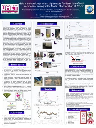
  • 1. RESEARCH POSTER PRESENTATION DESIGN © 2012 www.PosterPresentation s.com During the investigation, it was shown that gold nanoparticles of a larger size (around 0.02 M HAuCl4) and at a certain quantity are more efficient, when being used as substrates of analytes on lab filter paper strips, to enhance the detection of Adenine in SERS. In contrast, particles of a smaller size will have little to no effect on the reading, as show in Figure 6. As expected, the process of analyzing the strips on SERS took only a few seconds using commonly available and portable materials. Further work on this investigation should include testing the strips with other DNA components: Guanine, Cytosine, Thymine, as well as RNA component Uracil. In addition, the use of an Ink-Jet printer to quickly reproduce the sensor strips. • Pedro M. Fierro-Mercado and Samuel P. Hérnandez Rivera (2012) “Highly Sensitive Filter Paper Substrate for SERS Trace Explosives Detection” International Journal of Spectroscopy, Vol. 2012, 1-7. • Huixiang Li and Lewis Rothberg (2004) “DNA Sequence Detection Using Selective Fluorescence Quenching of Tagged Oligonucleotide Probes by Gold Nanoparticles” Analytical Chemistry, Vol. 76 (18), pp. 5414-5417. • http://www.semrock.com/Data/Sites/1/semrockimages/technote_images/fig-9- sers.gif • http://sbir.gsfc.nasa.gov/SBIR/successes/images/9-028pic.jpg • http://2.bp.blogspot.com/_WMdGdPSdn0U/TVEhvTNFl2I/AAAAAAAABIM/ UujDNgsBxWg/s400/gold_noparticle • http://upload.wikimedia.org/wikipedia/commons/thumb/e/e4/DNA_chemical_st ructure.svg/300px-DNA_chemical_structure.svg.png • AGMUS Institute of Mathematics • National Science Foundation • Universidad Metropolitana • Dr. Juan Arratia • Saturday Academy • Dr. Oliva M. Primera-Pedrozo • Dr. Samuel P. Hernandez-Rivera • Pedro Fierro • Marcos Rodríguez • Ricardo Lorenzana • Gold Nanoparticles have a wide range of applications in science, particularly for their absorption of light. • As sensors, they are particularly useful as substrates in the Surface Enhanced Raman Spectroscopy (SERS) to detect the energy in chemical bonds. • This technique allows for detection of specific molecules in analytes. • Raman Spectroscopy is a technique that uses photons at different wavelengths in order to determine the properties of compounds. • The atoms in the analytes scatter light from the Raman emission. This will result in a change in the energy of the photons, which can be used to characterize the chemical bonds of the atoms. • Surface Enhanced Raman Spectroscopy (SERS) is a technique that produces silver plasmons on the analyte by using a metal substrate so that it resonates with a metal tip. • This process greatly enhances the Raman emission, which makes it ideal to detect low concentrations of compounds. 1Escuela Secundaria de la Universidad de Puerto Rico, San Juan, Puerto Rico 2Colegio Luterano Resurrección, Inc., Carolina, Puerto Rico 3Nanomaterials Science Lab. Universidad Metropolitana, Recinto de Cupey, San Juan, Puerto Rico Ricardo Rodriguez-Garcia1, Stephanie Chan-Yau2, Marcos Rodriguez3, Ricardo Lorenzana3 , Oliva M. Primera-Pedrozo3 Gold nanoparticle printer-strip sensors for detection of DNA components using SERS: Model of adsorption at 785nm • Synthesize gold nanoparticles at two sizes and analyze their effect in the detection of DNA components using SERS. • Prepare lab-filter paper sensors using gold nanoparticles as substrates, which will efficiently detect DNA components in a short amount of time. • Determine what amount and size of gold nanoparticles is best for detecting DNA components. Figure 6: Gold Nanoparticles (HAuCl4 0.01 M) are tested in SERS in liquid using different concentrations of Adenine and 0.01 M NaCl. (A) First Vial: 1000µL AuNPs. (B) 1000 µL AuNPs with Adenine 1x10⁻² M and NaCl 0.01 M. (C) 1000µL AuNPs with Adenine 1x10⁻² M. (D) Second Vial: 1000µL AuNPs. (E) 1000µL AuNPs with Adenine 1x10⁻⁵ M. (F) 1000µL AuNPs with Adenine 1x10⁻⁵ M and NaCl 0.01 M. Figure 8: SERS test of strips with different amounts of AuNPs (.02 M HAuCl4) and 50µL of adenine. (A) no AuNPs. (B) 50µL AuNPs. (C) 100µL AuNPs. (D) 150µL AuNPs. (E) 200µL AuNPs. (F) 250µL AuNPs. Results Methodology Acknowledgements References Conclusions Objectives Introduction Abstract Gold Nanoparticles have a wide range of applications in biology, chemistry, and other related fields. These particles can strongly absorb light and dissipate it in their surroundings, thus making them great for manipulating heat. In addition, they are used to deliver therapeutic agents. Due to their density, they can be utilized as probes for transmission electron microscopy. As sensors, they are particularly useful as substrates in the Surface Enhanced Raman Spectroscopy (SERS) to detect the energy in chemical bonds. This technique allows for detection of specific molecules in analytes. Currently, gold nanoparticles are utilized as substrates in sensors to detect low concentrations of hazardous materials, especially those relating to explosives, through the use of Surface Enhanced Raman Spectroscopy (SERS). This process enhances the Raman emission through the use of metal (Gold) which will resonate with a metal tip. These properties allowed the development of lab filter paper strip-based sensors in order to detect DNA components in analytes. Gold nanoparticles were synthesized using 0.01M and 0.02M hydrogen tetrachloroaurate (III) trihydrate solution with sodium citrate tribasic dihydrate (TSC, 1%) as both a reducing agent and capping agent. For this investigation, the strips were tested with the DNA component adenine. After obtaining results from SERS, it can be concluded that the use of larger gold nanoparticles (0.02M of gold salt) is best to accurately detect adenine. Also, the strip sensors proved to be a fast and simple method to detect DNA components, which, if perfected, can be useful for medical examinations and the detection of biological agents. Further work on this investigation should include testing on other DNA components guanine, cytosine, and thymine, as well as RNA component Uracil. Figure 2: SERS test using vial or liquid Figure 3: SERS test using a flat surface. Synthesis: UHP H2O Add Sodium Citrate 1 .0 % (dropwise) Purification/Characterization: Add HAuCl4 trihydrate 0.01M/0.02 M Gold Nanoparticles 1 mL of AuNPs in each centrifuge tube Centrifuge at 10,000 rpm for 1.0 H Remove supernatant and re-disperse in 2-propanol UV-Visible Spectroscopy SERS Strips-substrate preparationLiquid 30 50 70 90 110 130 150 400 900 1400 1900 Counts Raman Shift /cm^-1 A B C D E F I 519nm I 523nm -0.02 0.18 0.38 0.58 0.78 0.98 1.18 1.38 1.58 1.78 1.98 400 500 600 700 800 Absorbance Wavelength (nm) A B Figure 5: UV-Visible Spectroscopy of AuNPs at different concentrations. (A) AuNPs using 0.01 M HAuCl4 trihydrate. (B) AuNPs using 0.02 M HAuCl4 trihydrate. Figure 7: SERS test of gold nanoparticles (0.02 M HAuCl4 trihydrate) using 1x10⁻² Adenine and 0.01 M NaCl. (A) Adenine Bulk. (B) Adenine 1x10⁻² M. (C) AuNPs .02 M. (D) NaCl .01 M. (E) AuNPs with Adenine 1x10⁻² M. (F) AuNPs with Adenine 1x10⁻² M and NaCl .01 M. Figure 1: Gold nanoparticles Figure 4: DNA molecule 722.813874 735.1245725 0 1000 2000 3000 4000 5000 6000 690 710 730 750 770 Counts Raman Shift/ cm-1 A B C D E F 734.2452369 730.7278945 0 200 400 600 800 1000 1200 1400 1600 1800 2000 640 660 680 700 720 740 760 780 800 Counts Raman Shift / cm-1 A B C D E F 0.02 M- Large nanoparticles 735 cm-1 Figure 9: Model of adsorption AdenineRaman laser