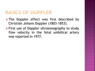
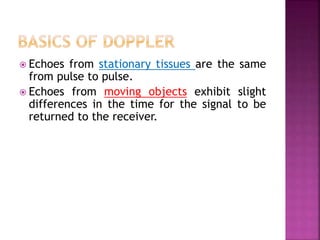
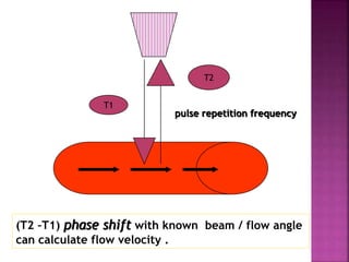
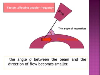
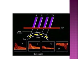
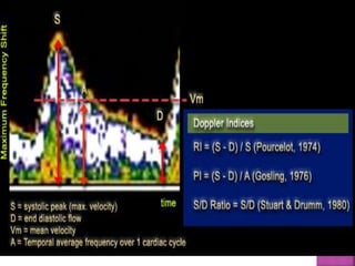
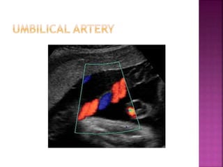
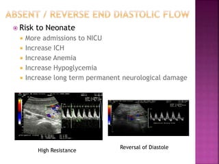
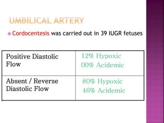
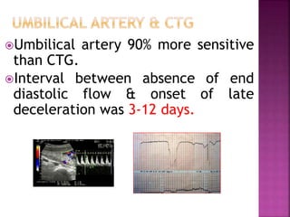
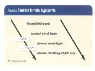
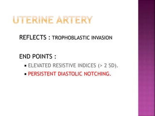
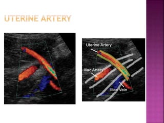
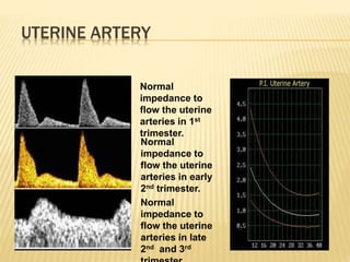
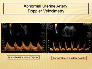
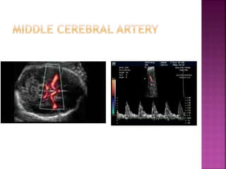
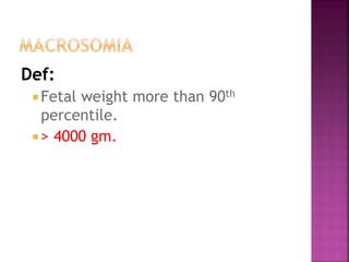
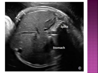
MAGDY ABDELRAHMAN MOHAMED
Lecturer of OB/GYN
2015
Intrauterine growth restriction.
Fetal doppler.
Macrosomia.
 Fetal weight below 10th percentile for
gestational age.
 Incidence: 3-5%.
 1st suspicion due to decrease fundal level
below expected from gestational age.
Types:
 Symetrical.
Asymetrical.
Etiology:
 Fetal.
Maternal.
 Placental.
 Fetal doppler.
 BPP.
 NST & contraction stress test.
WHY
Take decision of termination at proper time.
 The Doppler effect was first described by
Christian Johann Doppler (1803–1853).
 First use of Doppler ultrasonography to study
flow velocity in the fetal umbilical artery
was reported in 1977.
 Echoes from stationary tissues are the same
from pulse to pulse.
 Echoes from moving objects exhibit slight
differences in the time for the signal to be
returned to the receiver.
pulse repetition frequency
(T2 –T1) phase shift with known beam / flow angle
can calculate flow velocity .
T1
T2
the angle q between the beam and the
direction of flow becomes smaller.
q
The angle of insonation
Flow velocity
Factors affecting doppler frequency
Umbilical artery.
Uterine artery.
Middle cerebral artery.
Benefit of Umbilical Artery Evaluation
Less experienced operators can achieve
highly reproducible results with simple,
inexpensive continuous-wave equipment .
With advancing gestation,
umbilical arterial Doppler
waveforms demonstrate a
progressive rise in the
end-diastolic velocity and
a decrease in the
pulsatility index.
Umbilical artery
S/D ratio:
2-3 in 2nd & 3rd trimester
PI :
1.5 – 2.0 in 2nd trimester
1.0 – 1.5 in 3rd trimester
RI:
Decreases with gest. In
late 2nd and 3rd it is around
0.5
 Risk to Neonate
 More admissions to NICU
 Increase ICH
 Increase Anemia
 Increase Hypoglycemia
 Increase long term permanent neurological damage
High Resistance
Reversal of Diastole
 Cordocentesis was carried out in 39 IUGR fetuses
80% Hypoxic
46% Acidemic
Absent / Reverse
Diastolic Flow
12% Hypoxic
00% Acidemic
Positive Diastolic
Flow
Umbilical artery 90% more sensitive
than CTG.
Interval between absence of end
diastolic flow & onset of late
deceleration was 3-12 days.
REFLECTS : TROPHOBLASTIC INVASION
END POINTS :
 ELEVATED RESISTIVE INDICES (> 2 SD).
 PERSISTENT DIASTOLIC NOTCHING.
UTERINE ARTERY
Normal
impedance to
flow the uterine
arteries in 1st
trimester.
Normal
impedance to
flow the uterine
arteries in early
2nd trimester.
Normal
impedance to
flow the uterine
arteries in late
2nd and 3rd
Normal uterine artery Doppler Abnormal uterine artery Doppler
Abnormal Uterine Artery
Doppler Velocimetry
Peak systolic velocity can be used in
diagnosis of fetal anemia.
Def:
 Fetal weight more than 90th
percentile.
 > 4000 gm.
Risk factor:
 Diabetes.
 obesity.
 High parity.
 Prolonged pregnancy.
 Previous history of macrosomia.
 Excessive weight gain during pregnancy.
Diagnosis:
Estimated fetal weight.
AC > 90th percentile.
Increased S.C. fat thickness.
Fetal doppler & fetal growth

Fetal doppler & fetal growth