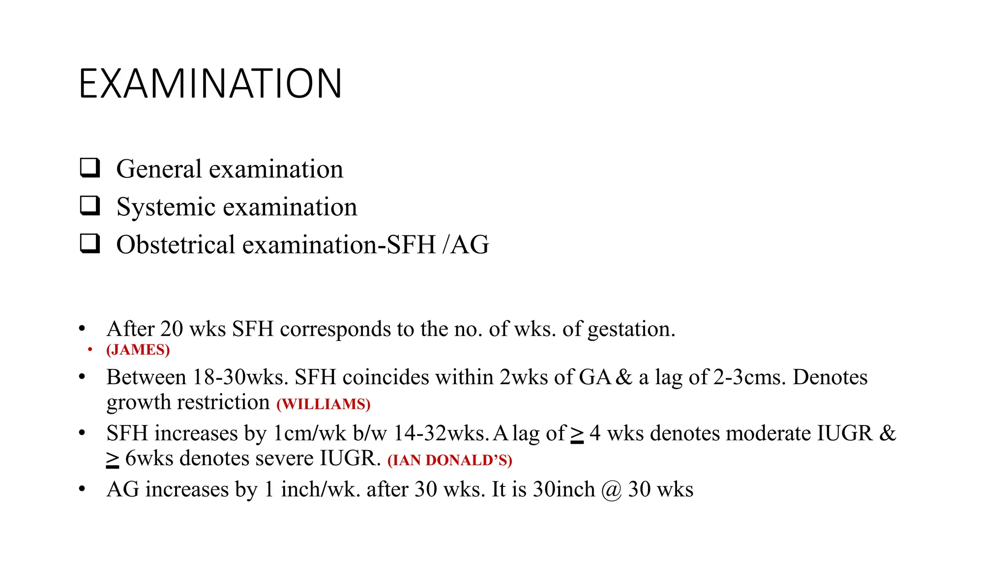
DIAGNOSIS OF IUGR
HISTORY
• Correct gestational age
• History of Previous IUGR baby
• History of disorders affecting placental function
• Obstetric history
• Dietary history
• Drug / Radiation exposure / Addiction
• Family history
• Socioeconomic status
EXAMINATION
 General examination
 Systemic examination
 Obstetrical examination-SFH /AG
• After 20 wks SFH corresponds to the no. of wks. of gestation.
• (JAMES)
• Between 18-30wks. SFH coincides within 2wks of GA& a lag of 2-3cms. Denotes
growth restriction (WILLIAMS)
• SFH increases by 1cm/wk b/w 14-32wks.Alag of > 4 wks denotes moderate IUGR &
> 6wks denotes severe IUGR. (IAN DONALD’S)
• AG increases by 1 inch/wk. after 30 wks. It is 30inch @ 30 wks
INVESTIGATION
 RoutineANC investigations
 UL
TRASOUND: Most useful inv.
• Gestation age determination- prior to 24 wks, but most
• accurate @10-12 wks
• CRL is the most accurate parameter(WILLIAMS)
• There is an error of around:
• 7days in 1sttrimester
• 10-11 days in 2ndtrimester
• 21 days in 3rdtrimester (JAMES)
• Determination of EFW:AC & EFW
• Determination of multiple gestation
• Determination of Fetal wellbeing
• Determination of Congenital anomalies:
• @ 16-20 wks of gestation (WILLIAMS)
• Determination of placenta:
• Assessment of fetal growth:
• Repeat @ 32-34 wks
• BPD(Bi Parietal Diameter)- Most accurate for
• dating in 2ndtrimester (14-26wks) [WILLIAMS]
Proximally-outer table, Distally-inner table
•
• @ THE LEVEL OFTHALAMUS & CA
VUM SEPTUMMEASURED PELLUCIDUM
• WILLIAMS 23rd EDITION
HC(Head Circumference): More accurate than
BPD in
Dolicocephalic or Brachycephalic head
CEPHALIC INDEX:
Ratio of BPD to Occipitofrontal diameter
• FL(Femur Length):
• Measured @ the level perpendicular to shaft excluding the
• epiphysis
• Correlates well with the BPD & Gestational age
• AC(Abdominal Circumference):
• Single best parameter for detection of IUGR because it is related to the liver
size which reflects fetal glycogen storage (JAMES)
• Its sensitivity is further inc. by serial measurements atleast 14 days apart (JAMES)
• We should not not label as growth restricted fetus unless AC is far below
normal or unless other parameters correlate. (JAMES)
ABDOMINAL CIRCUMFERENCE
TCD(Trans Cerebellar Diameter): Correlates well
with the gestation age
Relatively spared in mild to moderate
Uteroplacental dysfunction.
Upto 25 wks TCD in cms. = GA (IAN DONALD’S)
Age independent ratios
• HC/AC:
• Decreases linearly from 16-40wks. normally. Ratio>2SD is
suggestive of IUGR (IAN DONALD’S)
• FL/AC:
• Normal value = 22 + 2% in second half of preg. Ratio above23.5% is
abnormal (IAN DONALD’S)
• Determination ofAmniotic Fluid V
olume: Type II IUGR
causes Oligohydraminos Amniotic Fluid Index(AFI) = 5-
18 cms. Maximum Liquor Pocket = 2-8 cms.
• Doppler effect:
• Change in the apparent frequency due to relative motion
between the source & the observer.
•(Doppler probe & RBCs)
• When the sound wave strikes a moving target, the frequency
of sound waves reflected back is proportionate to the velocity
& direction of moving object.
• Used to determine the volume & rate of blood flow
through maternal vessels
Types:
• 1)CONTINUOUS W
A
VE DOPPLER:
• Two crystals are used, one transmits & other receives wave
• Used in M-mode echocardiography
• 1) PULSE W
A
VE DOPPLER:
Only one crystal that
• Transmits-wait-Receives-wait-Transmits
• Allows precise targeting & visualization of the vessel of interest
• Have software that displays blood flow-
• Towards transducer as RED
• Away from transducer as BLUE
• Angle of Insonation: Between doppler beam & direction of flow
• Higher the angle lesser the frequency & more the error
Frequency change relative to angle of insonation
• To minimize error we use RA
TIOS, to cancel off
the cosϴ
Arterial Doppler indices
 S/D Ratio: Peak systolic flow(S)
End diastolic flow(D)
 PULSA
TILITY INDEX : peak systolic velocity
end diastolic velocity
 RESISTANCE INDEX : S-D
S
• Venous Doppler indices
COLOUR DOPPLER
MATERNAL COMPARTMENT
• Uterine artery
FETAL COMPARTMENT
• Umbilical artery
• Middle Cerebral Artery
• Venous Doppler
Uterine artery Doppler:
• Its main use is in screening.
• Early diastolic notch in the uterine artery
@ 12-14 wks. suggest delayed trophoblasticinvasion (JAMES)
• Persistence of notch beyond 24 wks confirms & indicates an
increase risk of Pre-eclampsia,
• Placental abruption & Early onset IUGR. (JAMES)
• Increase impedence of flow in Uterine artery
@ 16-20 wks was predictive of superimposed
• pre-eclampsia developing inwomen with chronic hypertension.
(WILLIAMS)
Umbilical ARTERY DOPPLER
• UmbilicalA. Doppler indices should be measured
only after 23 wks (STUDD)
• It is useful adjunct in the management of pregnancies complicated by
fetal-growth restriction.(ACOG-2008)
• It is not recommended for screening of low-risk pregnancies or for
complications other than growth restriction. (WILLIAMS)
• Umbilical artery Doppler becomes abnormal when at least 30% of
the fetal villous structure is abnormal. (JAMES)
• In extreme cases of growth restriction, end-diastolic flow may become
absent or even reversed (AREDF).
• AREDF occurs when 60-70% of the fetal villous
• structure is abnormal.
• About ½ of the cases ofAREDF are associated with
• aneuploidy or a major anomaly (WILLIAMS)
• Fetuses of preeclamptic women who hadAREDF were more likely
to have hypoglycemia & polycythemia. (WILLIAMS)
ABNORMAL Umbilical ARTERY WAVEFORM
Perinatal mortality rate in AEDF- 9-41% (IAN
DONALD’S)
Reversal of the End Diastolic Flow
Perinatal mortality rate of REDF- 33-73% (IAN DONALD’S)
• Abnormal Umbilical artery flow pattern indicate an increased risk of
hypoxemia & acidemia proportionate to severity of Doppler abnormality.
(JAMES)
• Umbilical artery Doppler can also be used to distinguish b/w the high risk
small fetus that is truly growth restricted that needs inc. monitoring & the
low risk small fetus. (IAN DONALD’S)
• When Umbilical artery Doppler are incorporated into management
algorithm of growth restricted fetus, perinatal death is reduced as much
as 29%. (STUDD)
• In summary UADoppler in suspected IUGR pregnancies improves
perinatal outcome & should be used to monitor these fetuses
MIDDLE CEREBRAL ARTERY(MCA)
DOPPLER
• It was used for assessment of-
• FetalAnemia
• Growth restriction
 FETALANEMIA: (In Rh isoimmunisation)
• With increasing anemia  cardiac output increases
& blood viscosity decreases  increase flow to brain  Elevated
peak systolic velocity .WILLIAMS 23rd EDITION
Normal MCA WAVEFORM
MCA DOPPLER IN FETAL ANEMIA
 GROWTH RESTRICTION:
• It is involved in severely growth restricted fetus after involvement of
Umbilical artery.
• It is the progression of the Doppler finding & is due to the adaptive
compensatory mechanism in the fetus against increasing hypoxia
(Brain sparing effect)
Increasing hypoxia
Inc. blood flow to Vital
Organs(Brain, Heart&
Adrenals)
BRAIN SPARING EFFECT
Or
CEPHALISATION
Dec. blood flow to
Abdominal Organs(Liver &
Kidneys)
OLIGOHYDRAMINOS
MCADOPPLER- Inc.
Diastolic Flow
Dec. RI/PI/SD ratio & abn MCA-
PSV
MCA WAVEFORM IN IUGR
INCREASED FLOW DURING DIASTOLE
CEREBRO-PLACENTALRA
TIO(CPR):
MCAPulsatility Index
UmbilicalA. Pulsatility Index
It is more sensitive index for detecting poor
perinatal outcome than UAor MCADoppler
alone, but due to non standardized technique of
calculating CPR limit its clinical utility. (STUDD)
• Abnormal MCA reflects inc risk of adverse perinatal
outcome(PTL, Intrapartum acidemia & inc NICU
admission)
• Not superior to Umbilical artery Doppler
• High negative predictive value for adverse outcome
• Normal UA & MCA Doppler indices & normal AFI
in growth restricted fetus <32 wks have negative
predictive value of 97% for adverse outcome
SUMMARY OF MCA:
Despite the association of abn. MCA& adverse
perinatal outcome, there are no specific
interventions to improve outcome based on
abn. findings.
However abn. values should prompt more
frequent fetal survillence
Venous Doppler studies
• Reflects fetal cardiac function
• Most commonly used V
enous Doppler indices:
Ductus V
enosus
Inferior vena cava
Hepatic vein
Umbilical vein(Intra abdominal portion)
Ductus venosus doppler
PERINATALMORTALITY INABSENT OR REVERSE FLOW OF DV IS
63-100% (IAN DONALD’S)
HYPOXIA
INC BLOOD SHUNTING THROUGH
DV B/W UMBILICAL VEIN & IVC
INC. PULSA
TILITY
INDEX FOR VEINS (PIV)
REVERSED a W
A
VE
IN
DV PULSA
TION
PULSA
TIONS IN THE
UMBILICAL VEIN
REVERSAL OF FLOW IN
IVC DURINGA
TRIAL
CONTRACTION
ABNORMAL WAVEFORM IN UMBILICAL VEIN
UMBILICAL VEIN
Important points on venous Doppler
• Especially useful in early onset IUGR
Reason: In Term /near term fetuses
there is shorter interval & delivery is often
indicated
With advancing GAcardiac
activity becomes more efficient  slow
Steady decline in Doppler indices
• When DV & Umbilical vein Doppler- Sensitivity
inc to 70-80%.
Other investigations
 Amniocentesis
 Karyotyping
 Colour doppler
 TORCH test
 Antiphospholipid antibody
 Thrombophillia screen
 Thyroid function test
 Detailed level II ultrasound
 Biophysical Profile(BPP)
 Cardiotocography
Presumptive diagnosis of IUGR
• Symphysis Fundal Height not increasing at a normal rate
• Fetus with smallAC
• Flattening of growth curve on two consecutive occasion 14 days
apart
• Beyond 24 wks., an elevated umbilical artery Doppler index
• After 34 wks umbilical artery Doppler index may be normal
& a dec. CPR or MCADoppler index may be the only
supporting evidence of placental-based IUGR
Thank you….

DIAGNOSIS OF IUGR in pregnancycriteria in India.pptx

  • 1.
  • 2.
    HISTORY • Correct gestationalage • History of Previous IUGR baby • History of disorders affecting placental function • Obstetric history • Dietary history • Drug / Radiation exposure / Addiction • Family history • Socioeconomic status
  • 3.
    EXAMINATION  General examination Systemic examination  Obstetrical examination-SFH /AG • After 20 wks SFH corresponds to the no. of wks. of gestation. • (JAMES) • Between 18-30wks. SFH coincides within 2wks of GA& a lag of 2-3cms. Denotes growth restriction (WILLIAMS) • SFH increases by 1cm/wk b/w 14-32wks.Alag of > 4 wks denotes moderate IUGR & > 6wks denotes severe IUGR. (IAN DONALD’S) • AG increases by 1 inch/wk. after 30 wks. It is 30inch @ 30 wks
  • 4.
    INVESTIGATION  RoutineANC investigations UL TRASOUND: Most useful inv. • Gestation age determination- prior to 24 wks, but most • accurate @10-12 wks • CRL is the most accurate parameter(WILLIAMS) • There is an error of around: • 7days in 1sttrimester • 10-11 days in 2ndtrimester • 21 days in 3rdtrimester (JAMES) • Determination of EFW:AC & EFW • Determination of multiple gestation • Determination of Fetal wellbeing
  • 5.
    • Determination ofCongenital anomalies: • @ 16-20 wks of gestation (WILLIAMS) • Determination of placenta: • Assessment of fetal growth: • Repeat @ 32-34 wks • BPD(Bi Parietal Diameter)- Most accurate for • dating in 2ndtrimester (14-26wks) [WILLIAMS]
  • 6.
    Proximally-outer table, Distally-innertable • • @ THE LEVEL OFTHALAMUS & CA VUM SEPTUMMEASURED PELLUCIDUM • WILLIAMS 23rd EDITION
  • 7.
    HC(Head Circumference): Moreaccurate than BPD in Dolicocephalic or Brachycephalic head CEPHALIC INDEX: Ratio of BPD to Occipitofrontal diameter
  • 8.
    • FL(Femur Length): •Measured @ the level perpendicular to shaft excluding the • epiphysis • Correlates well with the BPD & Gestational age • AC(Abdominal Circumference): • Single best parameter for detection of IUGR because it is related to the liver size which reflects fetal glycogen storage (JAMES) • Its sensitivity is further inc. by serial measurements atleast 14 days apart (JAMES) • We should not not label as growth restricted fetus unless AC is far below normal or unless other parameters correlate. (JAMES)
  • 9.
  • 10.
    TCD(Trans Cerebellar Diameter):Correlates well with the gestation age Relatively spared in mild to moderate Uteroplacental dysfunction. Upto 25 wks TCD in cms. = GA (IAN DONALD’S)
  • 11.
    Age independent ratios •HC/AC: • Decreases linearly from 16-40wks. normally. Ratio>2SD is suggestive of IUGR (IAN DONALD’S) • FL/AC: • Normal value = 22 + 2% in second half of preg. Ratio above23.5% is abnormal (IAN DONALD’S)
  • 12.
    • Determination ofAmnioticFluid V olume: Type II IUGR causes Oligohydraminos Amniotic Fluid Index(AFI) = 5- 18 cms. Maximum Liquor Pocket = 2-8 cms.
  • 14.
    • Doppler effect: •Change in the apparent frequency due to relative motion between the source & the observer. •(Doppler probe & RBCs) • When the sound wave strikes a moving target, the frequency of sound waves reflected back is proportionate to the velocity & direction of moving object. • Used to determine the volume & rate of blood flow through maternal vessels
  • 15.
    Types: • 1)CONTINUOUS W A VEDOPPLER: • Two crystals are used, one transmits & other receives wave • Used in M-mode echocardiography • 1) PULSE W A VE DOPPLER: Only one crystal that • Transmits-wait-Receives-wait-Transmits • Allows precise targeting & visualization of the vessel of interest • Have software that displays blood flow- • Towards transducer as RED • Away from transducer as BLUE
  • 16.
    • Angle ofInsonation: Between doppler beam & direction of flow • Higher the angle lesser the frequency & more the error
  • 17.
    Frequency change relativeto angle of insonation
  • 18.
    • To minimizeerror we use RA TIOS, to cancel off the cosϴ Arterial Doppler indices  S/D Ratio: Peak systolic flow(S) End diastolic flow(D)
  • 19.
     PULSA TILITY INDEX: peak systolic velocity end diastolic velocity  RESISTANCE INDEX : S-D S • Venous Doppler indices
  • 20.
    COLOUR DOPPLER MATERNAL COMPARTMENT •Uterine artery FETAL COMPARTMENT • Umbilical artery • Middle Cerebral Artery • Venous Doppler
  • 21.
  • 22.
    • Its mainuse is in screening. • Early diastolic notch in the uterine artery @ 12-14 wks. suggest delayed trophoblasticinvasion (JAMES) • Persistence of notch beyond 24 wks confirms & indicates an increase risk of Pre-eclampsia, • Placental abruption & Early onset IUGR. (JAMES) • Increase impedence of flow in Uterine artery @ 16-20 wks was predictive of superimposed • pre-eclampsia developing inwomen with chronic hypertension. (WILLIAMS)
  • 24.
  • 25.
    • UmbilicalA. Dopplerindices should be measured only after 23 wks (STUDD) • It is useful adjunct in the management of pregnancies complicated by fetal-growth restriction.(ACOG-2008) • It is not recommended for screening of low-risk pregnancies or for complications other than growth restriction. (WILLIAMS) • Umbilical artery Doppler becomes abnormal when at least 30% of the fetal villous structure is abnormal. (JAMES)
  • 26.
    • In extremecases of growth restriction, end-diastolic flow may become absent or even reversed (AREDF). • AREDF occurs when 60-70% of the fetal villous • structure is abnormal. • About ½ of the cases ofAREDF are associated with • aneuploidy or a major anomaly (WILLIAMS) • Fetuses of preeclamptic women who hadAREDF were more likely to have hypoglycemia & polycythemia. (WILLIAMS)
  • 27.
  • 28.
    Perinatal mortality ratein AEDF- 9-41% (IAN DONALD’S)
  • 29.
    Reversal of theEnd Diastolic Flow Perinatal mortality rate of REDF- 33-73% (IAN DONALD’S)
  • 30.
    • Abnormal Umbilicalartery flow pattern indicate an increased risk of hypoxemia & acidemia proportionate to severity of Doppler abnormality. (JAMES) • Umbilical artery Doppler can also be used to distinguish b/w the high risk small fetus that is truly growth restricted that needs inc. monitoring & the low risk small fetus. (IAN DONALD’S) • When Umbilical artery Doppler are incorporated into management algorithm of growth restricted fetus, perinatal death is reduced as much as 29%. (STUDD) • In summary UADoppler in suspected IUGR pregnancies improves perinatal outcome & should be used to monitor these fetuses
  • 31.
    MIDDLE CEREBRAL ARTERY(MCA) DOPPLER •It was used for assessment of- • FetalAnemia • Growth restriction  FETALANEMIA: (In Rh isoimmunisation) • With increasing anemia  cardiac output increases & blood viscosity decreases  increase flow to brain  Elevated peak systolic velocity .WILLIAMS 23rd EDITION
  • 32.
  • 33.
    MCA DOPPLER INFETAL ANEMIA
  • 34.
     GROWTH RESTRICTION: •It is involved in severely growth restricted fetus after involvement of Umbilical artery. • It is the progression of the Doppler finding & is due to the adaptive compensatory mechanism in the fetus against increasing hypoxia (Brain sparing effect)
  • 35.
    Increasing hypoxia Inc. bloodflow to Vital Organs(Brain, Heart& Adrenals) BRAIN SPARING EFFECT Or CEPHALISATION Dec. blood flow to Abdominal Organs(Liver & Kidneys) OLIGOHYDRAMINOS MCADOPPLER- Inc. Diastolic Flow Dec. RI/PI/SD ratio & abn MCA- PSV
  • 36.
    MCA WAVEFORM INIUGR INCREASED FLOW DURING DIASTOLE
  • 37.
    CEREBRO-PLACENTALRA TIO(CPR): MCAPulsatility Index UmbilicalA. PulsatilityIndex It is more sensitive index for detecting poor perinatal outcome than UAor MCADoppler alone, but due to non standardized technique of calculating CPR limit its clinical utility. (STUDD)
  • 38.
    • Abnormal MCAreflects inc risk of adverse perinatal outcome(PTL, Intrapartum acidemia & inc NICU admission) • Not superior to Umbilical artery Doppler • High negative predictive value for adverse outcome • Normal UA & MCA Doppler indices & normal AFI in growth restricted fetus <32 wks have negative predictive value of 97% for adverse outcome
  • 39.
    SUMMARY OF MCA: Despitethe association of abn. MCA& adverse perinatal outcome, there are no specific interventions to improve outcome based on abn. findings. However abn. values should prompt more frequent fetal survillence
  • 40.
    Venous Doppler studies •Reflects fetal cardiac function • Most commonly used V enous Doppler indices: Ductus V enosus Inferior vena cava Hepatic vein Umbilical vein(Intra abdominal portion)
  • 41.
    Ductus venosus doppler PERINATALMORTALITYINABSENT OR REVERSE FLOW OF DV IS 63-100% (IAN DONALD’S)
  • 42.
    HYPOXIA INC BLOOD SHUNTINGTHROUGH DV B/W UMBILICAL VEIN & IVC INC. PULSA TILITY INDEX FOR VEINS (PIV) REVERSED a W A VE IN DV PULSA TION PULSA TIONS IN THE UMBILICAL VEIN REVERSAL OF FLOW IN IVC DURINGA TRIAL CONTRACTION
  • 43.
    ABNORMAL WAVEFORM INUMBILICAL VEIN UMBILICAL VEIN
  • 44.
    Important points onvenous Doppler • Especially useful in early onset IUGR Reason: In Term /near term fetuses there is shorter interval & delivery is often indicated With advancing GAcardiac activity becomes more efficient  slow Steady decline in Doppler indices • When DV & Umbilical vein Doppler- Sensitivity inc to 70-80%.
  • 45.
    Other investigations  Amniocentesis Karyotyping  Colour doppler  TORCH test  Antiphospholipid antibody  Thrombophillia screen  Thyroid function test  Detailed level II ultrasound  Biophysical Profile(BPP)  Cardiotocography
  • 46.
    Presumptive diagnosis ofIUGR • Symphysis Fundal Height not increasing at a normal rate • Fetus with smallAC • Flattening of growth curve on two consecutive occasion 14 days apart • Beyond 24 wks., an elevated umbilical artery Doppler index • After 34 wks umbilical artery Doppler index may be normal & a dec. CPR or MCADoppler index may be the only supporting evidence of placental-based IUGR
  • 47.