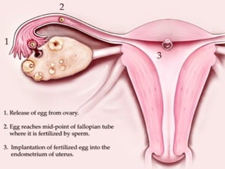
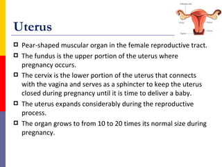
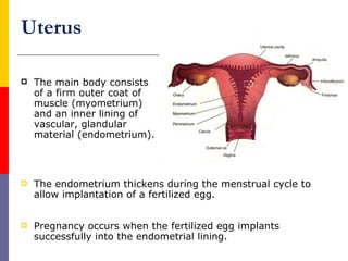
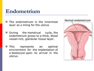
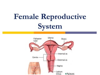
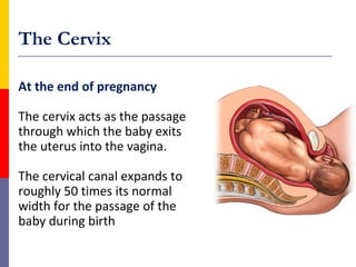
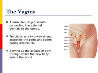
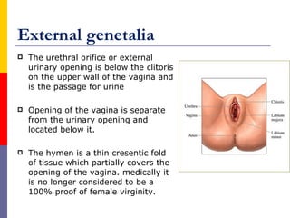
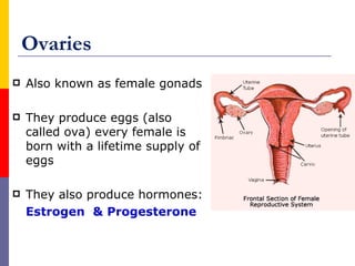
The female reproductive system produces eggs and sex hormones, supports embryo development, and enables childbirth. Major organs include the ovaries, fallopian tubes, uterus, cervix, and vagina. The ovaries produce eggs and hormones. The fallopian tubes catch eggs and allow fertilization. The uterus thickens its lining each month to potentially support a pregnancy, and expands greatly during pregnancy. The cervix acts as a passage for sperm and babies, and the vagina accepts sperm and births babies.
![Major Organs
Cervix
Vagina
Ovaries [gonads]
Uterine tubes [fallopian tubes]
Uterus](https://image.slidesharecdn.com/femalereproductivesystem-120329125448-phpapp02-221223182813-81c1b36f/85/femalereproductivesystem-120329125448-phpapp02-pdf-3-320.jpg)
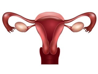
![Fallopian tubes [uterine tubes]
Stretch from the uterus to the ovaries and measure about 8
to 13 cm in length.
The ends of the fallopian tubes lying next to the ovaries
feather into ends called fimbria
Millions of tiny hair-like cilia line the fimbria and interior of
the fallopian tubes.
The cilia beat in waves hundreds of times a second catching
the egg at ovulation and moving it through the tube to the
uterine cavity.
Fertilization typically occurs in the fallopian tube](https://image.slidesharecdn.com/femalereproductivesystem-120329125448-phpapp02-221223182813-81c1b36f/85/femalereproductivesystem-120329125448-phpapp02-pdf-12-320.jpg)