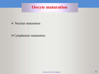
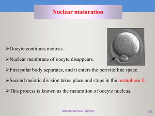
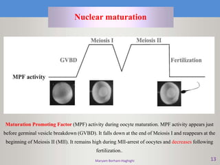
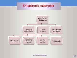
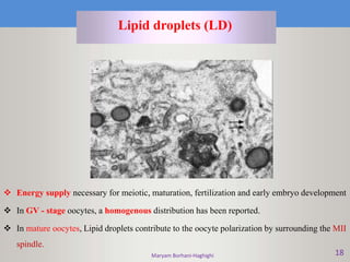
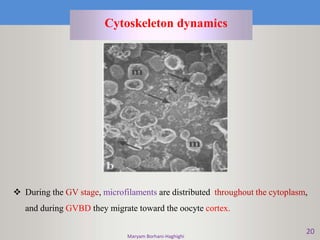
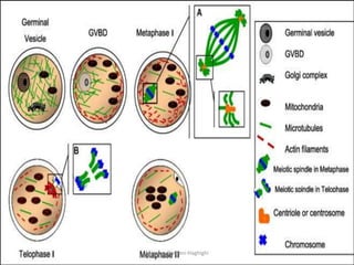
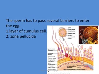
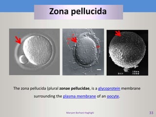
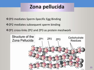
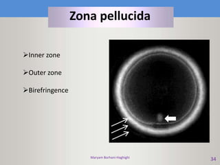
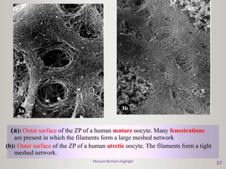
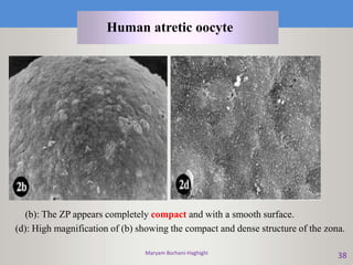
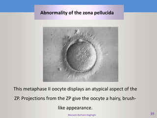
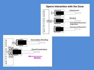
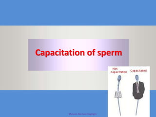
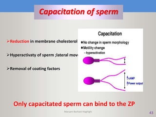
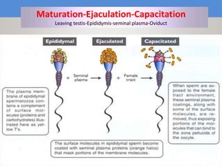
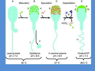
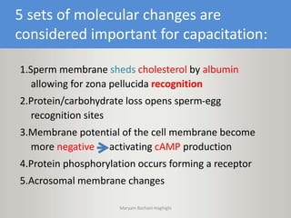
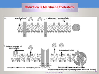
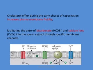
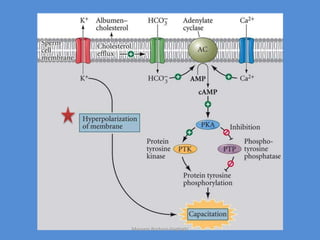
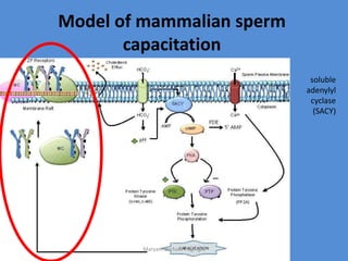
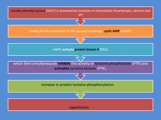
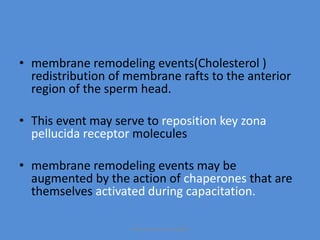
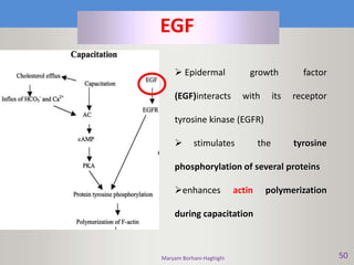
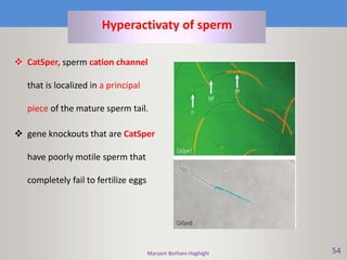
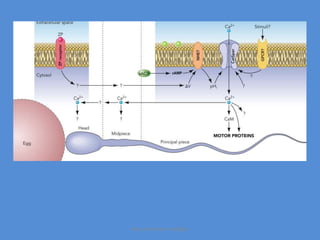
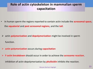
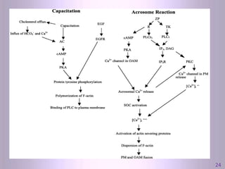
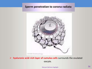
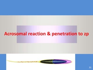
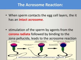
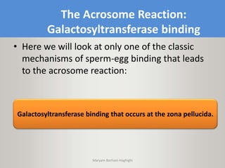
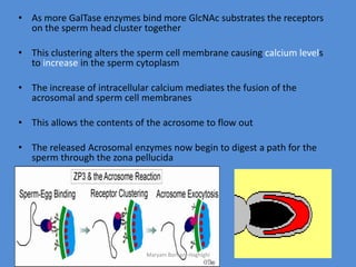
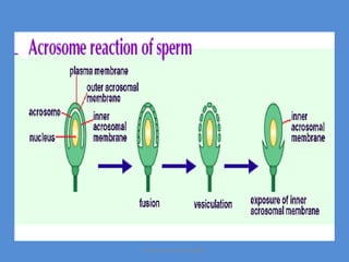
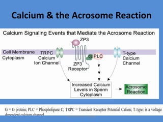
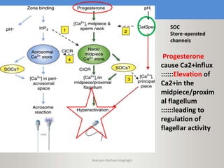
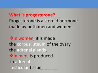
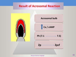
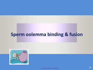
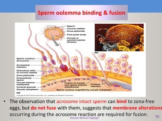
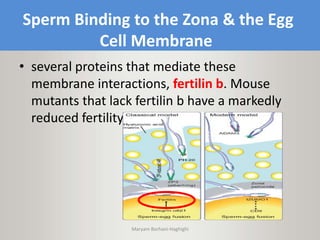
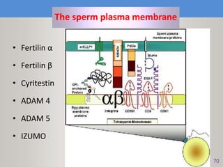
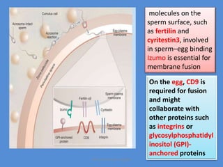
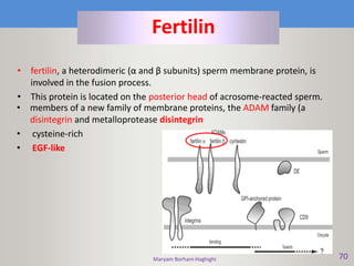
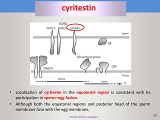
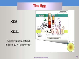
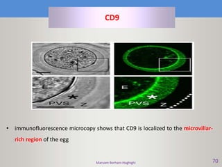
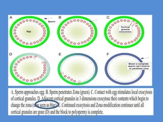
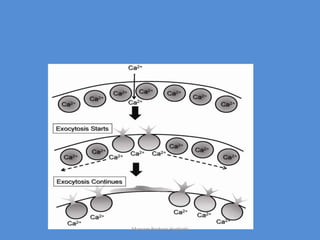
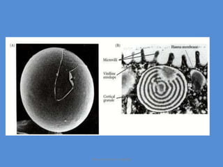
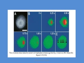
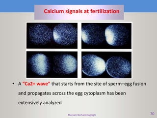
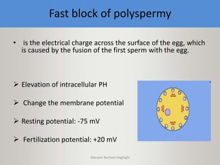
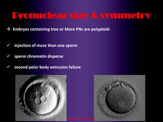
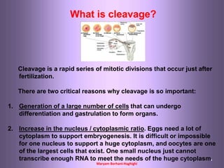
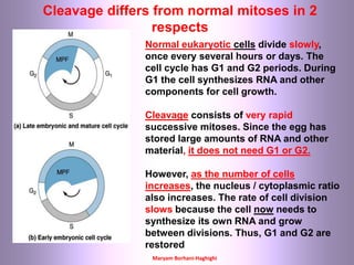
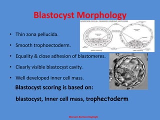
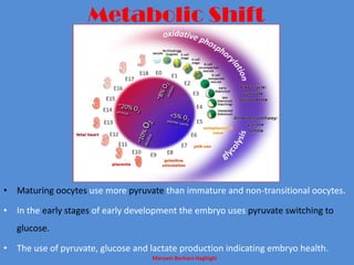
Maryam Borhani-Haghighi
PhD student
of Tehran Medical
University
Maryam Borhani-Haghighi 11
Oocyte maturation
 Nuclear maturation
Cytoplasmic maturation
Maryam Borhani-Haghighi 12
Oocyte continues meiosis.
Nuclear membrane of oocyte disappears.
First polar body separates, and it enters the perivitelline space.
Second meiotic division takes place and stops in the metaphase II.
This process is known as the maturation of oocyte nucleus.
Nuclear maturation
Maryam Borhani-Haghighi 13
Nuclear maturation
Maturation Promoting Factor (MPF) activity during oocyte maturation. MPF activity appears just
before germinal vesicle breakdown (GVBD). It falls down at the end of Meiosis I and reappears at the
beginning of Meiosis II (MII). It remains high during MII-arrest of oocytes and decreases following
fertilization..
Maryam Borhani-Haghighi
14
Cytoplasmic maturation
Cytoplasmic
maturation
Organelle
redistribution
Mitochondria
Endoplasmic
reticulum
(ER)
Cortical
granules lipid droplet
Protein
synthesis
Cytoskeleton
dynamics
Maryam Borhani-Haghighi 15
Mitochondria
 Before the beginning of oocyte maturation. Homogenous distribution of
mitochondria in the cell cortex is observed
 After nuclear maturation, mitochondria are dispersed throughout the cytoplasm
except the very central region of oocytes.
BB - Balbiani body
M - mitochondria
n - nucleus
Maryam Borhani-Haghighi 16
Endoplasmic reticulum (ER)
 ER :protein and lipid synthesis organelle
 store of Ca2+ oocyte activation in during fertilization.
 In mature oocytes, an accumulation of ER in the oocyte cortex,
specifically in the region with cortical granule exocytosis and sperm-egg fusion
Maryam Borhani-Haghighi 17
Cortical Granule (CG)
 Exocitosis of cortical granules plays an essential role in the block against
polyspermy.
 In immature oocytes, CGs are found in the whole cytoplasm (the cortex
cytoplasm and the inner cytoplasm)
 In mature oocytes, an asymmetric distribution of CGs in the cortex is observed.
Maryam Borhani-Haghighi 18
Lipid droplets (LD)
 Energy supply necessary for meiotic, maturation, fertilization and early embryo development
 In GV - stage oocytes, a homogenous distribution has been reported.
 In mature oocytes, Lipid droplets contribute to the oocyte polarization by surrounding the MII
spindle.
20
Cytoskeleton dynamics
 During the GV stage, microfilaments are distributed throughout the cytoplasm,
and during GVBD they migrate toward the oocyte cortex.
Maryam Borhani-Haghighi
The sperm has to pass several barriers to enter
the egg.
1.layer of cumulus cell.
2. zona pellucida
Maryam Borhani-Haghighi
Zona pellucida
Maryam Borhani-Haghighi 33
The zona pellucida (plural zonae pellucidae, is a glycoprotein membrane
surrounding the plasma membrane of an oocyte.
Zona pellucida
Maryam Borhani-Haghighi
35
ZP3 mediates Sperm-Specific Egg Binding
ZP2 mediates subsequent sperm binding
ZP1 cross-links ZP2 and ZP3 as protein meshwork
Zona pellucida
Maryam Borhani-Haghighi 40
 As a receptor
 Inducer of Acrosomal Reaction
 Blockade of heterospecific fertilization. Species-specific barrier
to sperm binding and penetration;
 Prevention of polyspermy
 Protection of the embryo before implantation
Functions of the ZP include
• ZP keeps sperm of foreign species out .( Not
100% effective; some cross-species fertilization
can occur (e.g., horse & donkey = mule; some
speciesof monkeys can cross fertilize) )
• Remove the ZP and other species sperm can
fertilize and egg (e.g., Hamster Test for Male
Fertility; human sperm can fertilize the zona-less
hamster egg)
Maryam Borhani-Haghighi
• The mutant mice which lacked ZP3 couldn't bind
the sperm to the zona pellucida and thus did not
get fertilized
• When their missing ZP3 gene was replaced with a
human ZP3 gene, the mice made the human ZP3
and it was incorporated into their zona pellucida.
With the human ZP3 in their zonas the eggs could
be fertilized by mouse sperm revealing that
human and mouse ZP3 can mediate the same
critical events.
Maryam Borhani-Haghighi
34
Zona pellucida
Inner zone
Outer zone
Birefringence
Maryam Borhani-Haghighi
37
(a): Outer surface of the ZP of a human mature oocyte. Many fenestrations
are present in which the filaments form a large meshed network
(b): Outer surface of the ZP of a human atretic oocyte. The filaments form a tight
meshed network.
Maryam Borhani-Haghighi
38
(b): The ZP appears completely compact and with a smooth surface.
(d): High magnification of (b) showing the compact and dense structure of the zona.
Human atretic oocyte
Maryam Borhani-Haghighi 39
This metaphase II oocyte displays an atypical aspect of the
ZP. Projections from the ZP give the oocyte a hairy, brush-
like appearance.
Abnormality of the zona pellucida
Maryam Borhani-Haghighi
Capacitation of sperm
43
Capacitation of sperm
Reduction in membrane cholesterol increase fludity
Hyperactivaty of sperm ;lateral movement of head
Removal of coating factors
Only capacitated sperm can bind to the ZP
Maryam Borhani-Haghighi
Maryam Borhani-Haghighi 52
Maturation-Ejaculation-Capacitation
Leaving testis-Epididymis-seminal plasma-Oviduct
Maryam Borhani-Haghighi
5 sets of molecular changes are
considered important for capacitation:
1.Sperm membrane sheds cholesterol by albumin
allowing for zona pellucida recognition
2.Protein/carbohydrate loss opens sperm-egg
recognition sites
3.Membrane potential of the cell membrane become
more negative activating cAMP production
4.Protein phosphorylation occurs forming a receptor
5.Acrosomal membrane changes
Maryam Borhani-Haghighi
Maryam Borhani-Haghighi 45
Reduction In Membrane Cholesterol
Cholesterol efflux during the early phases of capacitation
increases plasma membrane fluidity,
facilitating the entry of bicarbonate (HCO3-) and calcium ions
(Ca2+) into the sperm cytosol through specific membrane
channels.
Maryam Borhani-Haghighi
Model of mammalian sperm
capacitation
soluble
adenylyl
cyclase
(SACY)
Maryam Borhani-Haghighi
which then simultaneously inhibits the activity of tyrosine phosphatases (PTP) and
activates tyrosine kinases (PTK).
cAMP, activate protein kinase A (PKA),
leading to the production of the second messenger cyclic AMP (cAMP).
soluble adenylyl cyclase (SACY) is activated by increases in intracellular bicarbonate, calcium and
pH
increase in protein tyrosine phosphorylation
capacitation
Maryam Borhani-Haghighi
• membrane remodeling events(Cholesterol )
redistribution of membrane rafts to the anterior
region of the sperm head.
• This event may serve to reposition key zona
pellucida receptor molecules
• membrane remodeling events may be
augmented by the action of chaperones that are
themselves activated during capacitation.
Maryam Borhani-Haghighi
Maryam Borhani-Haghighi 50
EGF
 Epidermal growth factor
(EGF)interacts with its receptor
tyrosine kinase (EGFR)
 stimulates the tyrosine
phosphorylation of several proteins
enhances actin polymerization
during capacitation
Maryam Borhani-Haghighi 54
Hyperactivaty of sperm
 CatSper, sperm cation channel
that is localized in a principal
piece of the mature sperm tail.
 gene knockouts that are CatSper
have poorly motile sperm that
completely fail to fertilize eggs
Maryam Borhani-Haghighi
Maryam Borhani-Haghighi 23
Role of actin cytoskeleton in mammalian sperm
capacitation
• In human sperm the regions reported to contain actin include the acrosomal space,
the equatorial and post acrosomal regions, and the tail.
 actin polymerization and depolymerization might be involved in sperm
function.
 actin polymerization occurs during capacitation
 F-actin breakdown should occur in order to achieve the acrosome reaction.
inhibition of actin depolymerization by phalloidin inhibits the reaction.
Maryam Borhani-Haghighi
3124
Maryam Borhani-Haghighi 31
Sperm penetration to corona radiata
Maryam Borhani-Haghighi 56
 hyaluronic acid–rich layer of cumulus cells surrounds the ovulated
oocyte.
Sperm penetration to corona radiata
Maryam Borhani-Haghighi 31
Acrosomal reaction & penetration to zp
The Acrosome Reaction:
• When sperm contacts the egg cell layers, the it
has an intact acrosome.
• stimulation of the sperm by agents from the
corona radiata followed by binding to the
zona pellucida, leads to the acrosome reaction
Maryam Borhani-Haghighi
The Acrosome Reaction:
Galactosyltransferase binding
• Here we will look at only one of the classic
mechanisms of sperm-egg binding that leads
to the acrosome reaction:
Galactosyltransferase binding that occurs at the zona pellucida.
Maryam Borhani-Haghighi
Galactosyltransferase
• Each sperm has galactosyltransferase
(GalTase) enzymes on its head
• Galactosyltransferase is an enzyme that
transfers a sugar group from one molecule to
another
Maryam Borhani-Haghighi
Unlike normal enzyme reactions in which the enzyme binds its
substrate and produces products, this enzyme reaction cannot go
to completion. Since it can’t complete the reaction the enzyme
and the substrate remain attached to each other.
Maryam Borhani-Haghighi
• Each sperm binds N-acetylglucosamine, GlcNAc)
residues in ZP3 via galactosyltransferase (GalTase)
enzymes in the sperm cell membrane. Thus the
GlcNAc is the substrate for the GalTase enzyme.
• so the sperm remain attached via the sugar-enzyme
binding because the enzyme reaction cannot go to
completion .
Maryam Borhani-Haghighi
ZP3 and the Acrosome Reaction
Maryam Borhani-Haghighi
• As more GalTase enzymes bind more GlcNAc substrates the receptors
on the sperm head cluster together
• This clustering alters the sperm cell membrane causing calcium levels
to increase in the sperm cytoplasm
• The increase of intracellular calcium mediates the fusion of the
acrosomal and sperm cell membranes
• This allows the contents of the acrosome to flow out
• The released Acrosomal enzymes now begin to digest a path for the
sperm through the zona pellucida
Maryam Borhani-Haghighi
Calcium & the Acrosome Reaction
Maryam Borhani-Haghighi
In freshly ovulated eggs, ZP3 is in very close physical association with ZP2.
Acrosome-reacted sperm bind to ZP2 via their exposed inner acrosomal membranes, penetrate the
zona, fuse with the oocyte, and fertilize the egg.
Maryam Borhani-Haghighi
Immediately after fertilization, cortical granules release a ZP2-
specific protease and other enzymes into the perivitelline
space.
This cortical granule protease clips ZP2 and converts it into the
cleaved form (ZP2c) that no longer supports the binding of
acrosome-reacted sperm.
ZP3 dissociates from ZP2c, and undergoes a subtle modification that
converts it into a form (ZP3f) that lacks sperm receptor and acrosome-
inducing activity.
Maryam Borhani-Haghighi
Hyaluronidase
neuraminidase
acrosin
protease
trypsin-like enzyme
Arylsulfatase
PLC
Esterase
Maryam Borhani-Haghighi
 Ca2+-
Calcium is an important modulator for capacitation and AR
and is probably the key messenger in the information
exchange between sperm and egg .
 Progestrone
 Follicular fluid
 Coffein
 K+ and Na+
Acrosomal reaction
SOC
Store-operated
channels
Progesterone
cause Ca2+influx
::::::Elevation of
Ca2+in the
midpiece/proxim
al flagellum
::::::leading to
regulation of
flagellar activity
Maryam Borhani-Haghighi
What is progesterone?
Progesterone is a steroid hormone
made by both men and women.
In women, it is made
the corpus luteum of the ovary
the adrenal glands.
In men, is produced
in adrenal
testicular tissue.Maryam Borhani-Haghighi
Maryam Borhani-Haghighi 65
Result of Acrosomal Reaction
Acrosomal bulb
Ca / cAMP
Ph (7.1 7.5)
Zp Zp2f
Maryam Borhani-Haghighi 31
Sperm oolemma binding & fusion
Maryam Borhani-Haghighi 70
Sperm oolemma binding & fusion
• The observation that acrosome intact sperm can bind to zona-free
eggs, but do not fuse with them, suggests that membrane alterations
occurring during the acrosome reaction are required for fusion.
Sperm Binding to the Zona & the Egg
Cell Membrane
• several proteins that mediate these
membrane interactions, fertilin b. Mouse
mutants that lack fertilin b have a markedly
reduced fertility.
Maryam Borhani-Haghighi
Maryam Borhani-Haghighi 70
The sperm plasma membrane
• Fertilin α
• Fertilin β
• Cyritestin
• ADAM 4
• ADAM 5
• IZUMO
molecules on the
sperm surface, such
as fertilin and
cyritestin3, involved
in sperm–egg binding
Izumo is essential for
membrane fusion
On the egg, CD9 is
required for fusion
and might
collaborate with
other proteins such
as integrins or
glycosylphosphatidyl
inositol (GPI)-
anchored proteins
Maryam Borhani-Haghighi
Maryam Borhani-Haghighi 70
• members of a new family of membrane proteins, the ADAM family (a
disintegrin and metalloprotease disintegrin
• cysteine-rich
• EGF-like
Fertilin
• fertilin, a heterodimeric (α and β subunits) sperm membrane protein, is
involved in the fusion process.
• This protein is located on the posterior head of acrosome-reacted sperm.
Maryam Borhani-Haghighi 70
• Localization of cyritestin in the equatorial region is consistent with its
participation in sperm–egg fusion.
• Although both the equatorial regions and posterior head of the sperm
membrane fuse with the egg membrane,
cyritestin
Maryam Borhani-Haghighi 70
The Egg
.CD9
.CD81
Glycosylphosphatidyl
inositol (GPI)-anchored
Maryam Borhani-Haghighi 70
• immunofluorescence microcopy shows that CD9 is localized to the microvillar-
rich region of the egg
CD9
Maryam Borhani-Haghighi 70
CD81
• CD81, which fairly closely resembles CD9 in structure
and some functions, has a role in gamete fusion.
However, the results of deleting the CD81 gene are
less dramatic than those seen with the deletion of
CD9.
Maryam Borhani-Haghighi 70
Sperm oolema binding & fusion
The egg plasma membrane can be divided into two major regions.
1.The part of the membrane that directly overlies the metaphase
chromosomes has a smooth surface devoid of microvilli.
2. The remainder of the egg is rich in microvillar protrusions. This is the region
of the egg where sperm both bind and fuse.
Maryam Borhani-Haghighi
Maryam Borhani-Haghighi 70
Fertilization cone
Block to Polyspermy
• Special blocks to polyspermy exist:
• 1. Fast Block: electrical change in egg
membrane
2. Slow Block: modification of zona pellucida
Maryam Borhani-Haghighi
Maryam Borhani-Haghighi 70
Slow block to polyspermy
• Elavation of Ca2+
• Cortical granule reaction
Exocytosis Perivitteline space
 Zp2
 β-hexoaminidase
 Tyrosin
Slow block of polyspermy
The Cortical Reaction
• Calcium not only plays a role
in the acrosome reaction, it
also mediates the subsequent
event of corticalgranule
exocytosis in the egg.
• Ca2+ induces local exocytosis
of cortical granules
• Granules release to stimulate
adjacent cortical granules to
undergo exocytosis
• Wave of exocytosis occurs
around egg in 3 dimensions
from original site of sperm
entry Maryam Borhani-Haghighi
Maryam Borhani-Haghighi
Maryam Borhani-Haghighi 70
Calcium signals at fertilization
• A “Ca2+ wave” that starts from the site of sperm–egg fusion
and propagates across the egg cytoplasm has been
extensively analyzed
Fast block of polyspermy
• is the electrical charge across the surface of the egg, which
is caused by the fusion of the first sperm with the egg.
 Elevation of intracellular PH
 Change the membrane potential
 Resting potential: -75 mV
 Fertilization potential: +20 mV
Maryam Borhani-Haghighi
a) Glutathione
b) Heparin
c) Zn
Decondensation of sperm nucleus
From the following cell is seen the protamine
to histone transition where maternally derived
histones replace protamines resulting in the
decondensation of the sperm headMaryam Borhani-Haghighi
Human sperm nuclear decondensation in vivo involves
protamine disulfide bond reduction by glutathione
(GSH) and protamine/histone exchange, presumably
with heparan sulfate (HS) as the protamine acceptor
Maryam Borhani-Haghighi
A significant fraction of the zinc is lost
from sperm chromatin when the cysteine
thiols in protamine are oxidized into
disulfide bonds.
Extraction of zinc from the freshly
ejaculated spermatozoon allows
immediate decondensation
Maryam Borhani-Haghighi
• Male PN : 22.1 µm
• Female PN : 24.4 µm
• Male PN has more Nucleoli
• Female PN is close to polar body
The male pronucleus is colorized in blue;
female in pink.
Maryam Borhani-Haghighi
Maryam Borhani-Haghighi 70
Role of tail
sperm-aster
 A centrosome with astral rays in the cytoplasm of
an inseminated ovum;
 it is brought in by the penetrating spermatozoon
and gives rise to the mitotic spindle of the first-
cleavage division.
Proximal centriol
Maryam Borhani-Haghighi 31
syngamy
Maryam Borhani-Haghighi 70
Syngamy
Union of gametes resulting
in formation of a zygote
syngamy
Maryam Borhani-Haghighi 31
Singamy
At gamete fusion the sperm tail is incorporated into the ooplasm,
the centriolar region forms the sperm aster while the sperm head is
decondensing
; this aster acts to guide the female pronucleus towards the male pronucleus.
The centriole duplicates during the pronuclear stage,
and at syngamy centrioles are found at opposite poles of the first cleavage.
The centrosome has several implications for human infertility.
It is possible that immotile or nonprogressively motile spermatozoa may
possess centriolar abnormalities or an absence of centrioles.
Similarly, antisperm antibodies against centrioles may be responsible for
mitotic arrest.
One way of solving this problem would be the use of donor centrosomesMaryam Borhani-Haghighi
Maryam Borhani-Haghighi
Pronuclear scoring system
• 16-18 hours after fertilization
• Grading is based on:
– Pronuclear size, symmetry & position
– Size, number, equality & distribution of nucleoli
– Appearance of cytoplasm
Van Blerkom (1990)
Maryam Borhani-Haghighi
Pronuclear size & symmetry
 Embryos containing tree or More PNs are polyploid:
 injection of more than one sperm
 sperm chromatin disperse
 second polar body extrusion failure
Maryam Borhani-Haghighi
What is cleavage?
Cleavage is a rapid series of mitotic divisions that occur just after
fertilization.
There are two critical reasons why cleavage is so important:
1. Generation of a large number of cells that can undergo
differentiation and gastrulation to form organs.
2. Increase in the nucleus / cytoplasmic ratio. Eggs need a lot of
cytoplasm to support embryogenesis. It is difficult or impossible
for one nucleus to support a huge cytoplasm, and oocytes are one
of the largest cells that exist. One small nucleus just cannot
transcribe enough RNA to meet the needs of the huge cytoplasm.
Maryam Borhani-Haghighi
Cleavage differs from normal mitoses in 2
respects
Normal eukaryotic cells divide slowly,
once every several hours or days. The
cell cycle has G1 and G2 periods. During
G1 the cell synthesizes RNA and other
components for cell growth.
Cleavage consists of very rapid
successive mitoses. Since the egg has
stored large amounts of RNA and other
material, it does not need G1 or G2.
However, as the number of cells
increases, the nucleus / cytoplasmic ratio
also increases. The rate of cell division
slows because the cell now needs to
synthesize its own RNA and grow
between divisions. Thus, G1 and G2 are
restored
Maryam Borhani-Haghighi
Asynchronous cleavage: mammalian embryos are unusual in that they have
asynchronous cleavage. Not all blastomeres divide at the same time.
The first cleavage is meridional, and the second cleavage is rotational.
The 2 blastomeres divide in different planes (one is equatorial and one is meridional
Maryam Borhani-Haghighi
Cleaved embryo assessment
24-28 hours after insemination
2 cells
40-44 hours after insemination
4 cells
64-68 hours after insemination
8 cells
Maryam Borhani-Haghighi
•Morula stage in mammals begins when the embryo
consist of 16 blastomers , occurs 3 to 4 days after
fertilization, when embryo passes from oviduct into
the uterus
MORULA
Maryam Borhani-Haghighi
Compaction
During compaction each of the eight blastomeres undergo a polarization.
Polarization and the formation of tight junction allow the blastomers to
create an inner embryo environment that is different from the outside
environment
Tight junctions develop beneath ext. surfaces and gap junction form
between the internal surfaces
Maryam Borhani-Haghighi
Blastocyst Morphology
• Thin zona pellucida.
• Smooth trophoectoderm.
• Equality & close adhesion of blastomeres.
• Clearly visible blastocyst cavity.
• Well developed inner cell mass.
Blastocyst scoring is based on:
blastocyst, Inner cell mass, trophectoderm
Maryam Borhani-Haghighi
eomesoder
min
Cdx2
Trophobla
st
Oct4
Nanog
ICM
Maryam Borhani-Haghighi 70
The act of hatching involves the local digestion of the zona by an enzyme produced in
patch of trophoblast cells situated opposite the inner cell mass (this location
minimizes the risk of collateral enzymatic damage to the embryo
Having escape from the zona ,the blastocyst begins the process of implantation
Hatching
Maryam Borhani-Haghighi
Metabolic Shift
• Maturing oocytes use more pyruvate than immature and non-transitional oocytes.
• In the early stages of early development the embryo uses pyruvate switching to
glucose.
• The use of pyruvate, glucose and lactate production indicating embryo health.
Maryam Borhani-Haghighi
• Blastocyst with a glucose uptake >5µg/h develop
better in culture and give rise to more pregnancies.
• Morphologically normal blastocyst use more
glucose than degenerating blastocyst.
Thank you for your
attention
Thank you for your attentionMaryam Borhani-Haghighi
Maryam Borhani-Haghighi 70
Thank you for your
attention
Developing Embryo
Thank you for your attention
Maryam Borhani-Haghighi

Human fertilization

  • 1.
    Maryam Borhani-Haghighi PhD student ofTehran Medical University
  • 2.
    Maryam Borhani-Haghighi 11 Oocytematuration  Nuclear maturation Cytoplasmic maturation
  • 3.
    Maryam Borhani-Haghighi 12 Oocytecontinues meiosis. Nuclear membrane of oocyte disappears. First polar body separates, and it enters the perivitelline space. Second meiotic division takes place and stops in the metaphase II. This process is known as the maturation of oocyte nucleus. Nuclear maturation
  • 4.
    Maryam Borhani-Haghighi 13 Nuclearmaturation Maturation Promoting Factor (MPF) activity during oocyte maturation. MPF activity appears just before germinal vesicle breakdown (GVBD). It falls down at the end of Meiosis I and reappears at the beginning of Meiosis II (MII). It remains high during MII-arrest of oocytes and decreases following fertilization..
  • 5.
  • 6.
    Maryam Borhani-Haghighi 15 Mitochondria Before the beginning of oocyte maturation. Homogenous distribution of mitochondria in the cell cortex is observed  After nuclear maturation, mitochondria are dispersed throughout the cytoplasm except the very central region of oocytes. BB - Balbiani body M - mitochondria n - nucleus
  • 7.
    Maryam Borhani-Haghighi 16 Endoplasmicreticulum (ER)  ER :protein and lipid synthesis organelle  store of Ca2+ oocyte activation in during fertilization.  In mature oocytes, an accumulation of ER in the oocyte cortex, specifically in the region with cortical granule exocytosis and sperm-egg fusion
  • 8.
    Maryam Borhani-Haghighi 17 CorticalGranule (CG)  Exocitosis of cortical granules plays an essential role in the block against polyspermy.  In immature oocytes, CGs are found in the whole cytoplasm (the cortex cytoplasm and the inner cytoplasm)  In mature oocytes, an asymmetric distribution of CGs in the cortex is observed.
  • 9.
    Maryam Borhani-Haghighi 18 Lipiddroplets (LD)  Energy supply necessary for meiotic, maturation, fertilization and early embryo development  In GV - stage oocytes, a homogenous distribution has been reported.  In mature oocytes, Lipid droplets contribute to the oocyte polarization by surrounding the MII spindle.
  • 10.
    20 Cytoskeleton dynamics  Duringthe GV stage, microfilaments are distributed throughout the cytoplasm, and during GVBD they migrate toward the oocyte cortex. Maryam Borhani-Haghighi
  • 11.
  • 12.
    The sperm hasto pass several barriers to enter the egg. 1.layer of cumulus cell. 2. zona pellucida Maryam Borhani-Haghighi
  • 13.
  • 14.
    Maryam Borhani-Haghighi 33 Thezona pellucida (plural zonae pellucidae, is a glycoprotein membrane surrounding the plasma membrane of an oocyte. Zona pellucida
  • 15.
    Maryam Borhani-Haghighi 35 ZP3 mediatesSperm-Specific Egg Binding ZP2 mediates subsequent sperm binding ZP1 cross-links ZP2 and ZP3 as protein meshwork Zona pellucida
  • 16.
    Maryam Borhani-Haghighi 40 As a receptor  Inducer of Acrosomal Reaction  Blockade of heterospecific fertilization. Species-specific barrier to sperm binding and penetration;  Prevention of polyspermy  Protection of the embryo before implantation Functions of the ZP include
  • 17.
    • ZP keepssperm of foreign species out .( Not 100% effective; some cross-species fertilization can occur (e.g., horse & donkey = mule; some speciesof monkeys can cross fertilize) ) • Remove the ZP and other species sperm can fertilize and egg (e.g., Hamster Test for Male Fertility; human sperm can fertilize the zona-less hamster egg) Maryam Borhani-Haghighi
  • 18.
    • The mutantmice which lacked ZP3 couldn't bind the sperm to the zona pellucida and thus did not get fertilized • When their missing ZP3 gene was replaced with a human ZP3 gene, the mice made the human ZP3 and it was incorporated into their zona pellucida. With the human ZP3 in their zonas the eggs could be fertilized by mouse sperm revealing that human and mouse ZP3 can mediate the same critical events. Maryam Borhani-Haghighi
  • 19.
    Maryam Borhani-Haghighi 34 Zona pellucida Innerzone Outer zone Birefringence
  • 20.
    Maryam Borhani-Haghighi 37 (a): Outersurface of the ZP of a human mature oocyte. Many fenestrations are present in which the filaments form a large meshed network (b): Outer surface of the ZP of a human atretic oocyte. The filaments form a tight meshed network.
  • 21.
    Maryam Borhani-Haghighi 38 (b): TheZP appears completely compact and with a smooth surface. (d): High magnification of (b) showing the compact and dense structure of the zona. Human atretic oocyte
  • 22.
    Maryam Borhani-Haghighi 39 Thismetaphase II oocyte displays an atypical aspect of the ZP. Projections from the ZP give the oocyte a hairy, brush- like appearance. Abnormality of the zona pellucida
  • 23.
  • 24.
  • 25.
    43 Capacitation of sperm Reductionin membrane cholesterol increase fludity Hyperactivaty of sperm ;lateral movement of head Removal of coating factors Only capacitated sperm can bind to the ZP Maryam Borhani-Haghighi
  • 26.
  • 27.
  • 28.
    5 sets ofmolecular changes are considered important for capacitation: 1.Sperm membrane sheds cholesterol by albumin allowing for zona pellucida recognition 2.Protein/carbohydrate loss opens sperm-egg recognition sites 3.Membrane potential of the cell membrane become more negative activating cAMP production 4.Protein phosphorylation occurs forming a receptor 5.Acrosomal membrane changes Maryam Borhani-Haghighi
  • 29.
    Maryam Borhani-Haghighi 45 ReductionIn Membrane Cholesterol
  • 30.
    Cholesterol efflux duringthe early phases of capacitation increases plasma membrane fluidity, facilitating the entry of bicarbonate (HCO3-) and calcium ions (Ca2+) into the sperm cytosol through specific membrane channels. Maryam Borhani-Haghighi
  • 31.
  • 32.
    Model of mammaliansperm capacitation soluble adenylyl cyclase (SACY) Maryam Borhani-Haghighi
  • 33.
    which then simultaneouslyinhibits the activity of tyrosine phosphatases (PTP) and activates tyrosine kinases (PTK). cAMP, activate protein kinase A (PKA), leading to the production of the second messenger cyclic AMP (cAMP). soluble adenylyl cyclase (SACY) is activated by increases in intracellular bicarbonate, calcium and pH increase in protein tyrosine phosphorylation capacitation Maryam Borhani-Haghighi
  • 34.
    • membrane remodelingevents(Cholesterol ) redistribution of membrane rafts to the anterior region of the sperm head. • This event may serve to reposition key zona pellucida receptor molecules • membrane remodeling events may be augmented by the action of chaperones that are themselves activated during capacitation. Maryam Borhani-Haghighi
  • 35.
    Maryam Borhani-Haghighi 50 EGF Epidermal growth factor (EGF)interacts with its receptor tyrosine kinase (EGFR)  stimulates the tyrosine phosphorylation of several proteins enhances actin polymerization during capacitation
  • 36.
    Maryam Borhani-Haghighi 54 Hyperactivatyof sperm  CatSper, sperm cation channel that is localized in a principal piece of the mature sperm tail.  gene knockouts that are CatSper have poorly motile sperm that completely fail to fertilize eggs
  • 37.
  • 38.
    Maryam Borhani-Haghighi 23 Roleof actin cytoskeleton in mammalian sperm capacitation • In human sperm the regions reported to contain actin include the acrosomal space, the equatorial and post acrosomal regions, and the tail.  actin polymerization and depolymerization might be involved in sperm function.  actin polymerization occurs during capacitation  F-actin breakdown should occur in order to achieve the acrosome reaction. inhibition of actin depolymerization by phalloidin inhibits the reaction.
  • 39.
  • 40.
    Maryam Borhani-Haghighi 31 Spermpenetration to corona radiata
  • 41.
    Maryam Borhani-Haghighi 56 hyaluronic acid–rich layer of cumulus cells surrounds the ovulated oocyte. Sperm penetration to corona radiata
  • 42.
    Maryam Borhani-Haghighi 31 Acrosomalreaction & penetration to zp
  • 43.
    The Acrosome Reaction: •When sperm contacts the egg cell layers, the it has an intact acrosome. • stimulation of the sperm by agents from the corona radiata followed by binding to the zona pellucida, leads to the acrosome reaction Maryam Borhani-Haghighi
  • 44.
    The Acrosome Reaction: Galactosyltransferasebinding • Here we will look at only one of the classic mechanisms of sperm-egg binding that leads to the acrosome reaction: Galactosyltransferase binding that occurs at the zona pellucida. Maryam Borhani-Haghighi
  • 45.
    Galactosyltransferase • Each spermhas galactosyltransferase (GalTase) enzymes on its head • Galactosyltransferase is an enzyme that transfers a sugar group from one molecule to another Maryam Borhani-Haghighi
  • 46.
    Unlike normal enzymereactions in which the enzyme binds its substrate and produces products, this enzyme reaction cannot go to completion. Since it can’t complete the reaction the enzyme and the substrate remain attached to each other. Maryam Borhani-Haghighi
  • 47.
    • Each spermbinds N-acetylglucosamine, GlcNAc) residues in ZP3 via galactosyltransferase (GalTase) enzymes in the sperm cell membrane. Thus the GlcNAc is the substrate for the GalTase enzyme. • so the sperm remain attached via the sugar-enzyme binding because the enzyme reaction cannot go to completion . Maryam Borhani-Haghighi
  • 48.
    ZP3 and theAcrosome Reaction Maryam Borhani-Haghighi
  • 49.
    • As moreGalTase enzymes bind more GlcNAc substrates the receptors on the sperm head cluster together • This clustering alters the sperm cell membrane causing calcium levels to increase in the sperm cytoplasm • The increase of intracellular calcium mediates the fusion of the acrosomal and sperm cell membranes • This allows the contents of the acrosome to flow out • The released Acrosomal enzymes now begin to digest a path for the sperm through the zona pellucida Maryam Borhani-Haghighi
  • 50.
  • 51.
    Calcium & theAcrosome Reaction Maryam Borhani-Haghighi
  • 52.
  • 53.
    In freshly ovulatedeggs, ZP3 is in very close physical association with ZP2. Acrosome-reacted sperm bind to ZP2 via their exposed inner acrosomal membranes, penetrate the zona, fuse with the oocyte, and fertilize the egg. Maryam Borhani-Haghighi
  • 54.
    Immediately after fertilization,cortical granules release a ZP2- specific protease and other enzymes into the perivitelline space. This cortical granule protease clips ZP2 and converts it into the cleaved form (ZP2c) that no longer supports the binding of acrosome-reacted sperm. ZP3 dissociates from ZP2c, and undergoes a subtle modification that converts it into a form (ZP3f) that lacks sperm receptor and acrosome- inducing activity. Maryam Borhani-Haghighi
  • 55.
  • 56.
  • 57.
    Maryam Borhani-Haghighi  Ca2+- Calciumis an important modulator for capacitation and AR and is probably the key messenger in the information exchange between sperm and egg .  Progestrone  Follicular fluid  Coffein  K+ and Na+ Acrosomal reaction
  • 58.
    SOC Store-operated channels Progesterone cause Ca2+influx ::::::Elevation of Ca2+inthe midpiece/proxim al flagellum ::::::leading to regulation of flagellar activity Maryam Borhani-Haghighi
  • 59.
    What is progesterone? Progesteroneis a steroid hormone made by both men and women. In women, it is made the corpus luteum of the ovary the adrenal glands. In men, is produced in adrenal testicular tissue.Maryam Borhani-Haghighi
  • 60.
    Maryam Borhani-Haghighi 65 Resultof Acrosomal Reaction Acrosomal bulb Ca / cAMP Ph (7.1 7.5) Zp Zp2f
  • 61.
    Maryam Borhani-Haghighi 31 Spermoolemma binding & fusion
  • 62.
    Maryam Borhani-Haghighi 70 Spermoolemma binding & fusion • The observation that acrosome intact sperm can bind to zona-free eggs, but do not fuse with them, suggests that membrane alterations occurring during the acrosome reaction are required for fusion.
  • 63.
    Sperm Binding tothe Zona & the Egg Cell Membrane • several proteins that mediate these membrane interactions, fertilin b. Mouse mutants that lack fertilin b have a markedly reduced fertility. Maryam Borhani-Haghighi
  • 64.
    Maryam Borhani-Haghighi 70 Thesperm plasma membrane • Fertilin α • Fertilin β • Cyritestin • ADAM 4 • ADAM 5 • IZUMO
  • 65.
    molecules on the spermsurface, such as fertilin and cyritestin3, involved in sperm–egg binding Izumo is essential for membrane fusion On the egg, CD9 is required for fusion and might collaborate with other proteins such as integrins or glycosylphosphatidyl inositol (GPI)- anchored proteins Maryam Borhani-Haghighi
  • 66.
    Maryam Borhani-Haghighi 70 •members of a new family of membrane proteins, the ADAM family (a disintegrin and metalloprotease disintegrin • cysteine-rich • EGF-like Fertilin • fertilin, a heterodimeric (α and β subunits) sperm membrane protein, is involved in the fusion process. • This protein is located on the posterior head of acrosome-reacted sperm.
  • 67.
    Maryam Borhani-Haghighi 70 •Localization of cyritestin in the equatorial region is consistent with its participation in sperm–egg fusion. • Although both the equatorial regions and posterior head of the sperm membrane fuse with the egg membrane, cyritestin
  • 68.
    Maryam Borhani-Haghighi 70 TheEgg .CD9 .CD81 Glycosylphosphatidyl inositol (GPI)-anchored
  • 69.
    Maryam Borhani-Haghighi 70 •immunofluorescence microcopy shows that CD9 is localized to the microvillar- rich region of the egg CD9
  • 70.
    Maryam Borhani-Haghighi 70 CD81 •CD81, which fairly closely resembles CD9 in structure and some functions, has a role in gamete fusion. However, the results of deleting the CD81 gene are less dramatic than those seen with the deletion of CD9.
  • 71.
    Maryam Borhani-Haghighi 70 Spermoolema binding & fusion The egg plasma membrane can be divided into two major regions. 1.The part of the membrane that directly overlies the metaphase chromosomes has a smooth surface devoid of microvilli. 2. The remainder of the egg is rich in microvillar protrusions. This is the region of the egg where sperm both bind and fuse.
  • 72.
  • 73.
  • 74.
    Block to Polyspermy •Special blocks to polyspermy exist: • 1. Fast Block: electrical change in egg membrane 2. Slow Block: modification of zona pellucida Maryam Borhani-Haghighi
  • 75.
  • 76.
    Maryam Borhani-Haghighi 70 Slowblock to polyspermy • Elavation of Ca2+ • Cortical granule reaction Exocytosis Perivitteline space  Zp2  β-hexoaminidase  Tyrosin
  • 77.
    Slow block ofpolyspermy The Cortical Reaction • Calcium not only plays a role in the acrosome reaction, it also mediates the subsequent event of corticalgranule exocytosis in the egg. • Ca2+ induces local exocytosis of cortical granules • Granules release to stimulate adjacent cortical granules to undergo exocytosis • Wave of exocytosis occurs around egg in 3 dimensions from original site of sperm entry Maryam Borhani-Haghighi
  • 78.
  • 79.
  • 80.
  • 81.
  • 82.
    Maryam Borhani-Haghighi 70 Calciumsignals at fertilization • A “Ca2+ wave” that starts from the site of sperm–egg fusion and propagates across the egg cytoplasm has been extensively analyzed
  • 83.
    Fast block ofpolyspermy • is the electrical charge across the surface of the egg, which is caused by the fusion of the first sperm with the egg.  Elevation of intracellular PH  Change the membrane potential  Resting potential: -75 mV  Fertilization potential: +20 mV Maryam Borhani-Haghighi
  • 84.
    Maryam Borhani-Haghighi a) Glutathione b)Heparin c) Zn Decondensation of sperm nucleus
  • 85.
    From the followingcell is seen the protamine to histone transition where maternally derived histones replace protamines resulting in the decondensation of the sperm headMaryam Borhani-Haghighi
  • 86.
    Human sperm nucleardecondensation in vivo involves protamine disulfide bond reduction by glutathione (GSH) and protamine/histone exchange, presumably with heparan sulfate (HS) as the protamine acceptor Maryam Borhani-Haghighi
  • 87.
    A significant fractionof the zinc is lost from sperm chromatin when the cysteine thiols in protamine are oxidized into disulfide bonds. Extraction of zinc from the freshly ejaculated spermatozoon allows immediate decondensation Maryam Borhani-Haghighi
  • 88.
    • Male PN: 22.1 µm • Female PN : 24.4 µm • Male PN has more Nucleoli • Female PN is close to polar body The male pronucleus is colorized in blue; female in pink. Maryam Borhani-Haghighi
  • 89.
    Maryam Borhani-Haghighi 70 Roleof tail sperm-aster  A centrosome with astral rays in the cytoplasm of an inseminated ovum;  it is brought in by the penetrating spermatozoon and gives rise to the mitotic spindle of the first- cleavage division. Proximal centriol
  • 90.
  • 91.
    Maryam Borhani-Haghighi 70 Syngamy Unionof gametes resulting in formation of a zygote syngamy
  • 92.
  • 93.
    At gamete fusionthe sperm tail is incorporated into the ooplasm, the centriolar region forms the sperm aster while the sperm head is decondensing ; this aster acts to guide the female pronucleus towards the male pronucleus. The centriole duplicates during the pronuclear stage, and at syngamy centrioles are found at opposite poles of the first cleavage. The centrosome has several implications for human infertility. It is possible that immotile or nonprogressively motile spermatozoa may possess centriolar abnormalities or an absence of centrioles. Similarly, antisperm antibodies against centrioles may be responsible for mitotic arrest. One way of solving this problem would be the use of donor centrosomesMaryam Borhani-Haghighi
  • 94.
    Maryam Borhani-Haghighi Pronuclear scoringsystem • 16-18 hours after fertilization • Grading is based on: – Pronuclear size, symmetry & position – Size, number, equality & distribution of nucleoli – Appearance of cytoplasm Van Blerkom (1990)
  • 95.
    Maryam Borhani-Haghighi Pronuclear size& symmetry  Embryos containing tree or More PNs are polyploid:  injection of more than one sperm  sperm chromatin disperse  second polar body extrusion failure
  • 96.
    Maryam Borhani-Haghighi What iscleavage? Cleavage is a rapid series of mitotic divisions that occur just after fertilization. There are two critical reasons why cleavage is so important: 1. Generation of a large number of cells that can undergo differentiation and gastrulation to form organs. 2. Increase in the nucleus / cytoplasmic ratio. Eggs need a lot of cytoplasm to support embryogenesis. It is difficult or impossible for one nucleus to support a huge cytoplasm, and oocytes are one of the largest cells that exist. One small nucleus just cannot transcribe enough RNA to meet the needs of the huge cytoplasm.
  • 97.
    Maryam Borhani-Haghighi Cleavage differsfrom normal mitoses in 2 respects Normal eukaryotic cells divide slowly, once every several hours or days. The cell cycle has G1 and G2 periods. During G1 the cell synthesizes RNA and other components for cell growth. Cleavage consists of very rapid successive mitoses. Since the egg has stored large amounts of RNA and other material, it does not need G1 or G2. However, as the number of cells increases, the nucleus / cytoplasmic ratio also increases. The rate of cell division slows because the cell now needs to synthesize its own RNA and grow between divisions. Thus, G1 and G2 are restored
  • 98.
    Maryam Borhani-Haghighi Asynchronous cleavage:mammalian embryos are unusual in that they have asynchronous cleavage. Not all blastomeres divide at the same time. The first cleavage is meridional, and the second cleavage is rotational. The 2 blastomeres divide in different planes (one is equatorial and one is meridional
  • 99.
    Maryam Borhani-Haghighi Cleaved embryoassessment 24-28 hours after insemination 2 cells 40-44 hours after insemination 4 cells 64-68 hours after insemination 8 cells
  • 100.
    Maryam Borhani-Haghighi •Morula stagein mammals begins when the embryo consist of 16 blastomers , occurs 3 to 4 days after fertilization, when embryo passes from oviduct into the uterus MORULA
  • 101.
    Maryam Borhani-Haghighi Compaction During compactioneach of the eight blastomeres undergo a polarization. Polarization and the formation of tight junction allow the blastomers to create an inner embryo environment that is different from the outside environment Tight junctions develop beneath ext. surfaces and gap junction form between the internal surfaces
  • 102.
    Maryam Borhani-Haghighi Blastocyst Morphology •Thin zona pellucida. • Smooth trophoectoderm. • Equality & close adhesion of blastomeres. • Clearly visible blastocyst cavity. • Well developed inner cell mass. Blastocyst scoring is based on: blastocyst, Inner cell mass, trophectoderm
  • 103.
  • 104.
    Maryam Borhani-Haghighi 70 Theact of hatching involves the local digestion of the zona by an enzyme produced in patch of trophoblast cells situated opposite the inner cell mass (this location minimizes the risk of collateral enzymatic damage to the embryo Having escape from the zona ,the blastocyst begins the process of implantation Hatching
  • 105.
    Maryam Borhani-Haghighi Metabolic Shift •Maturing oocytes use more pyruvate than immature and non-transitional oocytes. • In the early stages of early development the embryo uses pyruvate switching to glucose. • The use of pyruvate, glucose and lactate production indicating embryo health.
  • 106.
    Maryam Borhani-Haghighi • Blastocystwith a glucose uptake >5µg/h develop better in culture and give rise to more pregnancies. • Morphologically normal blastocyst use more glucose than degenerating blastocyst.
  • 107.
    Thank you foryour attention Thank you for your attentionMaryam Borhani-Haghighi
  • 108.
    Maryam Borhani-Haghighi 70 Thankyou for your attention
  • 109.
    Developing Embryo Thank youfor your attention Maryam Borhani-Haghighi
  • 110.