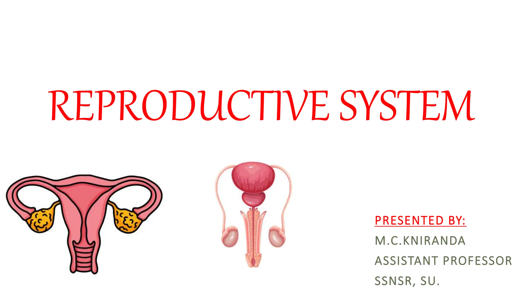
REPRODUCTIVE SYSTEM
PRESENTED BY:
M.C.KNIRANDA
ASSISTANT PROFESSOR
SSNSR, SU.
INTRODUCTION
 One of the essential features of life is the power of reproduction.
Both, females and males have a combination of external and internal
organs that serve as the means of reproduction.
 A woman’s reproductive phase is the period of time when she has
the chance to get pregnant and give birth.
Mons Pubis
The mons pubis is an inverted triangular area of fatty tissue present in front of
pubic symphysis. It is formed by accumulation of subcutaneous fat. It is covered
by pubic hair.
Labia Majora
These are the outer folds of skin of the external female genitalia. The labia majora
(singular: labium majus) are two well known areas where the skin folds that extend
downward and backward from the mons pubis to the perineum.
Labia Minora
The labia minora are comparatively thin and delicate folds of
hairless skin that are present inside the labia majora and cover the
opening of vagina and urethra.
Clitoris
A little erectile component that is small, sensitive, and located on
the outside of the body in front of the opening of the vagina. It is
an analogue of the male penis.
Vestibule of Vagina
The vestibule of the vagina is the area between the two labia
minora folds.
It has urethral opening, a vaginal opening on the vestibule’s back,
a thin membrane called a hymen in virgins, and Bartholin glands
(greater vestibular glands).
BLOOD AND NERVE SUPPLY -
Arterial supply to the vulva is by branches of external and internal
pudendal arteries.
Nerve supply: Ilioinguinal nerve, Genitofemoral nerve, Perineal nerve.
vestibule
INTERNALGENITALORGANS
FEMALE INTERNAL GENITALORGANS
Vagina
The vagina, a fibromuscular canal that runs from the vulva to the uterus, is
about 10 cm long. It receives the penis during sex and serves as the female
copulatory organ.
It is situated anterior to the rectum and behind the bladder and urethra.
There are anterior and posterior walls in the vagina. The lower section of the
cervix is enclosed(surrounded) by the upper portion of the vagina.
The space of the vagina around cervix is called fornix, which can be further
divided into anterior, posterior, medial, and lateral part.
Uterus
 The uterus commonly called as womb is a hollow, pear-shaped organ that
holds the developing fetus.
 The uterus is about 30–40 gm in weight and measures about 7–8 cm long,
5–7 cm wide and 2–3 cm thick in non- pregnant healthy women.
Parts of Uterus
Body
Cervix
Body of Uterus
The main upper expanded part of the uterus is called as body of the uterus.
The body has a fundus, two surfaces (anterior and posterior) and two borders
(lateral).
Cervix of Uterus
The lower, cylindrical portion of the body known as the cervix opens into the
vagina. It is 2.5 cm long.
Structure of Uterus
• The wall of uterus has 3 layers –
1. Perimetrium: The outermost, protective
layer.
2. Myometrium: The highly muscular
middle layer. This is what expands
during pregnancy and contracts to push
the baby out.
3. Endometrium: inner most mucosal
lining. This layer shed during menstrual
cycle.
Ovary
 The ovaries are small, oval-shaped glands that are located on
either side of the uterus.
 The ovaries produce eggs and hormones.
 Each ovary is about 4 cm long, 2 cm wide, and 1 cm thick.
Functions
The functions of the ovaries are as follows:
1. Production of oocytes (female gametes):
During the reproductive period of females, in the middle of
menstrual cycle, ovaries release a mature ovum by rupture of
mature graafian follicle (ovulation). (Graafian follicle is a mature fluid-filled cavity
present inside the ovary which contains the female gamete/ovum)
2. Production of hormones:
 The ovaries produce two hormones—progesterone and estrogen
Fallopian Tube
The fallopian tubes, also known as uterine tubes, are a pair of small tubes
that are joined to the top of the uterus and measure around 10 cm (4
inches) in length.
They are used to transport ova from the ovaries to the uterus. Each
tube lies in the upper free margin of the broad ligaments of the uterus.
Fertilization of an egg by a sperm normally occurs in
the fallopian tubes.
The fertilized egg then moves to the uterus, where it
implants to the uterine lining.
STRUCTURE OF UTERUS
Parts of fallopian tube
From lateral to medial end the parts are infundibulum, ampulla, isthmus, and
intramural.
1. Infundibulum:
 It is (1 cm long) the funnel-shaped and extends beyond the broad ligament of
uterus. Numerous irregular structures known as fimbriae are present on this
end.
 The term “ovarian fimbria” refers to one of the fimbriae that is longer than
the others. It detects when ovulation will occur and helps the ovum enter the
fallopian tube.
2) Ampulla: The part just medial to infundibulum is the ampulla (5–6 cm
long). It is thin walled, tortuous (twists and bends, not clear) and widest part
of the tube. It is the site of fertilization of ovum.
3) Isthmus: It is the narrow cord like part having length 2.5– 3 cm.
4) Intramural (Interstitial): This part (1 cm) lies within the wall of the
uterus.
female reproductive system.pptx

female reproductive system.pptx

  • 1.
  • 2.
    INTRODUCTION  One ofthe essential features of life is the power of reproduction. Both, females and males have a combination of external and internal organs that serve as the means of reproduction.  A woman’s reproductive phase is the period of time when she has the chance to get pregnant and give birth.
  • 8.
    Mons Pubis The monspubis is an inverted triangular area of fatty tissue present in front of pubic symphysis. It is formed by accumulation of subcutaneous fat. It is covered by pubic hair. Labia Majora These are the outer folds of skin of the external female genitalia. The labia majora (singular: labium majus) are two well known areas where the skin folds that extend downward and backward from the mons pubis to the perineum.
  • 9.
    Labia Minora The labiaminora are comparatively thin and delicate folds of hairless skin that are present inside the labia majora and cover the opening of vagina and urethra. Clitoris A little erectile component that is small, sensitive, and located on the outside of the body in front of the opening of the vagina. It is an analogue of the male penis.
  • 10.
    Vestibule of Vagina Thevestibule of the vagina is the area between the two labia minora folds. It has urethral opening, a vaginal opening on the vestibule’s back, a thin membrane called a hymen in virgins, and Bartholin glands (greater vestibular glands).
  • 11.
    BLOOD AND NERVESUPPLY - Arterial supply to the vulva is by branches of external and internal pudendal arteries. Nerve supply: Ilioinguinal nerve, Genitofemoral nerve, Perineal nerve.
  • 12.
  • 13.
  • 14.
    Vagina The vagina, afibromuscular canal that runs from the vulva to the uterus, is about 10 cm long. It receives the penis during sex and serves as the female copulatory organ. It is situated anterior to the rectum and behind the bladder and urethra. There are anterior and posterior walls in the vagina. The lower section of the cervix is enclosed(surrounded) by the upper portion of the vagina. The space of the vagina around cervix is called fornix, which can be further divided into anterior, posterior, medial, and lateral part.
  • 15.
    Uterus  The uteruscommonly called as womb is a hollow, pear-shaped organ that holds the developing fetus.  The uterus is about 30–40 gm in weight and measures about 7–8 cm long, 5–7 cm wide and 2–3 cm thick in non- pregnant healthy women. Parts of Uterus Body Cervix
  • 16.
    Body of Uterus Themain upper expanded part of the uterus is called as body of the uterus. The body has a fundus, two surfaces (anterior and posterior) and two borders (lateral). Cervix of Uterus The lower, cylindrical portion of the body known as the cervix opens into the vagina. It is 2.5 cm long.
  • 17.
    Structure of Uterus •The wall of uterus has 3 layers – 1. Perimetrium: The outermost, protective layer. 2. Myometrium: The highly muscular middle layer. This is what expands during pregnancy and contracts to push the baby out. 3. Endometrium: inner most mucosal lining. This layer shed during menstrual cycle.
  • 18.
    Ovary  The ovariesare small, oval-shaped glands that are located on either side of the uterus.  The ovaries produce eggs and hormones.  Each ovary is about 4 cm long, 2 cm wide, and 1 cm thick.
  • 19.
    Functions The functions ofthe ovaries are as follows: 1. Production of oocytes (female gametes): During the reproductive period of females, in the middle of menstrual cycle, ovaries release a mature ovum by rupture of mature graafian follicle (ovulation). (Graafian follicle is a mature fluid-filled cavity present inside the ovary which contains the female gamete/ovum) 2. Production of hormones:  The ovaries produce two hormones—progesterone and estrogen
  • 20.
    Fallopian Tube The fallopiantubes, also known as uterine tubes, are a pair of small tubes that are joined to the top of the uterus and measure around 10 cm (4 inches) in length. They are used to transport ova from the ovaries to the uterus. Each tube lies in the upper free margin of the broad ligaments of the uterus. Fertilization of an egg by a sperm normally occurs in the fallopian tubes. The fertilized egg then moves to the uterus, where it implants to the uterine lining.
  • 21.
  • 22.
    Parts of fallopiantube From lateral to medial end the parts are infundibulum, ampulla, isthmus, and intramural. 1. Infundibulum:  It is (1 cm long) the funnel-shaped and extends beyond the broad ligament of uterus. Numerous irregular structures known as fimbriae are present on this end.  The term “ovarian fimbria” refers to one of the fimbriae that is longer than the others. It detects when ovulation will occur and helps the ovum enter the fallopian tube.
  • 23.
    2) Ampulla: Thepart just medial to infundibulum is the ampulla (5–6 cm long). It is thin walled, tortuous (twists and bends, not clear) and widest part of the tube. It is the site of fertilization of ovum. 3) Isthmus: It is the narrow cord like part having length 2.5– 3 cm. 4) Intramural (Interstitial): This part (1 cm) lies within the wall of the uterus.