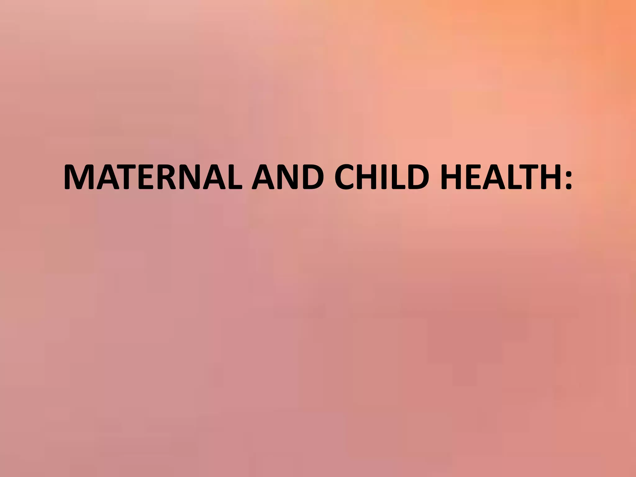
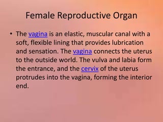
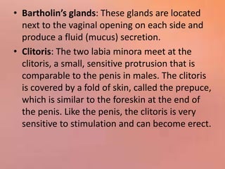
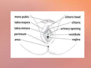
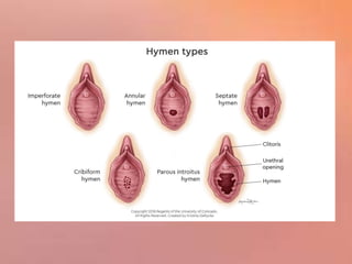
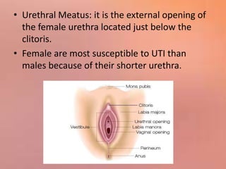
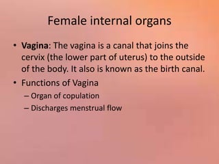
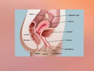
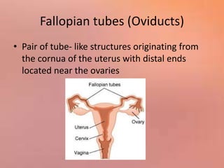
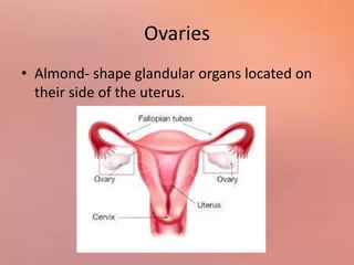
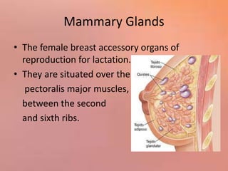
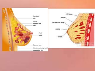
1) The document describes the key external and internal female reproductive organs including the vagina, labia, clitoris, uterus, fallopian tubes, ovaries, and mammary glands.
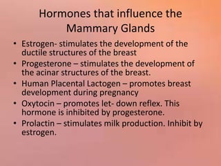
2) It explains the functions of these organs such as facilitating sexual intercourse, menstruation, fertilization, pregnancy, and lactation.
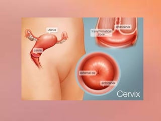
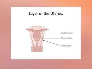
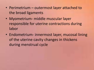
3) The structures and roles of each organ are defined in detail, such as the various parts of the uterus that allow it to accommodate a growing fetus and aid in childbirth.


![ONFH[AVN HIP] -TRIPLE REGIME -A NOVAL SURGICAL CONCEPT .pptx](https://cdn.slidesharecdn.com/ss_thumbnails/onfhavnhip2026koaconcalicutdrgokuldevdrmashraf-260210064517-213ec005-thumbnail.jpg?width=640&height=640&fit=bounds)


