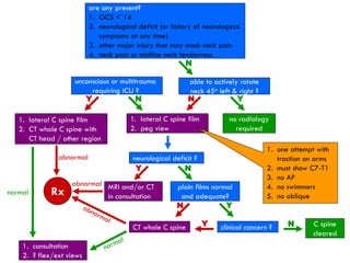
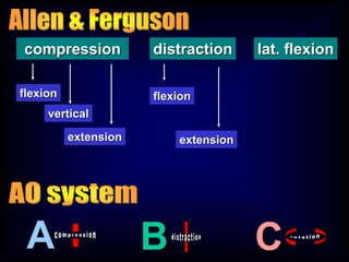
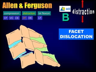
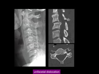
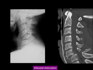
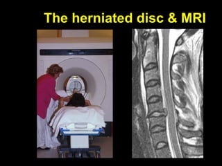
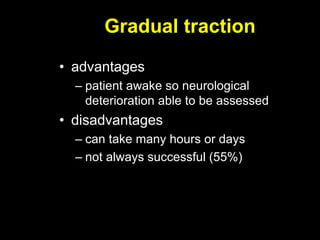
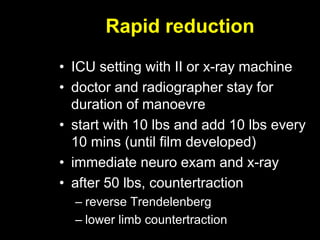
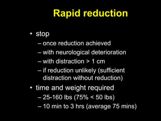
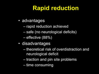
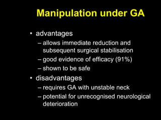
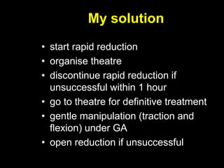
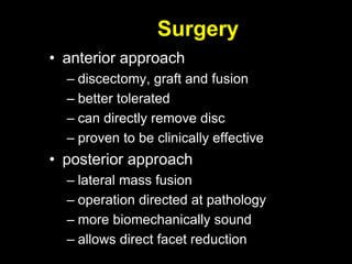
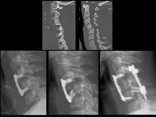
Cervical spine trauma
Initial management of
facet dislocation
Paul Licina
Brisbane
evaluation
history
examination
imaging
•mechanism
•neurological symptoms
•neck
•neurology
•other injuries•x-ray
•CT
•MRI
are any present?
1. GCS < 14
2. neurological deficit (or history of neurological
symptoms at any time)
3. other major injury that may mask neck pain
4. neck pain or midline neck tenderness
N
able to actively rotate
neck 45o left & right ?
N Y
1. lateral C spine film
2. peg view
no radiology
required
neurological deficit ?
N
plain films normal
and adequate?
N Y
CT whole C spine clinical concern ?Y N C spine
cleared
1. consultation
2. ? flex/ext views
Rx
1. one attempt with
traction on arms
2. must show C7-T1
3. no AP
4. no swimmers
5. no oblique
Y
1. lateral C spine film
2. CT whole C spine with
CT head / other region
1. consultation
2. ? flex/ext views
normal
abnormal
unconscious or multitrauma
requiring ICU ?
Y
Y
MRI and/or CT
in consultation
abnormal
N
classification
0
1
2
3
4
5
6
7
upper cervical spine
lower cervical spine
•‘atypical’ vertebrae
•distinct injury patterns
•separate classifications
•‘typical’ vertebrae
•complex injury patterns
•classified together
compression distraction lat. flexion
flexion
extension
flexion
vertical
extension
A CB
DF DECF VC CE LF
compression distraction lat flexion
DF
distraction
AO
B
FACET
DISLOCATION
unifacetal dislocation
bifacetal dislocation
MRI
surgery
reduction
DECISIONS
The herniated disc & MRI
• incidence of herniated disc
– varies from 0% to 50%
• significance of herniated disc
– reduction may lead to further
displacement of disc into canal
• clinical evidence
– case reports of catastrophic neurologic
deterioration with herniated disc found
– deterioration occurred after reduction
– reduction (open or closed) under GA
The herniated disc & MRI
• questions
– which patients should have MRI ?
– when should it be performed ?
– what should be done for a herniated
disc ?
• answers
– everyone should have an MRI before
reduction
– a herniated disc should be removed
before reduction
Contentions
• neurological deterioration during
closed reduction rare
– ? significance of disc protrusion
– canal size increased with reduction
• ? is delay to obtain MRI before
reduction justified
• ? need for MRI at all if routine
anterior discectomy and fusion
My solution
• plain x-ray and CT scan
• if neurologically intact, no need for MRI
• if neurologically complete, obtain MRI
– only if established defect (days old)
– if early, treat as incomplete below
• if neurologically incomplete, initiate
rapid reduction
– delay for MRI not justified
– reduction will increase space for cord
• proceed to theatre for definitive
treatment
Gradual traction, rapid reduction,
manipulation or open reduction?
Gradual traction
• traditional technique
• skull tongs applied
• conscious patient
• 5-10 lb added every 30 min – 2 hrs
• neuro exam and x-ray
• maximum weight 25-50 lbs
• continued until reduction achieved or
success unlikely (72 hrs)
Gradual traction
• advantages
– patient awake so neurological
deterioration able to be assessed
• disadvantages
– can take many hours or days
– not always successful (55%)
Rapid reduction
• ICU setting with II or x-ray machine
• doctor and radiographer stay for
duration of manoevre
• start with 10 lbs and add 10 lbs every
10 mins (until film developed)
• immediate neuro exam and x-ray
• after 50 lbs, countertraction
– reverse Trendelenberg
– lower limb countertraction
Rapid reduction
• stop
– once reduction achieved
– with neurological deterioration
– with distraction > 1 cm
– if reduction unlikely (sufficient
distraction without reduction)
• time and weight required
– 25-160 lbs (75% < 50 lbs)
– 10 min to 3 hrs (average 75 mins)
Rapid reduction
• advantages
– rapid reduction achieved
– safe (no neurological deficits)
– effective (88%)
• disadvantages
– theoretical risk of overdistraction and
neurological deficit
– traction and pin site problems
– time consuming
Manipulation under GA
• advantages
– allows immediate reduction and
subsequent surgical stabilisation
– good evidence of efficacy (91%)
– shown to be safe
• disadvantages
– requires GA with unstable neck
– potential for unrecognised neurological
deterioration
My solution
• start rapid reduction
• organise theatre
• discontinue rapid reduction if
unsuccessful within 1 hour
• go to theatre for definitive treatment
• gentle manipulation (traction and
flexion) under GA
• open reduction if unsuccessful
Surgery
Surgery
• anterior approach
– discectomy, graft and fusion
– better tolerated
– can directly remove disc
– proven to be clinically effective
• posterior approach
– lateral mass fusion
– operation directed at pathology
– more biomechanically sound
– allows direct facet reduction
Facet dislocation management

Facet dislocation management

  • 1.
    Cervical spine trauma Initialmanagement of facet dislocation Paul Licina Brisbane
  • 2.
  • 3.
  • 4.
    are any present? 1.GCS < 14 2. neurological deficit (or history of neurological symptoms at any time) 3. other major injury that may mask neck pain 4. neck pain or midline neck tenderness N able to actively rotate neck 45o left & right ? N Y 1. lateral C spine film 2. peg view no radiology required neurological deficit ? N plain films normal and adequate? N Y CT whole C spine clinical concern ?Y N C spine cleared 1. consultation 2. ? flex/ext views Rx 1. one attempt with traction on arms 2. must show C7-T1 3. no AP 4. no swimmers 5. no oblique Y 1. lateral C spine film 2. CT whole C spine with CT head / other region 1. consultation 2. ? flex/ext views normal abnormal unconscious or multitrauma requiring ICU ? Y Y MRI and/or CT in consultation abnormal N
  • 5.
  • 6.
    0 1 2 3 4 5 6 7 upper cervical spine lowercervical spine •‘atypical’ vertebrae •distinct injury patterns •separate classifications •‘typical’ vertebrae •complex injury patterns •classified together
  • 7.
    compression distraction lat.flexion flexion extension flexion vertical extension A CB
  • 9.
    DF DECF VCCE LF compression distraction lat flexion DF distraction AO B FACET DISLOCATION
  • 10.
  • 11.
  • 12.
  • 13.
  • 14.
    The herniated disc& MRI • incidence of herniated disc – varies from 0% to 50% • significance of herniated disc – reduction may lead to further displacement of disc into canal • clinical evidence – case reports of catastrophic neurologic deterioration with herniated disc found – deterioration occurred after reduction – reduction (open or closed) under GA
  • 15.
    The herniated disc& MRI • questions – which patients should have MRI ? – when should it be performed ? – what should be done for a herniated disc ? • answers – everyone should have an MRI before reduction – a herniated disc should be removed before reduction
  • 16.
    Contentions • neurological deteriorationduring closed reduction rare – ? significance of disc protrusion – canal size increased with reduction • ? is delay to obtain MRI before reduction justified • ? need for MRI at all if routine anterior discectomy and fusion
  • 17.
    My solution • plainx-ray and CT scan • if neurologically intact, no need for MRI • if neurologically complete, obtain MRI – only if established defect (days old) – if early, treat as incomplete below • if neurologically incomplete, initiate rapid reduction – delay for MRI not justified – reduction will increase space for cord • proceed to theatre for definitive treatment
  • 18.
    Gradual traction, rapidreduction, manipulation or open reduction?
  • 19.
    Gradual traction • traditionaltechnique • skull tongs applied • conscious patient • 5-10 lb added every 30 min – 2 hrs • neuro exam and x-ray • maximum weight 25-50 lbs • continued until reduction achieved or success unlikely (72 hrs)
  • 20.
    Gradual traction • advantages –patient awake so neurological deterioration able to be assessed • disadvantages – can take many hours or days – not always successful (55%)
  • 21.
    Rapid reduction • ICUsetting with II or x-ray machine • doctor and radiographer stay for duration of manoevre • start with 10 lbs and add 10 lbs every 10 mins (until film developed) • immediate neuro exam and x-ray • after 50 lbs, countertraction – reverse Trendelenberg – lower limb countertraction
  • 22.
    Rapid reduction • stop –once reduction achieved – with neurological deterioration – with distraction > 1 cm – if reduction unlikely (sufficient distraction without reduction) • time and weight required – 25-160 lbs (75% < 50 lbs) – 10 min to 3 hrs (average 75 mins)
  • 23.
    Rapid reduction • advantages –rapid reduction achieved – safe (no neurological deficits) – effective (88%) • disadvantages – theoretical risk of overdistraction and neurological deficit – traction and pin site problems – time consuming
  • 24.
    Manipulation under GA •advantages – allows immediate reduction and subsequent surgical stabilisation – good evidence of efficacy (91%) – shown to be safe • disadvantages – requires GA with unstable neck – potential for unrecognised neurological deterioration
  • 25.
    My solution • startrapid reduction • organise theatre • discontinue rapid reduction if unsuccessful within 1 hour • go to theatre for definitive treatment • gentle manipulation (traction and flexion) under GA • open reduction if unsuccessful
  • 26.
  • 27.
    Surgery • anterior approach –discectomy, graft and fusion – better tolerated – can directly remove disc – proven to be clinically effective • posterior approach – lateral mass fusion – operation directed at pathology – more biomechanically sound – allows direct facet reduction