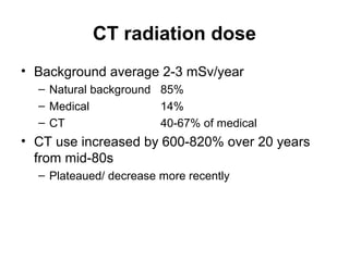
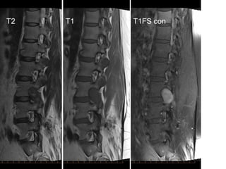
SlideShare a Scribd company logo
Some issues in spine imaging
Dr Greg Cowderoy
Some issues in spine imaging
• Radiation exposure: is it important?
• Role of X-ray, bone scan and other modalities
• CT vs MRI for back pain
• Guidelines for imaging of low back pain
• Imaging appearances of disc herniation and spinal st
• The young athlete with back pain
• Indications for vertebroplasty
• A few cases
Some issues in spine imaging
• Radiation exposure: is it important?
• Role of X-ray, bone scan and other modalities
• CT vs MRI for back pain
• Guidelines for imaging of low back pain
• Imaging appearances of disc herniation and
spinal stenosis
• The young athlete with back pain
• Indications for vertebroplasty
• A few cases
CT radiation dose
• Background average 2-3 mSv/year
– Natural background 85%
– Medical 14%
– CT 40-67% of medical
• CT use increased by 600-820% over 20 years
from mid-80s
– Plateaued/ decrease more recently
CT dose reduction strategies
• Only use CT where appropriate (US, MRI)
• Scan parameters: pitch, kvp, mAs
– Paediatrics
– Built-in protocols
– Automatic tube current modulation
– Iterative image reconstruction
• Minimize phases
– No pre-contrast for trauma, oncology follow-up
• Minimize coverage
– L3 to S1 in most cases
Radiation doses
Imaging test Effective
dose (mSv)
CXRs Background
exposure
Flying hours
CXR 0.02 1 3 days 4
Lumbar X-ray 1.5 75 6/12 300
Lumbar CT 2-10 100-500 8/12 - 3 years 400 - 1800
Bone scan 6 300 2 years 1200
CT risk controversies
• Validity of linear, no threshold model
• Variable literature
– Increased cancer risk in some
– Beneficial effect of low level radiation in others
• Children more radiosensitive and at greater risk
for decades
• Triple risk secondary tumours
– Leukaemia 50mGy
– Brain tumour 60mGy
• Lancet 2012;380:499-505
Some issues in spine imaging
• Radiation exposure: is it important?
• Role of X-ray, bone scan and other modalities
• CT vs MRI for back pain
• Guidelines for imaging of low back pain
• Imaging appearances of disc herniation and
spinal stenosis
• The young athlete with back pain
• Indications for vertebroplasty
• A few cases
Imaging modalities
• Radiographs (X-rays)
• Scintigraphy (bone scan)
• CT
• MRI
Radiographs
What you see
• Bony anatomy and
alignment
• Disc height
Radiographs
What you see
• Bony anatomy and
alignment
• Disc height
Disadvantages
• Radiation
• Nonspecific
– OA changes in most
adults
• Insensitive
– No direct visualisation
of neural and other
nonbony structures
Bone scan
What you see
• Bone pathology
– Osteoblastic activity
Bone scan
What you see
• Bone pathology
– Osteoblastic activity
Disadvantages
• Radiation
• Very nonspecific
• Relatively poor
anatomical resolution
– (Improved with
SPECT; SPECT/CT)
– No direct visualisation
of neural and other
nonbony structures
SPECT-CT
SPECT-CT
SPECT-CT
SPECT-CT
SPECT-CT
SPECT-CT
CT
What you see
• Bony anatomy and
alignment
• Cross sectional view
of spinal canal and
foramina
• Disc, thecal sac,
nerve roots
CT
What you see
• Bony anatomy and
alignment
• Cross sectional view
of spinal canal and
foramina
• Disc, thecal sac,
nerve roots
Disadvantages
• Nonspecific
– Most adults have
‘findings’
• Poor visualisation of
individual neural
structures and disc
anatomy
• Radiation
MRI
What you see
• Bony anatomy and
alignment
• Bone pathology
• Multiplanar view of
spinal canal and
foramina
• Disc: hydration and
structure
• Neural structures:
cord, nerve roots
MRI
What you see
• Bony anatomy and
alignment
• Bone pathology
• Multiplanar view of
spinal canal and
foramina
• Disc: hydration and
structure
• Neural structures:
cord, nerve roots
Disadvantages
• Nonspecific
– Most adults have
‘findings’
• Availability
• Expense
– Rebate
GP rebatable MRI: children
GP rebatable MRI: adults
Some issues in spine imaging
• Radiation exposure: is it important?
• Role of X-ray, bone scan and other modalities
• CT vs MRI for back pain
• Guidelines for imaging of low back pain
• Imaging appearances of disc herniation and
spinal stenosis
• The young athlete with back pain
• Indications for vertebroplasty
• A few cases
T1
T2
T2 T1
MRI: T1
– Fat bright
• Bone marrow
– Bone cortex black
– Anatomy
MRI: T2
– Bone cortex black
– Anatomy
– Fluid bright
– Fat bright
• Bone marrow
– Oedema bright
• Difficult to
differentiate
MRI: STIR or T2FS
– Fat ‘saturated out’
• Bone marrow black
– Fluid bright
– Differentiate oedema
from marrow
T1 T2 STIR
MRI: T1FS-Gd
– Fat ‘saturated out’
• Bone marrow and other
fat black
– Non-fat T1 bright
• Haemorrhage
• Movement
• Enhancement
Pathology
Veins
Nerve root ganglia
T1 T1FS-Gd
T1FS-Gd
Some issues in spine imaging
• Radiation exposure: is it important?
• Role of X-ray, bone scan and other modalities
• CT vs MRI for back pain
• Guidelines for imaging of low back pain
• Imaging appearances of disc herniation and
spinal stenosis
• The young athlete with back pain
• Indications for vertebroplasty
• A few cases
Ineffectiveness of imaging for
nonspecific LBP
• Favourable natural Hx
– Most improve by 4 weeks; unaffected by imaging
• Nonspecificity: loose association between findings
and symptoms
– ‘Abnormalities’ or normal aging?
• Potential harms:
– Radiation
– ‘Labelling’
– Incidental findings
Ann Intern Med 2011;154:181-190
Degenerative changes on imaging
• Ubiquitous and nonspecific
Brinjikji AJNR 2015;36:811
Imaging Finding
Age (yr)
20 30 40 50 60 70 80
Disk degeneration 37% 52% 68% 80% 88% 93% 96%
Disk signal loss 17% 33% 54% 73% 86% 94% 97%
Disk height loss 24% 34% 45% 56% 67% 76% 84%
Disk bulge 30% 40% 50% 60% 69% 77% 84%
Disk protrusion 29% 31% 33% 36% 38% 40% 43%
Annular fissure 19% 20% 22% 23% 25% 27% 29%
Facet degeneration 4% 9% 18% 32% 50% 69% 83%
Spondylolisthesis 3% 5% 8% 14% 23% 35% 50%
Appropriate imaging for back
pain
• Clinical presentations: classification into 3 broad
categories
1. Nonspecific low back pain
2. Back pain associated with radiculopathy
3. Back pain associated with a specific cause requiring
prompt evaluation
Back pain categories
1. Nonspecific (mechanical) low back pain
– Acute: < 12 weeks
– Chronic: > 12 weeks
– Ligament/ muscle strain/ tear
– Intervertebral disc degeneration
– Osteoarthritis
– Facet joints
– SI joints
– Spondylolysis/ spondylolisthesis
Back pain categories
2. Back pain associated with radiculopathy
a) Unilateral acute nerve root compression (sciatica)
– Leg pain >> back pain
– Disc herniation
a) Unilateral chronic nerve root compression
– Disc herniation or spinal stenosis
a) Bilateral chronic nerve root compression
– Spinal stenosis
– DD vascular claudication
a) Bilateral acute nerve root compression = ‘cauda equina
syndrome’
Cauda equina syndrome
• Bilateral acute nerve root compression
– Massive disc protrusion/ sequestration
• Sudden onset bilateral leg pain
• Saddle anaesthesia
• Rapidly progressive or severe neurological
deficits
– Motor deficits at >1 level
– Faecal incontinence
– Urinary retention
Back pain categories
3. Back pain associated with a specific cause requiring
prompt evaluation
− Cauda equina syndrome
− Cancer
− Vertebral infection
− Vertebral compression fracture
− Ankylosing spondylitis (inflammatory
spondyloarthropathy)
LOW BACK PAIN GUIDELINES
Diagnostic triage
1. Non-specific LBP
2. Radiculopathy
3. Specific LBP
• ‘Red flags’
‘Red Flags’
• Cauda equina syndrome
• Known 10
tumour
• Weight loss
• Severe symptoms, not
settling
• Fever
• Recent infection or Sx
• Osteoporosis
• Steroid use
• Non-mechanical pain
• Child*
LOW BACK PAIN GUIDELINES
1.Focused Hx and examination to place patients
into 1 of 3 categories
2.No imaging for nonspecific LBP
3.Imaging for LBP + severe or progressive
neurological deficits OR risk factors for specific
cause
4.Imaging for LBP and radiculopathy if candidates
for surgery or epidural injection
LOW BACK PAIN GUIDELINES
• American College of Physicians & American
Pain Society Recommendations
– Ann Intern Med 2007;147:478-491
• Choosing Wisely Australia
– www.choosingwisely.org.au
• National Institute for Clinical Excellence (NICE)
UK
• ACR Appropriateness Criteria
www.imagingpathways.health.wa.gov.au
Diagnostic work-up
Possible cause Imaging Additional studies
Nonspecific LBP None None
Radiculopathy MRI (CT)
Cauda equina MRI
Cancer MRI for known 10
X-ray for other eg wt loss
Staging: bone scan; PSMA
ESR
Vertebral infection MRI ESR, CRP
Vertebral compression # X-ray
MRI pre vertebroplasty
Ankylosing spondylitis X-ray, incl pelvis (MRI) HLA-B27; ESR, CRP
Ann Intern Med 2007;147:478-491
Some issues in spine imaging
• Radiation exposure: is it important?
• Role of X-ray, bone scan and other modalities
• CT vs MRI for back pain
• Guidelines for imaging of low back pain
• Imaging appearances of disc herniation and
spinal stenosis
• The young athlete with back pain
• Indications for vertebroplasty
• A few cases
NOMENCLATURE
• Consistent
• Reflect common usage where appropriate
• Surgically relevant
• ‘Able to visualize over the phone’
• 2 morphological characteristics:
– Nature of disc pathology
– Location
• Able to add further descriptors
– Neural structures
– Clinical context
• www.asnr.org/spine_nomenclature/reporting
• Spine Journal 2014;14:2525-2545
Annular tear/ fissure
• Annular fissure = degeneration
• Annular tear: outdated
– When ‘tear’ obvious result of trauma use the term
‘rupture’
• Annular high intensity zone (HIZ)
– Not synonymous with ‘fissure’
– Does not imply trauma
– Does not imply pain generator
Disc bulge
• Extension of disc tissue beyond intervertebral
disc space = displacement of annulus
• >25% circumference (>900
)
• Relatively short distance, <3mm
• Normal at L5/S1
Herniated disc
• Localised displacement of disc material beyond
intervertebral disc space (ie disrupted annulus) OR break in
vertebral end plate (Schmorl’s node)
• ‘Localised’ = <25% circumference (<900
)
– No longer divide into ‘broad based’ and ‘focal’
• ‘Herniation’ or ‘protrusion’
• Disc between but not beyond osteophyte OR adaptive to
subluxation/ listhesis is NOT herniation
• ‘HNP’ not accurate
– Herniation may include NP, cartilage, annulus, bone
• ‘Rupture’ tends to refer to trauma/ acute event
• ‘Prolapse’ and ‘bulging disc’ outdated
Protrusion vs extrusion
• Based on appearance
• Extrusion = greatest distance in any
plane between edges > base
OR
• Protrusion: contained
• Extrusion: uncontained = ruptured PLL
• Presence or absence of containment
more clinically relevant:
– Surgical approach
– Prediction of resorption
Sequestered disc
• Extruded disc material that has no continuity
with the disc of origin
• = free fragment
• Migrated disc:
– Disc material displaced away from site of extrusion
T2 T2
T1
Location of herniation
• Anatomic system that correlates with surgery
• Landmarks, transverse plane:
– Sagittal and coronal planes at centre of disc
– Medial edge of articular facet
– Medial, lateral borders of pedicles
Location of herniation
• Locations, transverse plane:
– ‘Central’ = midline
– ‘Right central’ & ‘left central’ =
paracentral/ posterolateral
– ‘Subarticular’ = lateral recess
– ‘Foraminal’
– ‘Extraforaminal’ = far lateral
Location of herniation
• Locations, transverse plane:
– ‘Central’ = midline
– ‘Right central’ & ‘left central’ =
paracentral/ posterolateral
– ‘Subarticular’ = lateral recess
– ‘Foraminal’
– ‘Extraforaminal’ = far lateral
Location of herniation
• Locations, transverse plane:
– ‘Central’ = midline
– ‘Right central’ & ‘left central’ =
paracentral/ posterolateral
– ‘Subarticular’ = lateral recess
– ‘Foraminal’
– ‘Extraforaminal’ = far lateral
Location of herniation
• Locations, transverse plane:
– ‘Central’ = midline
– ‘Right central’ & ‘left central’ =
paracentral/ posterolateral
– ‘Subarticular’ = lateral recess
– ‘Foraminal’
– ‘Extraforaminal’ = far lateral
Location of herniation
• Locations, transverse plane:
– ‘Central’ = midline
– ‘Right central’ & ‘left central’ =
paracentral/ posterolateral
– ‘Subarticular’ = lateral recess
– ‘Foraminal’
– ‘Extraforaminal’ = far lateral
Volume: degree of canal
compromise
• X-sectional area at site of maximal narrowing
• ‘Mild’: <1/3
• ‘Moderate’: 1/3 – 2/3
• ‘Severe’: > 2/3
• Correlation with fluid around cauda and
‘crowding’ of neural structures
• Other descriptors such as compression of
specific neural structures
Mild Moderate Severe
Modic 1
• Vascularised bone marrow
• Oedema
• Overlap with inflammatory
changes
T2 T1
Modic 2
• Proliferation of fatty tissue
• Most common form T2 T1
Modic 3
• Sclerotic bone
• Long standing
degenerative change
T2 T1
Nomenclature: summary
• No consensus for cervical and thoracic
• Cannot date disc pathology without serial
imaging
• Definitions based on morphology and pathology
• No implication of aetiology
• No distinction between symptomatic and
asymptomatic findings
Some issues in spine imaging
• Radiation exposure: is it important?
• Role of X-ray, bone scan and other modalities
• CT vs MRI for back pain
• Guidelines for imaging of low back pain
• Imaging appearances of disc herniation and
spinal stenosis
• The young athlete with back pain
• Indications for vertebroplasty
• A few cases
Pars protocol
 LBP in 40% children and adolescents
 Structural causes 12-26%
 Pars defect = spondylolysis
 Common cause
 Pars interarticularis (interarticular
part) = weakest part of neural arch
 Often accelerated by athletic
activity
 2 key concepts:
 Spectrum of pars pathology
 DD: other causes of pain
12F
Pars pathology
 Developmental deficiency
 Asymmetry of posterior elements: laminae and facet
joints
 Traumatic fracture: uncommon
 Stress injury
 Chronic repetitive low grade trauma
 Oedema early
 Sclerosis or fracture/ defect later
Pars stress injury
 Lumbar, esp L5
 Often bilateral
 Spondylolisthesis
 Disc pathology
 Unilateral defect →
contralateral stress
 Symptoms:
 Often asymptomatic
 LBP
 Hamstring tightness
 Increased by activity
Imaging of LBP in children
 Radiography
 CT
 SPECT/ SPECT-CT
 RadioGraphics 2015;35:819-834
 MRI
 Radiation dose in
children
 Triple risk secondary
tumours
 Leukaemia 50mGy
 Brain tumour 60mGy
 Lancet 2012;380:499-505
Imaging of LBP in children
Radiography CT SPECT-CT MRI
Defect
•Recent
•Old
+/-
+/-
+
+
+
+
+
+
Stress
•Oedema
•Sclerosis
-
+
-
+
+
+
+
+
Spondylolisthesis + + + +
Disc changes
•Dehydration
•Narrowing/ end-
plate deformity
-
+
-
+
-
+
+
+
T1
GE FS
STIR T2
T1 GEFS STIR
11F gymnast, back pain
Fracture + oedema = acute
T1
STIR
18M
LBP
Fracture, corticated, no oedema = chronic
T1 GEFS STIR
16F Unilateral LBP
Incomplete defect + oedema = acute stress reaction/
partial defect
15M Bilateral pars defects, assess healing at 6 months
T1 STIR
T1 STIR
Oedema resolved, right unhealed, left healed
LBP in children: DD
 Pedicle fracture
 Unilateral
 Vertical
 Often contralateral pars
 Spinous process avulsion
 Stress fracture
 Transverse process
 Spinous process
 Sacrum
LBP in children: DD
 Disc degeneration
 Discitis
 Tumour eg osteoid osteoma
T2
T1
17F Left LBP, acute on chronic
T1 GEFS T2
Multiple findings:
Left pars defect + oedema
Degenerate discs L4/5, L5/S1
Muscle tear or denervation left multifidus
Some issues in spine imaging
• Radiation exposure: is it important?
• Role of X-ray, bone scan and other modalities
• CT vs MRI for back pain
• Guidelines for imaging of low back pain
• Imaging appearances of disc herniation and
spinal stenosis
• The young athlete with back pain
• Vertebroplasty
• A few cases
Percutaneous vertebroplasty
Indications
• Painful crush fracture
– Osteoporosis
– Acute: 4-6 weeks
• Malignant crush fracture
– +/- biopsy
• Haemangioma
– Galibert Neurochirurgie 1987;33:166-8
Patient selection = key to
success
• Back pain
– Sudden onset
– May radiate anteriorly
– NOT sciatica
– Mechanical
– Restricted activity
– Poor sleep
• Local tenderness
• Imaging
24/3/2012
24/3/2012 16/12/2011
Imaging techniques
• MRI:
– Confirm presence of crush fracture
– Confirm that crush fracture is acute
– Diagnose other acute levels
– Integrity of spinal canal
– Accurately localise level
MRI
2
3
2
3
T1 STIR
Needle placement: Thoracic
Needle placement: Lumbar
Cement injection
Post procedure care
• Lie prone for 20 minutes
• Bed rest for 2-3 hours
• Discharge if well
– Post-sedation instructions
– Rest 24 hours
– Mobilize according to pain
• Advise re muscle pain
• Follow-up phone call(s)
Complications: rare
• Mild fever; nausea for 24 hours
• Rib fracture
• Foraminal leak
• Spinal canal leak
• Venous emboli
My results
• Audit of first 250 patients, 2001 to 2006
• Complete or near complete response
– No or minimal pain
– Good return of activity level
– 83.0 %
• Moderate response
– Still suffer pain, though noticeably reduced
– Some return of activity, though still restricted
– 12.0 %
• No response
– 5.0 %
Percutaneous vertebroplasty
Keys to success
• Patient selection
– Early referral
– MRI
• High quality fluoroscopy
– Accurate needle placement
– Cement injection
• Nursing care
– Cement preparation
– Patient care: pre and post
MBS funding September 2005
So, what happened?
• Buchbinder NEJM 2009;361:557-68
– Multicentre, randomized, double blind
– Vertebroplasty vs placebo ‘sham’ procedure
– N = 78: 38 vertebroplasty, 40 sham
– No difference in pain scales or quality of life
• MJA (Editorial) 2009;191:476-7
– ‘Percutaneous vertebroplasty is not an effective
treatment for acute osteoporotic vertebral fractures’
• Patient selection
– Up to 12 months pain
• Recruitment
– Majority of eligible
patients not recruited
• Technique
– Up to 3ml cement
– Stopped injection if
leaking
MBS funding withdrawn 2011
Where are we now?
• Uncommon in most places
• Ongoing studies
– Clark Lancet 2016;388:1408-1416
– ‘Vertebroplasty is superior to placebo for pain
reduction in acute osteoporotic spinal fractures of less
than 6 weeks' in duration. These findings will allow
patients with acute painful fractures to have an
additional means of pain management that is known
to be effective’.
• Included in appropriateness guidelines in UK
and USA
• No Medicare rebate
• Our cost: 1200 + day bed about 700
Some issues in spine imaging
• Radiation exposure: is it important?
• Role of X-ray, bone scan and other modalities
• CT vs MRI for back pain
• Guidelines for imaging of low back pain
• Imaging appearances of disc herniation and
spinal stenosis
• The young athlete with back pain
• Vertebroplasty
• A few cases
• 68M
• Sudden onset bilateral leg pain and weakness
• Urinary retention
• Dx: Cauda equina syndrome
• Cause: massive sequestration
• Other causes:
– Tumour
• Primary of lower cord: ependymoma
• Primary of nerve: BPNST
• Primary of dura: meningioma
• Primary of vertebral body: chordoma, giant cell
tumour
• Secondary
– Trauma
Cauda equina syndrome
30M 60F 70M
T2
30M
T2 T1 T1FS con
T1 T1FS con
60F
T2 T1 T1FS con
T1 T1FS con
70M
T2 T1 T1FS con
T1 T1FS con
T2 T1 T1FS con
76M CRC
• 62 year old male
• Severe low back pain of rapid onset
• Febrile and unwell
• 4 weeks ago underwent abdominal surgery for
perforated diverticulitis
T2 T1 T1FS con
T2 T1FS con
• 45 year old male
• 2 weeks post discectomy L4/5
• Recurrent bilateral leg pain
T2 T1
T2
T1FS con
T2
T1FS con
• Dx: recurrent disc:
– Central herniation + huge sequestration virtually filling
the spinal canal
• Note peripheral enhancement pattern
• DD: fibrosis
• 51 year old female
• Left sciatica
– Intermittent pain and paraesthesia
T2 T1 T1FS con
What is the most likely diagnosis?
1. Massive disc sequestration
2. Discitis complicated by abscess
3. Synovial cyst
4. Benign peripheral nerve sheath tumour
T2 T1 T1FS con
• Dx: benign peripheral nerve sheath tumour
(BPNST) of left L3 nerve root
– Many clinicians use the term ‘neuroma’
• Pathologically imprecise term
– Most are benign
• Schwannoma or neurofibroma
• Difficult (impossible) to differentiate on imaging
– BPNST is probably the best terminology
– Associated with NF1 and ‘NF2’ (MISME)
• 66 year old female
• Severe lower back pain on and off for years
• More recent (2 months) development of right
sciatica
What is the most likely diagnosis?
1. Massive disc sequestration
2. Discitis complicated by abscess
3. Synovial cyst
4. Benign peripheral nerve sheath tumour
L4/5
• Severe OA of facet (zygoapophyseal) joints
• Round heterogeneous lesion projecting into right
spinal canal
• Note: close relationship to facet joint
• Dx: synovial cyst
Synovial cyst lumbar facet joint
• Fairly common
• Key is relationship to degenerate facet joint
• Density may vary from pure cyst to varying levels of
calcification and heterogeneity
• Usually present clinically with intractable sciatica
• May respond to aspiration and steroid injection, but
usually treated surgically
T2 T1
T2 T1
Imaging for back pain

More Related Content

What's hot

MRI of Shoulder anatomy
MRI of Shoulder anatomyMRI of Shoulder anatomy
MRI of Shoulder anatomy
Dr. Muhammad Bin Zulfiqar
 
MRI KNEE JOINT ANATOMY
MRI KNEE JOINT ANATOMYMRI KNEE JOINT ANATOMY
MRI KNEE JOINT ANATOMY
Nikhil Bansal
 
Spine radiography
Spine radiographySpine radiography
Spine radiography
Dr. Muhammad Bin Zulfiqar
 
Presentation1.pptx, ultrasound examination of the wrist joint.
Presentation1.pptx, ultrasound examination of the wrist joint.Presentation1.pptx, ultrasound examination of the wrist joint.
Presentation1.pptx, ultrasound examination of the wrist joint.Abdellah Nazeer
 
Presentation2.pptx wrist joint.
Presentation2.pptx wrist joint.Presentation2.pptx wrist joint.
Presentation2.pptx wrist joint.Abdellah Nazeer
 
Mr imaging of spine
Mr imaging of spineMr imaging of spine
Mr imaging of spine
Dr. Muhammad Bin Zulfiqar
 
Basics of msk ultrasound By Dr. Raham Bacha
Basics of msk ultrasound  By Dr. Raham BachaBasics of msk ultrasound  By Dr. Raham Bacha
Basics of msk ultrasound By Dr. Raham Bacha
Medical Ultrasound
 
Presentation1.pptx, radiological anatomy of the thigh and leg.
Presentation1.pptx, radiological anatomy of the thigh and leg.Presentation1.pptx, radiological anatomy of the thigh and leg.
Presentation1.pptx, radiological anatomy of the thigh and leg.Abdellah Nazeer
 
Shear wave Elastography of Liver
Shear wave Elastography of Liver Shear wave Elastography of Liver
Shear wave Elastography of Liver
Dr. Manoj Krishnan Sarojam
 
Ultrasound - Knee
Ultrasound - KneeUltrasound - Knee
Post-graduate Certifcate Musculoskeletal Ultrasound - The Shoulder
Post-graduate Certifcate Musculoskeletal Ultrasound - The ShoulderPost-graduate Certifcate Musculoskeletal Ultrasound - The Shoulder
Post-graduate Certifcate Musculoskeletal Ultrasound - The Shoulder
Dr. Peter Resteghini
 
Doppler ultrasound of the portal system - Normal findings
Doppler ultrasound of the portal system - Normal findingsDoppler ultrasound of the portal system - Normal findings
Doppler ultrasound of the portal system - Normal findingsSamir Haffar
 
lower limb doppler examination -The essentials
lower limb doppler examination -The essentialslower limb doppler examination -The essentials
lower limb doppler examination -The essentials
Ahmed Bahnassy
 
Presentation1.pptx, radiological imaging of metabolic bone diseases.
Presentation1.pptx, radiological imaging of metabolic bone diseases.Presentation1.pptx, radiological imaging of metabolic bone diseases.
Presentation1.pptx, radiological imaging of metabolic bone diseases.
Abdellah Nazeer
 
Shoulder labral tears MRI
Shoulder labral tears MRIShoulder labral tears MRI
Shoulder labral tears MRI
Dr. Mohit Goel
 
Role of ultrasound in clinical evaluation of shoulder Dr. Muhammad Bin Zulfiqar
Role of ultrasound in clinical evaluation of shoulder Dr. Muhammad Bin ZulfiqarRole of ultrasound in clinical evaluation of shoulder Dr. Muhammad Bin Zulfiqar
Role of ultrasound in clinical evaluation of shoulder Dr. Muhammad Bin Zulfiqar
Dr. Muhammad Bin Zulfiqar
 
Doppler ultrasound of visceral arteries
Doppler ultrasound of visceral arteriesDoppler ultrasound of visceral arteries
Doppler ultrasound of visceral arteries
Samir Haffar
 
Neonatal spine ultrasound...normal and abnormal findings
Neonatal spine ultrasound...normal and abnormal findingsNeonatal spine ultrasound...normal and abnormal findings
Neonatal spine ultrasound...normal and abnormal findings
Ahmed Bahnassy
 
Mri knee trauma
Mri knee traumaMri knee trauma
Mri knee trauma
Anish Choudhary
 

What's hot (20)

MRI of Shoulder anatomy
MRI of Shoulder anatomyMRI of Shoulder anatomy
MRI of Shoulder anatomy
 
MRI KNEE JOINT ANATOMY
MRI KNEE JOINT ANATOMYMRI KNEE JOINT ANATOMY
MRI KNEE JOINT ANATOMY
 
Spine radiography
Spine radiographySpine radiography
Spine radiography
 
Presentation1.pptx, ultrasound examination of the wrist joint.
Presentation1.pptx, ultrasound examination of the wrist joint.Presentation1.pptx, ultrasound examination of the wrist joint.
Presentation1.pptx, ultrasound examination of the wrist joint.
 
Presentation2.pptx wrist joint.
Presentation2.pptx wrist joint.Presentation2.pptx wrist joint.
Presentation2.pptx wrist joint.
 
Mr imaging of spine
Mr imaging of spineMr imaging of spine
Mr imaging of spine
 
Basics of msk ultrasound By Dr. Raham Bacha
Basics of msk ultrasound  By Dr. Raham BachaBasics of msk ultrasound  By Dr. Raham Bacha
Basics of msk ultrasound By Dr. Raham Bacha
 
Presentation1.pptx, radiological anatomy of the thigh and leg.
Presentation1.pptx, radiological anatomy of the thigh and leg.Presentation1.pptx, radiological anatomy of the thigh and leg.
Presentation1.pptx, radiological anatomy of the thigh and leg.
 
Shear wave Elastography of Liver
Shear wave Elastography of Liver Shear wave Elastography of Liver
Shear wave Elastography of Liver
 
Ultrasound - Knee
Ultrasound - KneeUltrasound - Knee
Ultrasound - Knee
 
Post-graduate Certifcate Musculoskeletal Ultrasound - The Shoulder
Post-graduate Certifcate Musculoskeletal Ultrasound - The ShoulderPost-graduate Certifcate Musculoskeletal Ultrasound - The Shoulder
Post-graduate Certifcate Musculoskeletal Ultrasound - The Shoulder
 
Doppler ultrasound of the portal system - Normal findings
Doppler ultrasound of the portal system - Normal findingsDoppler ultrasound of the portal system - Normal findings
Doppler ultrasound of the portal system - Normal findings
 
lower limb doppler examination -The essentials
lower limb doppler examination -The essentialslower limb doppler examination -The essentials
lower limb doppler examination -The essentials
 
Presentation1.pptx, radiological imaging of metabolic bone diseases.
Presentation1.pptx, radiological imaging of metabolic bone diseases.Presentation1.pptx, radiological imaging of metabolic bone diseases.
Presentation1.pptx, radiological imaging of metabolic bone diseases.
 
Knee mri
Knee mriKnee mri
Knee mri
 
Shoulder labral tears MRI
Shoulder labral tears MRIShoulder labral tears MRI
Shoulder labral tears MRI
 
Role of ultrasound in clinical evaluation of shoulder Dr. Muhammad Bin Zulfiqar
Role of ultrasound in clinical evaluation of shoulder Dr. Muhammad Bin ZulfiqarRole of ultrasound in clinical evaluation of shoulder Dr. Muhammad Bin Zulfiqar
Role of ultrasound in clinical evaluation of shoulder Dr. Muhammad Bin Zulfiqar
 
Doppler ultrasound of visceral arteries
Doppler ultrasound of visceral arteriesDoppler ultrasound of visceral arteries
Doppler ultrasound of visceral arteries
 
Neonatal spine ultrasound...normal and abnormal findings
Neonatal spine ultrasound...normal and abnormal findingsNeonatal spine ultrasound...normal and abnormal findings
Neonatal spine ultrasound...normal and abnormal findings
 
Mri knee trauma
Mri knee traumaMri knee trauma
Mri knee trauma
 

Similar to Imaging for back pain

Appropriate imaging for back pain
Appropriate imaging for back painAppropriate imaging for back pain
Appropriate imaging for back pain
SpinePlus
 
Appropriate imaging for back pain
Appropriate imaging for back painAppropriate imaging for back pain
Appropriate imaging for back pain
SpinePlus
 
Appropriate imaging for back pain
Appropriate imaging for back painAppropriate imaging for back pain
Appropriate imaging for back pain
SpinePlus
 
Shoulder injury and back pain
Shoulder injury and back pain Shoulder injury and back pain
Shoulder injury and back pain
ABHISHEK SIRSIKAR
 
Low Back Pain abc A review of anatomy and treatment options for low back pain.
Low Back Pain abc A review of anatomy and treatment options for low back pain.Low Back Pain abc A review of anatomy and treatment options for low back pain.
Low Back Pain abc A review of anatomy and treatment options for low back pain.
raedalimd
 
Osteopenia and stress fractures
Osteopenia and stress fracturesOsteopenia and stress fractures
Osteopenia and stress fractures
JA Larson
 
Foot ankle trauma 2013
Foot ankle trauma 2013Foot ankle trauma 2013
Foot ankle trauma 2013
Winston Rennie
 
avascular-necrosis-hip3820.pptx
avascular-necrosis-hip3820.pptxavascular-necrosis-hip3820.pptx
avascular-necrosis-hip3820.pptx
Husain91
 
Scans and imaging
Scans and imagingScans and imaging
Scans and imaging
CommsEditors
 
back pain algorithm ravi
back pain algorithm raviback pain algorithm ravi
back pain algorithm ravi
Dr Ravi Shankar Sharma
 
Bone Status and Navy Stress Fractures
Bone Status and Navy Stress FracturesBone Status and Navy Stress Fractures
Bone Status and Navy Stress Fractures
JA Larson
 
Clavicle Fractures & ACJ Injuries
Clavicle Fractures & ACJ InjuriesClavicle Fractures & ACJ Injuries
Clavicle Fractures & ACJ InjuriesHiren Divecha
 
BWT spinal.pptx
BWT spinal.pptxBWT spinal.pptx
BWT spinal.pptx
SelwynRichards2
 
Adult Degenerative Scoliosis 2008
Adult Degenerative Scoliosis 2008Adult Degenerative Scoliosis 2008
Adult Degenerative Scoliosis 2008
Sohail Bajammal
 
Musculoskeletal Health Concerns of the Aging Population
Musculoskeletal Health Concerns of the Aging PopulationMusculoskeletal Health Concerns of the Aging Population
Musculoskeletal Health Concerns of the Aging Population
Allan Corpuz
 
Rotator Cuff Tendinopathy
Rotator Cuff TendinopathyRotator Cuff Tendinopathy
Rotator Cuff Tendinopathy
The Arm Clinic
 
Avascular necrosis hip
Avascular necrosis hipAvascular necrosis hip
Avascular necrosis hip
vinod naneria
 
Degenerative disease of the spine
Degenerative disease of the spineDegenerative disease of the spine
Degenerative disease of the spine
mohamedrafi112
 
Metrics in spinal deformity
Metrics in spinal deformityMetrics in spinal deformity
Metrics in spinal deformity
Shashank Gandhi
 
back_pain.pptx
back_pain.pptxback_pain.pptx
back_pain.pptx
compengwaelalahmar
 

Similar to Imaging for back pain (20)

Appropriate imaging for back pain
Appropriate imaging for back painAppropriate imaging for back pain
Appropriate imaging for back pain
 
Appropriate imaging for back pain
Appropriate imaging for back painAppropriate imaging for back pain
Appropriate imaging for back pain
 
Appropriate imaging for back pain
Appropriate imaging for back painAppropriate imaging for back pain
Appropriate imaging for back pain
 
Shoulder injury and back pain
Shoulder injury and back pain Shoulder injury and back pain
Shoulder injury and back pain
 
Low Back Pain abc A review of anatomy and treatment options for low back pain.
Low Back Pain abc A review of anatomy and treatment options for low back pain.Low Back Pain abc A review of anatomy and treatment options for low back pain.
Low Back Pain abc A review of anatomy and treatment options for low back pain.
 
Osteopenia and stress fractures
Osteopenia and stress fracturesOsteopenia and stress fractures
Osteopenia and stress fractures
 
Foot ankle trauma 2013
Foot ankle trauma 2013Foot ankle trauma 2013
Foot ankle trauma 2013
 
avascular-necrosis-hip3820.pptx
avascular-necrosis-hip3820.pptxavascular-necrosis-hip3820.pptx
avascular-necrosis-hip3820.pptx
 
Scans and imaging
Scans and imagingScans and imaging
Scans and imaging
 
back pain algorithm ravi
back pain algorithm raviback pain algorithm ravi
back pain algorithm ravi
 
Bone Status and Navy Stress Fractures
Bone Status and Navy Stress FracturesBone Status and Navy Stress Fractures
Bone Status and Navy Stress Fractures
 
Clavicle Fractures & ACJ Injuries
Clavicle Fractures & ACJ InjuriesClavicle Fractures & ACJ Injuries
Clavicle Fractures & ACJ Injuries
 
BWT spinal.pptx
BWT spinal.pptxBWT spinal.pptx
BWT spinal.pptx
 
Adult Degenerative Scoliosis 2008
Adult Degenerative Scoliosis 2008Adult Degenerative Scoliosis 2008
Adult Degenerative Scoliosis 2008
 
Musculoskeletal Health Concerns of the Aging Population
Musculoskeletal Health Concerns of the Aging PopulationMusculoskeletal Health Concerns of the Aging Population
Musculoskeletal Health Concerns of the Aging Population
 
Rotator Cuff Tendinopathy
Rotator Cuff TendinopathyRotator Cuff Tendinopathy
Rotator Cuff Tendinopathy
 
Avascular necrosis hip
Avascular necrosis hipAvascular necrosis hip
Avascular necrosis hip
 
Degenerative disease of the spine
Degenerative disease of the spineDegenerative disease of the spine
Degenerative disease of the spine
 
Metrics in spinal deformity
Metrics in spinal deformityMetrics in spinal deformity
Metrics in spinal deformity
 
back_pain.pptx
back_pain.pptxback_pain.pptx
back_pain.pptx
 

More from SpinePlus

Some WorkCover fusions work
Some WorkCover fusions workSome WorkCover fusions work
Some WorkCover fusions work
SpinePlus
 
Operative management for common back conditions
Operative management for common back conditionsOperative management for common back conditions
Operative management for common back conditions
SpinePlus
 
Pain management
Pain managementPain management
Pain management
SpinePlus
 
Non-operative management for common back conditions
Non-operative management for common back conditionsNon-operative management for common back conditions
Non-operative management for common back conditions
SpinePlus
 
Patient cases
Patient casesPatient cases
Patient cases
SpinePlus
 
Surgery for common lumbar conditions
Surgery for common lumbar conditionsSurgery for common lumbar conditions
Surgery for common lumbar conditions
SpinePlus
 
Pain management
Pain managementPain management
Pain management
SpinePlus
 
Common conditions of the lumbar spine
Common conditions of the lumbar spineCommon conditions of the lumbar spine
Common conditions of the lumbar spine
SpinePlus
 
Non-operative treatment for common back conditions
Non-operative treatment for common back conditions Non-operative treatment for common back conditions
Non-operative treatment for common back conditions
SpinePlus
 
Case Presentation
Case Presentation Case Presentation
Case Presentation
SpinePlus
 
The role of surgery in common lumbar conditions
The role of surgery in common lumbar conditionsThe role of surgery in common lumbar conditions
The role of surgery in common lumbar conditions
SpinePlus
 
Lumbar Stretching
Lumbar StretchingLumbar Stretching
Lumbar Stretching
SpinePlus
 
Lumbar Stabilisation
Lumbar StabilisationLumbar Stabilisation
Lumbar Stabilisation
SpinePlus
 
Discectomy Activity Guide
Discectomy Activity GuideDiscectomy Activity Guide
Discectomy Activity Guide
SpinePlus
 
6 week discectomy
6 week discectomy6 week discectomy
6 week discectomy
SpinePlus
 
3 week discectomy
3 week discectomy3 week discectomy
3 week discectomy
SpinePlus
 
Initial Discectomy
Initial DiscectomyInitial Discectomy
Initial Discectomy
SpinePlus
 
Pain diagram and questionnaire
Pain diagram and questionnairePain diagram and questionnaire
Pain diagram and questionnaire
SpinePlus
 
Rehabilitation for back pain
Rehabilitation for back painRehabilitation for back pain
Rehabilitation for back pain
SpinePlus
 
Interventional Procedures and Opioids
Interventional Procedures and OpioidsInterventional Procedures and Opioids
Interventional Procedures and Opioids
SpinePlus
 

More from SpinePlus (20)

Some WorkCover fusions work
Some WorkCover fusions workSome WorkCover fusions work
Some WorkCover fusions work
 
Operative management for common back conditions
Operative management for common back conditionsOperative management for common back conditions
Operative management for common back conditions
 
Pain management
Pain managementPain management
Pain management
 
Non-operative management for common back conditions
Non-operative management for common back conditionsNon-operative management for common back conditions
Non-operative management for common back conditions
 
Patient cases
Patient casesPatient cases
Patient cases
 
Surgery for common lumbar conditions
Surgery for common lumbar conditionsSurgery for common lumbar conditions
Surgery for common lumbar conditions
 
Pain management
Pain managementPain management
Pain management
 
Common conditions of the lumbar spine
Common conditions of the lumbar spineCommon conditions of the lumbar spine
Common conditions of the lumbar spine
 
Non-operative treatment for common back conditions
Non-operative treatment for common back conditions Non-operative treatment for common back conditions
Non-operative treatment for common back conditions
 
Case Presentation
Case Presentation Case Presentation
Case Presentation
 
The role of surgery in common lumbar conditions
The role of surgery in common lumbar conditionsThe role of surgery in common lumbar conditions
The role of surgery in common lumbar conditions
 
Lumbar Stretching
Lumbar StretchingLumbar Stretching
Lumbar Stretching
 
Lumbar Stabilisation
Lumbar StabilisationLumbar Stabilisation
Lumbar Stabilisation
 
Discectomy Activity Guide
Discectomy Activity GuideDiscectomy Activity Guide
Discectomy Activity Guide
 
6 week discectomy
6 week discectomy6 week discectomy
6 week discectomy
 
3 week discectomy
3 week discectomy3 week discectomy
3 week discectomy
 
Initial Discectomy
Initial DiscectomyInitial Discectomy
Initial Discectomy
 
Pain diagram and questionnaire
Pain diagram and questionnairePain diagram and questionnaire
Pain diagram and questionnaire
 
Rehabilitation for back pain
Rehabilitation for back painRehabilitation for back pain
Rehabilitation for back pain
 
Interventional Procedures and Opioids
Interventional Procedures and OpioidsInterventional Procedures and Opioids
Interventional Procedures and Opioids
 

Recently uploaded

Phone Us ❤85270-49040❤ #ℂall #gIRLS In Surat By Surat @ℂall @Girls Hotel With...
Phone Us ❤85270-49040❤ #ℂall #gIRLS In Surat By Surat @ℂall @Girls Hotel With...Phone Us ❤85270-49040❤ #ℂall #gIRLS In Surat By Surat @ℂall @Girls Hotel With...
Phone Us ❤85270-49040❤ #ℂall #gIRLS In Surat By Surat @ℂall @Girls Hotel With...
Savita Shen $i11
 
heat stroke and heat exhaustion in children
heat stroke and heat exhaustion in childrenheat stroke and heat exhaustion in children
heat stroke and heat exhaustion in children
SumeraAhmad5
 
ANATOMY AND PHYSIOLOGY OF URINARY SYSTEM.pptx
ANATOMY AND PHYSIOLOGY OF URINARY SYSTEM.pptxANATOMY AND PHYSIOLOGY OF URINARY SYSTEM.pptx
ANATOMY AND PHYSIOLOGY OF URINARY SYSTEM.pptx
Swetaba Besh
 
Surat @ℂall @Girls ꧁❤8527049040❤꧂@ℂall @Girls Service Vip Top Model Safe
Surat @ℂall @Girls ꧁❤8527049040❤꧂@ℂall @Girls Service Vip Top Model SafeSurat @ℂall @Girls ꧁❤8527049040❤꧂@ℂall @Girls Service Vip Top Model Safe
Surat @ℂall @Girls ꧁❤8527049040❤꧂@ℂall @Girls Service Vip Top Model Safe
Savita Shen $i11
 
TEST BANK for Operations Management, 14th Edition by William J. Stevenson, Ve...
TEST BANK for Operations Management, 14th Edition by William J. Stevenson, Ve...TEST BANK for Operations Management, 14th Edition by William J. Stevenson, Ve...
TEST BANK for Operations Management, 14th Edition by William J. Stevenson, Ve...
kevinkariuki227
 
Charaka Samhita Sutra Sthana 9 Chapter khuddakachatuspadadhyaya
Charaka Samhita Sutra Sthana 9 Chapter khuddakachatuspadadhyayaCharaka Samhita Sutra Sthana 9 Chapter khuddakachatuspadadhyaya
Charaka Samhita Sutra Sthana 9 Chapter khuddakachatuspadadhyaya
Dr KHALID B.M
 
Couples presenting to the infertility clinic- Do they really have infertility...
Couples presenting to the infertility clinic- Do they really have infertility...Couples presenting to the infertility clinic- Do they really have infertility...
Couples presenting to the infertility clinic- Do they really have infertility...
Sujoy Dasgupta
 
Cervical & Brachial Plexus By Dr. RIG.pptx
Cervical & Brachial Plexus By Dr. RIG.pptxCervical & Brachial Plexus By Dr. RIG.pptx
Cervical & Brachial Plexus By Dr. RIG.pptx
Dr. Rabia Inam Gandapore
 
BRACHYTHERAPY OVERVIEW AND APPLICATORS
BRACHYTHERAPY OVERVIEW  AND  APPLICATORSBRACHYTHERAPY OVERVIEW  AND  APPLICATORS
BRACHYTHERAPY OVERVIEW AND APPLICATORS
Krishan Murari
 
Triangles of Neck and Clinical Correlation by Dr. RIG.pptx
Triangles of Neck and Clinical Correlation by Dr. RIG.pptxTriangles of Neck and Clinical Correlation by Dr. RIG.pptx
Triangles of Neck and Clinical Correlation by Dr. RIG.pptx
Dr. Rabia Inam Gandapore
 
POST OPERATIVE OLIGURIA and its management
POST OPERATIVE OLIGURIA and its managementPOST OPERATIVE OLIGURIA and its management
POST OPERATIVE OLIGURIA and its management
touseefaziz1
 
Surgical Site Infections, pathophysiology, and prevention.pptx
Surgical Site Infections, pathophysiology, and prevention.pptxSurgical Site Infections, pathophysiology, and prevention.pptx
Surgical Site Infections, pathophysiology, and prevention.pptx
jval Landero
 
Are There Any Natural Remedies To Treat Syphilis.pdf
Are There Any Natural Remedies To Treat Syphilis.pdfAre There Any Natural Remedies To Treat Syphilis.pdf
Are There Any Natural Remedies To Treat Syphilis.pdf
Little Cross Family Clinic
 
Superficial & Deep Fascia of the NECK.pptx
Superficial & Deep Fascia of the NECK.pptxSuperficial & Deep Fascia of the NECK.pptx
Superficial & Deep Fascia of the NECK.pptx
Dr. Rabia Inam Gandapore
 
Physiology of Chemical Sensation of smell.pdf
Physiology of Chemical Sensation of smell.pdfPhysiology of Chemical Sensation of smell.pdf
Physiology of Chemical Sensation of smell.pdf
MedicoseAcademics
 
micro teaching on communication m.sc nursing.pdf
micro teaching on communication m.sc nursing.pdfmicro teaching on communication m.sc nursing.pdf
micro teaching on communication m.sc nursing.pdf
Anurag Sharma
 
Report Back from SGO 2024: What’s the Latest in Cervical Cancer?
Report Back from SGO 2024: What’s the Latest in Cervical Cancer?Report Back from SGO 2024: What’s the Latest in Cervical Cancer?
Report Back from SGO 2024: What’s the Latest in Cervical Cancer?
bkling
 
MANAGEMENT OF ATRIOVENTRICULAR CONDUCTION BLOCK.pdf
MANAGEMENT OF ATRIOVENTRICULAR CONDUCTION BLOCK.pdfMANAGEMENT OF ATRIOVENTRICULAR CONDUCTION BLOCK.pdf
MANAGEMENT OF ATRIOVENTRICULAR CONDUCTION BLOCK.pdf
Jim Jacob Roy
 
Lung Cancer: Artificial Intelligence, Synergetics, Complex System Analysis, S...
Lung Cancer: Artificial Intelligence, Synergetics, Complex System Analysis, S...Lung Cancer: Artificial Intelligence, Synergetics, Complex System Analysis, S...
Lung Cancer: Artificial Intelligence, Synergetics, Complex System Analysis, S...
Oleg Kshivets
 
Tom Selleck Health: A Comprehensive Look at the Iconic Actor’s Wellness Journey
Tom Selleck Health: A Comprehensive Look at the Iconic Actor’s Wellness JourneyTom Selleck Health: A Comprehensive Look at the Iconic Actor’s Wellness Journey
Tom Selleck Health: A Comprehensive Look at the Iconic Actor’s Wellness Journey
greendigital
 

Recently uploaded (20)

Phone Us ❤85270-49040❤ #ℂall #gIRLS In Surat By Surat @ℂall @Girls Hotel With...
Phone Us ❤85270-49040❤ #ℂall #gIRLS In Surat By Surat @ℂall @Girls Hotel With...Phone Us ❤85270-49040❤ #ℂall #gIRLS In Surat By Surat @ℂall @Girls Hotel With...
Phone Us ❤85270-49040❤ #ℂall #gIRLS In Surat By Surat @ℂall @Girls Hotel With...
 
heat stroke and heat exhaustion in children
heat stroke and heat exhaustion in childrenheat stroke and heat exhaustion in children
heat stroke and heat exhaustion in children
 
ANATOMY AND PHYSIOLOGY OF URINARY SYSTEM.pptx
ANATOMY AND PHYSIOLOGY OF URINARY SYSTEM.pptxANATOMY AND PHYSIOLOGY OF URINARY SYSTEM.pptx
ANATOMY AND PHYSIOLOGY OF URINARY SYSTEM.pptx
 
Surat @ℂall @Girls ꧁❤8527049040❤꧂@ℂall @Girls Service Vip Top Model Safe
Surat @ℂall @Girls ꧁❤8527049040❤꧂@ℂall @Girls Service Vip Top Model SafeSurat @ℂall @Girls ꧁❤8527049040❤꧂@ℂall @Girls Service Vip Top Model Safe
Surat @ℂall @Girls ꧁❤8527049040❤꧂@ℂall @Girls Service Vip Top Model Safe
 
TEST BANK for Operations Management, 14th Edition by William J. Stevenson, Ve...
TEST BANK for Operations Management, 14th Edition by William J. Stevenson, Ve...TEST BANK for Operations Management, 14th Edition by William J. Stevenson, Ve...
TEST BANK for Operations Management, 14th Edition by William J. Stevenson, Ve...
 
Charaka Samhita Sutra Sthana 9 Chapter khuddakachatuspadadhyaya
Charaka Samhita Sutra Sthana 9 Chapter khuddakachatuspadadhyayaCharaka Samhita Sutra Sthana 9 Chapter khuddakachatuspadadhyaya
Charaka Samhita Sutra Sthana 9 Chapter khuddakachatuspadadhyaya
 
Couples presenting to the infertility clinic- Do they really have infertility...
Couples presenting to the infertility clinic- Do they really have infertility...Couples presenting to the infertility clinic- Do they really have infertility...
Couples presenting to the infertility clinic- Do they really have infertility...
 
Cervical & Brachial Plexus By Dr. RIG.pptx
Cervical & Brachial Plexus By Dr. RIG.pptxCervical & Brachial Plexus By Dr. RIG.pptx
Cervical & Brachial Plexus By Dr. RIG.pptx
 
BRACHYTHERAPY OVERVIEW AND APPLICATORS
BRACHYTHERAPY OVERVIEW  AND  APPLICATORSBRACHYTHERAPY OVERVIEW  AND  APPLICATORS
BRACHYTHERAPY OVERVIEW AND APPLICATORS
 
Triangles of Neck and Clinical Correlation by Dr. RIG.pptx
Triangles of Neck and Clinical Correlation by Dr. RIG.pptxTriangles of Neck and Clinical Correlation by Dr. RIG.pptx
Triangles of Neck and Clinical Correlation by Dr. RIG.pptx
 
POST OPERATIVE OLIGURIA and its management
POST OPERATIVE OLIGURIA and its managementPOST OPERATIVE OLIGURIA and its management
POST OPERATIVE OLIGURIA and its management
 
Surgical Site Infections, pathophysiology, and prevention.pptx
Surgical Site Infections, pathophysiology, and prevention.pptxSurgical Site Infections, pathophysiology, and prevention.pptx
Surgical Site Infections, pathophysiology, and prevention.pptx
 
Are There Any Natural Remedies To Treat Syphilis.pdf
Are There Any Natural Remedies To Treat Syphilis.pdfAre There Any Natural Remedies To Treat Syphilis.pdf
Are There Any Natural Remedies To Treat Syphilis.pdf
 
Superficial & Deep Fascia of the NECK.pptx
Superficial & Deep Fascia of the NECK.pptxSuperficial & Deep Fascia of the NECK.pptx
Superficial & Deep Fascia of the NECK.pptx
 
Physiology of Chemical Sensation of smell.pdf
Physiology of Chemical Sensation of smell.pdfPhysiology of Chemical Sensation of smell.pdf
Physiology of Chemical Sensation of smell.pdf
 
micro teaching on communication m.sc nursing.pdf
micro teaching on communication m.sc nursing.pdfmicro teaching on communication m.sc nursing.pdf
micro teaching on communication m.sc nursing.pdf
 
Report Back from SGO 2024: What’s the Latest in Cervical Cancer?
Report Back from SGO 2024: What’s the Latest in Cervical Cancer?Report Back from SGO 2024: What’s the Latest in Cervical Cancer?
Report Back from SGO 2024: What’s the Latest in Cervical Cancer?
 
MANAGEMENT OF ATRIOVENTRICULAR CONDUCTION BLOCK.pdf
MANAGEMENT OF ATRIOVENTRICULAR CONDUCTION BLOCK.pdfMANAGEMENT OF ATRIOVENTRICULAR CONDUCTION BLOCK.pdf
MANAGEMENT OF ATRIOVENTRICULAR CONDUCTION BLOCK.pdf
 
Lung Cancer: Artificial Intelligence, Synergetics, Complex System Analysis, S...
Lung Cancer: Artificial Intelligence, Synergetics, Complex System Analysis, S...Lung Cancer: Artificial Intelligence, Synergetics, Complex System Analysis, S...
Lung Cancer: Artificial Intelligence, Synergetics, Complex System Analysis, S...
 
Tom Selleck Health: A Comprehensive Look at the Iconic Actor’s Wellness Journey
Tom Selleck Health: A Comprehensive Look at the Iconic Actor’s Wellness JourneyTom Selleck Health: A Comprehensive Look at the Iconic Actor’s Wellness Journey
Tom Selleck Health: A Comprehensive Look at the Iconic Actor’s Wellness Journey
 

Imaging for back pain

  • 1. Some issues in spine imaging Dr Greg Cowderoy
  • 2. Some issues in spine imaging • Radiation exposure: is it important? • Role of X-ray, bone scan and other modalities • CT vs MRI for back pain • Guidelines for imaging of low back pain • Imaging appearances of disc herniation and spinal st • The young athlete with back pain • Indications for vertebroplasty • A few cases
  • 3. Some issues in spine imaging • Radiation exposure: is it important? • Role of X-ray, bone scan and other modalities • CT vs MRI for back pain • Guidelines for imaging of low back pain • Imaging appearances of disc herniation and spinal stenosis • The young athlete with back pain • Indications for vertebroplasty • A few cases
  • 4. CT radiation dose • Background average 2-3 mSv/year – Natural background 85% – Medical 14% – CT 40-67% of medical • CT use increased by 600-820% over 20 years from mid-80s – Plateaued/ decrease more recently
  • 5. CT dose reduction strategies • Only use CT where appropriate (US, MRI) • Scan parameters: pitch, kvp, mAs – Paediatrics – Built-in protocols – Automatic tube current modulation – Iterative image reconstruction • Minimize phases – No pre-contrast for trauma, oncology follow-up • Minimize coverage – L3 to S1 in most cases
  • 6. Radiation doses Imaging test Effective dose (mSv) CXRs Background exposure Flying hours CXR 0.02 1 3 days 4 Lumbar X-ray 1.5 75 6/12 300 Lumbar CT 2-10 100-500 8/12 - 3 years 400 - 1800 Bone scan 6 300 2 years 1200
  • 7. CT risk controversies • Validity of linear, no threshold model • Variable literature – Increased cancer risk in some – Beneficial effect of low level radiation in others • Children more radiosensitive and at greater risk for decades • Triple risk secondary tumours – Leukaemia 50mGy – Brain tumour 60mGy • Lancet 2012;380:499-505
  • 8.
  • 9. Some issues in spine imaging • Radiation exposure: is it important? • Role of X-ray, bone scan and other modalities • CT vs MRI for back pain • Guidelines for imaging of low back pain • Imaging appearances of disc herniation and spinal stenosis • The young athlete with back pain • Indications for vertebroplasty • A few cases
  • 10. Imaging modalities • Radiographs (X-rays) • Scintigraphy (bone scan) • CT • MRI
  • 11. Radiographs What you see • Bony anatomy and alignment • Disc height
  • 12. Radiographs What you see • Bony anatomy and alignment • Disc height Disadvantages • Radiation • Nonspecific – OA changes in most adults • Insensitive – No direct visualisation of neural and other nonbony structures
  • 13. Bone scan What you see • Bone pathology – Osteoblastic activity
  • 14. Bone scan What you see • Bone pathology – Osteoblastic activity Disadvantages • Radiation • Very nonspecific • Relatively poor anatomical resolution – (Improved with SPECT; SPECT/CT) – No direct visualisation of neural and other nonbony structures
  • 21. CT What you see • Bony anatomy and alignment • Cross sectional view of spinal canal and foramina • Disc, thecal sac, nerve roots
  • 22. CT What you see • Bony anatomy and alignment • Cross sectional view of spinal canal and foramina • Disc, thecal sac, nerve roots Disadvantages • Nonspecific – Most adults have ‘findings’ • Poor visualisation of individual neural structures and disc anatomy • Radiation
  • 23. MRI What you see • Bony anatomy and alignment • Bone pathology • Multiplanar view of spinal canal and foramina • Disc: hydration and structure • Neural structures: cord, nerve roots
  • 24. MRI What you see • Bony anatomy and alignment • Bone pathology • Multiplanar view of spinal canal and foramina • Disc: hydration and structure • Neural structures: cord, nerve roots Disadvantages • Nonspecific – Most adults have ‘findings’ • Availability • Expense – Rebate
  • 25. GP rebatable MRI: children
  • 27.
  • 28. Some issues in spine imaging • Radiation exposure: is it important? • Role of X-ray, bone scan and other modalities • CT vs MRI for back pain • Guidelines for imaging of low back pain • Imaging appearances of disc herniation and spinal stenosis • The young athlete with back pain • Indications for vertebroplasty • A few cases
  • 29.
  • 30.
  • 31.
  • 32.
  • 33. T1
  • 34. T2
  • 35. T2 T1
  • 36.
  • 37. MRI: T1 – Fat bright • Bone marrow – Bone cortex black – Anatomy
  • 38. MRI: T2 – Bone cortex black – Anatomy – Fluid bright – Fat bright • Bone marrow – Oedema bright • Difficult to differentiate
  • 39. MRI: STIR or T2FS – Fat ‘saturated out’ • Bone marrow black – Fluid bright – Differentiate oedema from marrow
  • 41. MRI: T1FS-Gd – Fat ‘saturated out’ • Bone marrow and other fat black – Non-fat T1 bright • Haemorrhage • Movement • Enhancement Pathology Veins Nerve root ganglia T1 T1FS-Gd
  • 43.
  • 44. Some issues in spine imaging • Radiation exposure: is it important? • Role of X-ray, bone scan and other modalities • CT vs MRI for back pain • Guidelines for imaging of low back pain • Imaging appearances of disc herniation and spinal stenosis • The young athlete with back pain • Indications for vertebroplasty • A few cases
  • 45. Ineffectiveness of imaging for nonspecific LBP • Favourable natural Hx – Most improve by 4 weeks; unaffected by imaging • Nonspecificity: loose association between findings and symptoms – ‘Abnormalities’ or normal aging? • Potential harms: – Radiation – ‘Labelling’ – Incidental findings Ann Intern Med 2011;154:181-190
  • 46. Degenerative changes on imaging • Ubiquitous and nonspecific Brinjikji AJNR 2015;36:811 Imaging Finding Age (yr) 20 30 40 50 60 70 80 Disk degeneration 37% 52% 68% 80% 88% 93% 96% Disk signal loss 17% 33% 54% 73% 86% 94% 97% Disk height loss 24% 34% 45% 56% 67% 76% 84% Disk bulge 30% 40% 50% 60% 69% 77% 84% Disk protrusion 29% 31% 33% 36% 38% 40% 43% Annular fissure 19% 20% 22% 23% 25% 27% 29% Facet degeneration 4% 9% 18% 32% 50% 69% 83% Spondylolisthesis 3% 5% 8% 14% 23% 35% 50%
  • 47. Appropriate imaging for back pain • Clinical presentations: classification into 3 broad categories 1. Nonspecific low back pain 2. Back pain associated with radiculopathy 3. Back pain associated with a specific cause requiring prompt evaluation
  • 48. Back pain categories 1. Nonspecific (mechanical) low back pain – Acute: < 12 weeks – Chronic: > 12 weeks – Ligament/ muscle strain/ tear – Intervertebral disc degeneration – Osteoarthritis – Facet joints – SI joints – Spondylolysis/ spondylolisthesis
  • 49. Back pain categories 2. Back pain associated with radiculopathy a) Unilateral acute nerve root compression (sciatica) – Leg pain >> back pain – Disc herniation a) Unilateral chronic nerve root compression – Disc herniation or spinal stenosis a) Bilateral chronic nerve root compression – Spinal stenosis – DD vascular claudication a) Bilateral acute nerve root compression = ‘cauda equina syndrome’
  • 50. Cauda equina syndrome • Bilateral acute nerve root compression – Massive disc protrusion/ sequestration • Sudden onset bilateral leg pain • Saddle anaesthesia • Rapidly progressive or severe neurological deficits – Motor deficits at >1 level – Faecal incontinence – Urinary retention
  • 51. Back pain categories 3. Back pain associated with a specific cause requiring prompt evaluation − Cauda equina syndrome − Cancer − Vertebral infection − Vertebral compression fracture − Ankylosing spondylitis (inflammatory spondyloarthropathy)
  • 52. LOW BACK PAIN GUIDELINES Diagnostic triage 1. Non-specific LBP 2. Radiculopathy 3. Specific LBP • ‘Red flags’ ‘Red Flags’ • Cauda equina syndrome • Known 10 tumour • Weight loss • Severe symptoms, not settling • Fever • Recent infection or Sx • Osteoporosis • Steroid use • Non-mechanical pain • Child*
  • 53. LOW BACK PAIN GUIDELINES 1.Focused Hx and examination to place patients into 1 of 3 categories 2.No imaging for nonspecific LBP 3.Imaging for LBP + severe or progressive neurological deficits OR risk factors for specific cause 4.Imaging for LBP and radiculopathy if candidates for surgery or epidural injection
  • 54. LOW BACK PAIN GUIDELINES • American College of Physicians & American Pain Society Recommendations – Ann Intern Med 2007;147:478-491 • Choosing Wisely Australia – www.choosingwisely.org.au • National Institute for Clinical Excellence (NICE) UK • ACR Appropriateness Criteria
  • 56. Diagnostic work-up Possible cause Imaging Additional studies Nonspecific LBP None None Radiculopathy MRI (CT) Cauda equina MRI Cancer MRI for known 10 X-ray for other eg wt loss Staging: bone scan; PSMA ESR Vertebral infection MRI ESR, CRP Vertebral compression # X-ray MRI pre vertebroplasty Ankylosing spondylitis X-ray, incl pelvis (MRI) HLA-B27; ESR, CRP Ann Intern Med 2007;147:478-491
  • 57.
  • 58. Some issues in spine imaging • Radiation exposure: is it important? • Role of X-ray, bone scan and other modalities • CT vs MRI for back pain • Guidelines for imaging of low back pain • Imaging appearances of disc herniation and spinal stenosis • The young athlete with back pain • Indications for vertebroplasty • A few cases
  • 59. NOMENCLATURE • Consistent • Reflect common usage where appropriate • Surgically relevant • ‘Able to visualize over the phone’ • 2 morphological characteristics: – Nature of disc pathology – Location • Able to add further descriptors – Neural structures – Clinical context • www.asnr.org/spine_nomenclature/reporting • Spine Journal 2014;14:2525-2545
  • 60. Annular tear/ fissure • Annular fissure = degeneration • Annular tear: outdated – When ‘tear’ obvious result of trauma use the term ‘rupture’ • Annular high intensity zone (HIZ) – Not synonymous with ‘fissure’ – Does not imply trauma – Does not imply pain generator
  • 61.
  • 62. Disc bulge • Extension of disc tissue beyond intervertebral disc space = displacement of annulus • >25% circumference (>900 ) • Relatively short distance, <3mm • Normal at L5/S1
  • 63.
  • 64. Herniated disc • Localised displacement of disc material beyond intervertebral disc space (ie disrupted annulus) OR break in vertebral end plate (Schmorl’s node) • ‘Localised’ = <25% circumference (<900 ) – No longer divide into ‘broad based’ and ‘focal’ • ‘Herniation’ or ‘protrusion’ • Disc between but not beyond osteophyte OR adaptive to subluxation/ listhesis is NOT herniation • ‘HNP’ not accurate – Herniation may include NP, cartilage, annulus, bone • ‘Rupture’ tends to refer to trauma/ acute event • ‘Prolapse’ and ‘bulging disc’ outdated
  • 65.
  • 66. Protrusion vs extrusion • Based on appearance • Extrusion = greatest distance in any plane between edges > base OR • Protrusion: contained • Extrusion: uncontained = ruptured PLL • Presence or absence of containment more clinically relevant: – Surgical approach – Prediction of resorption
  • 67. Sequestered disc • Extruded disc material that has no continuity with the disc of origin • = free fragment • Migrated disc: – Disc material displaced away from site of extrusion
  • 69. Location of herniation • Anatomic system that correlates with surgery • Landmarks, transverse plane: – Sagittal and coronal planes at centre of disc – Medial edge of articular facet – Medial, lateral borders of pedicles
  • 70. Location of herniation • Locations, transverse plane: – ‘Central’ = midline – ‘Right central’ & ‘left central’ = paracentral/ posterolateral – ‘Subarticular’ = lateral recess – ‘Foraminal’ – ‘Extraforaminal’ = far lateral
  • 71. Location of herniation • Locations, transverse plane: – ‘Central’ = midline – ‘Right central’ & ‘left central’ = paracentral/ posterolateral – ‘Subarticular’ = lateral recess – ‘Foraminal’ – ‘Extraforaminal’ = far lateral
  • 72. Location of herniation • Locations, transverse plane: – ‘Central’ = midline – ‘Right central’ & ‘left central’ = paracentral/ posterolateral – ‘Subarticular’ = lateral recess – ‘Foraminal’ – ‘Extraforaminal’ = far lateral
  • 73. Location of herniation • Locations, transverse plane: – ‘Central’ = midline – ‘Right central’ & ‘left central’ = paracentral/ posterolateral – ‘Subarticular’ = lateral recess – ‘Foraminal’ – ‘Extraforaminal’ = far lateral
  • 74. Location of herniation • Locations, transverse plane: – ‘Central’ = midline – ‘Right central’ & ‘left central’ = paracentral/ posterolateral – ‘Subarticular’ = lateral recess – ‘Foraminal’ – ‘Extraforaminal’ = far lateral
  • 75. Volume: degree of canal compromise • X-sectional area at site of maximal narrowing • ‘Mild’: <1/3 • ‘Moderate’: 1/3 – 2/3 • ‘Severe’: > 2/3 • Correlation with fluid around cauda and ‘crowding’ of neural structures • Other descriptors such as compression of specific neural structures
  • 77.
  • 78. Modic 1 • Vascularised bone marrow • Oedema • Overlap with inflammatory changes T2 T1
  • 79. Modic 2 • Proliferation of fatty tissue • Most common form T2 T1
  • 80. Modic 3 • Sclerotic bone • Long standing degenerative change T2 T1
  • 81. Nomenclature: summary • No consensus for cervical and thoracic • Cannot date disc pathology without serial imaging • Definitions based on morphology and pathology • No implication of aetiology • No distinction between symptomatic and asymptomatic findings
  • 82.
  • 83. Some issues in spine imaging • Radiation exposure: is it important? • Role of X-ray, bone scan and other modalities • CT vs MRI for back pain • Guidelines for imaging of low back pain • Imaging appearances of disc herniation and spinal stenosis • The young athlete with back pain • Indications for vertebroplasty • A few cases
  • 84. Pars protocol  LBP in 40% children and adolescents  Structural causes 12-26%  Pars defect = spondylolysis  Common cause  Pars interarticularis (interarticular part) = weakest part of neural arch  Often accelerated by athletic activity  2 key concepts:  Spectrum of pars pathology  DD: other causes of pain 12F
  • 85. Pars pathology  Developmental deficiency  Asymmetry of posterior elements: laminae and facet joints  Traumatic fracture: uncommon  Stress injury  Chronic repetitive low grade trauma  Oedema early  Sclerosis or fracture/ defect later
  • 86. Pars stress injury  Lumbar, esp L5  Often bilateral  Spondylolisthesis  Disc pathology  Unilateral defect → contralateral stress  Symptoms:  Often asymptomatic  LBP  Hamstring tightness  Increased by activity
  • 87. Imaging of LBP in children  Radiography  CT  SPECT/ SPECT-CT  RadioGraphics 2015;35:819-834  MRI  Radiation dose in children  Triple risk secondary tumours  Leukaemia 50mGy  Brain tumour 60mGy  Lancet 2012;380:499-505
  • 88. Imaging of LBP in children Radiography CT SPECT-CT MRI Defect •Recent •Old +/- +/- + + + + + + Stress •Oedema •Sclerosis - + - + + + + + Spondylolisthesis + + + + Disc changes •Dehydration •Narrowing/ end- plate deformity - + - + - + + +
  • 89. T1
  • 90. GE FS
  • 92. T1 GEFS STIR 11F gymnast, back pain Fracture + oedema = acute
  • 94. T1 GEFS STIR 16F Unilateral LBP Incomplete defect + oedema = acute stress reaction/ partial defect
  • 95. 15M Bilateral pars defects, assess healing at 6 months T1 STIR
  • 96. T1 STIR Oedema resolved, right unhealed, left healed
  • 97. LBP in children: DD  Pedicle fracture  Unilateral  Vertical  Often contralateral pars  Spinous process avulsion  Stress fracture  Transverse process  Spinous process  Sacrum
  • 98. LBP in children: DD  Disc degeneration  Discitis  Tumour eg osteoid osteoma T2 T1
  • 99. 17F Left LBP, acute on chronic T1 GEFS T2 Multiple findings: Left pars defect + oedema Degenerate discs L4/5, L5/S1 Muscle tear or denervation left multifidus
  • 100.
  • 101. Some issues in spine imaging • Radiation exposure: is it important? • Role of X-ray, bone scan and other modalities • CT vs MRI for back pain • Guidelines for imaging of low back pain • Imaging appearances of disc herniation and spinal stenosis • The young athlete with back pain • Vertebroplasty • A few cases
  • 103. Indications • Painful crush fracture – Osteoporosis – Acute: 4-6 weeks • Malignant crush fracture – +/- biopsy • Haemangioma – Galibert Neurochirurgie 1987;33:166-8
  • 104. Patient selection = key to success • Back pain – Sudden onset – May radiate anteriorly – NOT sciatica – Mechanical – Restricted activity – Poor sleep • Local tenderness • Imaging
  • 105.
  • 108. Imaging techniques • MRI: – Confirm presence of crush fracture – Confirm that crush fracture is acute – Diagnose other acute levels – Integrity of spinal canal – Accurately localise level
  • 111.
  • 113.
  • 115. Post procedure care • Lie prone for 20 minutes • Bed rest for 2-3 hours • Discharge if well – Post-sedation instructions – Rest 24 hours – Mobilize according to pain • Advise re muscle pain • Follow-up phone call(s)
  • 116. Complications: rare • Mild fever; nausea for 24 hours • Rib fracture • Foraminal leak • Spinal canal leak • Venous emboli
  • 117. My results • Audit of first 250 patients, 2001 to 2006 • Complete or near complete response – No or minimal pain – Good return of activity level – 83.0 % • Moderate response – Still suffer pain, though noticeably reduced – Some return of activity, though still restricted – 12.0 % • No response – 5.0 %
  • 118. Percutaneous vertebroplasty Keys to success • Patient selection – Early referral – MRI • High quality fluoroscopy – Accurate needle placement – Cement injection • Nursing care – Cement preparation – Patient care: pre and post
  • 120. So, what happened? • Buchbinder NEJM 2009;361:557-68 – Multicentre, randomized, double blind – Vertebroplasty vs placebo ‘sham’ procedure – N = 78: 38 vertebroplasty, 40 sham – No difference in pain scales or quality of life • MJA (Editorial) 2009;191:476-7 – ‘Percutaneous vertebroplasty is not an effective treatment for acute osteoporotic vertebral fractures’
  • 121. • Patient selection – Up to 12 months pain • Recruitment – Majority of eligible patients not recruited • Technique – Up to 3ml cement – Stopped injection if leaking
  • 123. Where are we now? • Uncommon in most places • Ongoing studies – Clark Lancet 2016;388:1408-1416 – ‘Vertebroplasty is superior to placebo for pain reduction in acute osteoporotic spinal fractures of less than 6 weeks' in duration. These findings will allow patients with acute painful fractures to have an additional means of pain management that is known to be effective’. • Included in appropriateness guidelines in UK and USA • No Medicare rebate • Our cost: 1200 + day bed about 700
  • 124.
  • 125. Some issues in spine imaging • Radiation exposure: is it important? • Role of X-ray, bone scan and other modalities • CT vs MRI for back pain • Guidelines for imaging of low back pain • Imaging appearances of disc herniation and spinal stenosis • The young athlete with back pain • Vertebroplasty • A few cases
  • 126. • 68M • Sudden onset bilateral leg pain and weakness • Urinary retention
  • 127.
  • 128.
  • 129. • Dx: Cauda equina syndrome • Cause: massive sequestration • Other causes: – Tumour • Primary of lower cord: ependymoma • Primary of nerve: BPNST • Primary of dura: meningioma • Primary of vertebral body: chordoma, giant cell tumour • Secondary – Trauma
  • 131. 30M T2 T1 T1FS con T1 T1FS con
  • 132. 60F T2 T1 T1FS con T1 T1FS con
  • 133. 70M T2 T1 T1FS con T1 T1FS con
  • 134. T2 T1 T1FS con 76M CRC
  • 135. • 62 year old male • Severe low back pain of rapid onset • Febrile and unwell • 4 weeks ago underwent abdominal surgery for perforated diverticulitis
  • 136. T2 T1 T1FS con
  • 138. • 45 year old male • 2 weeks post discectomy L4/5 • Recurrent bilateral leg pain
  • 139. T2 T1
  • 142. • Dx: recurrent disc: – Central herniation + huge sequestration virtually filling the spinal canal • Note peripheral enhancement pattern • DD: fibrosis
  • 143. • 51 year old female • Left sciatica – Intermittent pain and paraesthesia
  • 144. T2 T1 T1FS con
  • 145. What is the most likely diagnosis? 1. Massive disc sequestration 2. Discitis complicated by abscess 3. Synovial cyst 4. Benign peripheral nerve sheath tumour
  • 146. T2 T1 T1FS con
  • 147. • Dx: benign peripheral nerve sheath tumour (BPNST) of left L3 nerve root – Many clinicians use the term ‘neuroma’ • Pathologically imprecise term – Most are benign • Schwannoma or neurofibroma • Difficult (impossible) to differentiate on imaging – BPNST is probably the best terminology – Associated with NF1 and ‘NF2’ (MISME)
  • 148. • 66 year old female • Severe lower back pain on and off for years • More recent (2 months) development of right sciatica
  • 149.
  • 150. What is the most likely diagnosis? 1. Massive disc sequestration 2. Discitis complicated by abscess 3. Synovial cyst 4. Benign peripheral nerve sheath tumour
  • 151. L4/5
  • 152. • Severe OA of facet (zygoapophyseal) joints • Round heterogeneous lesion projecting into right spinal canal • Note: close relationship to facet joint • Dx: synovial cyst
  • 153. Synovial cyst lumbar facet joint • Fairly common • Key is relationship to degenerate facet joint • Density may vary from pure cyst to varying levels of calcification and heterogeneity • Usually present clinically with intractable sciatica • May respond to aspiration and steroid injection, but usually treated surgically
  • 154. T2 T1
  • 155. T2 T1