Download to read offline
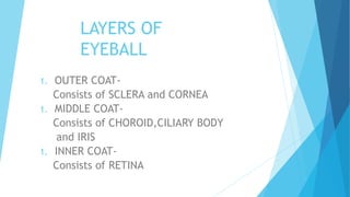
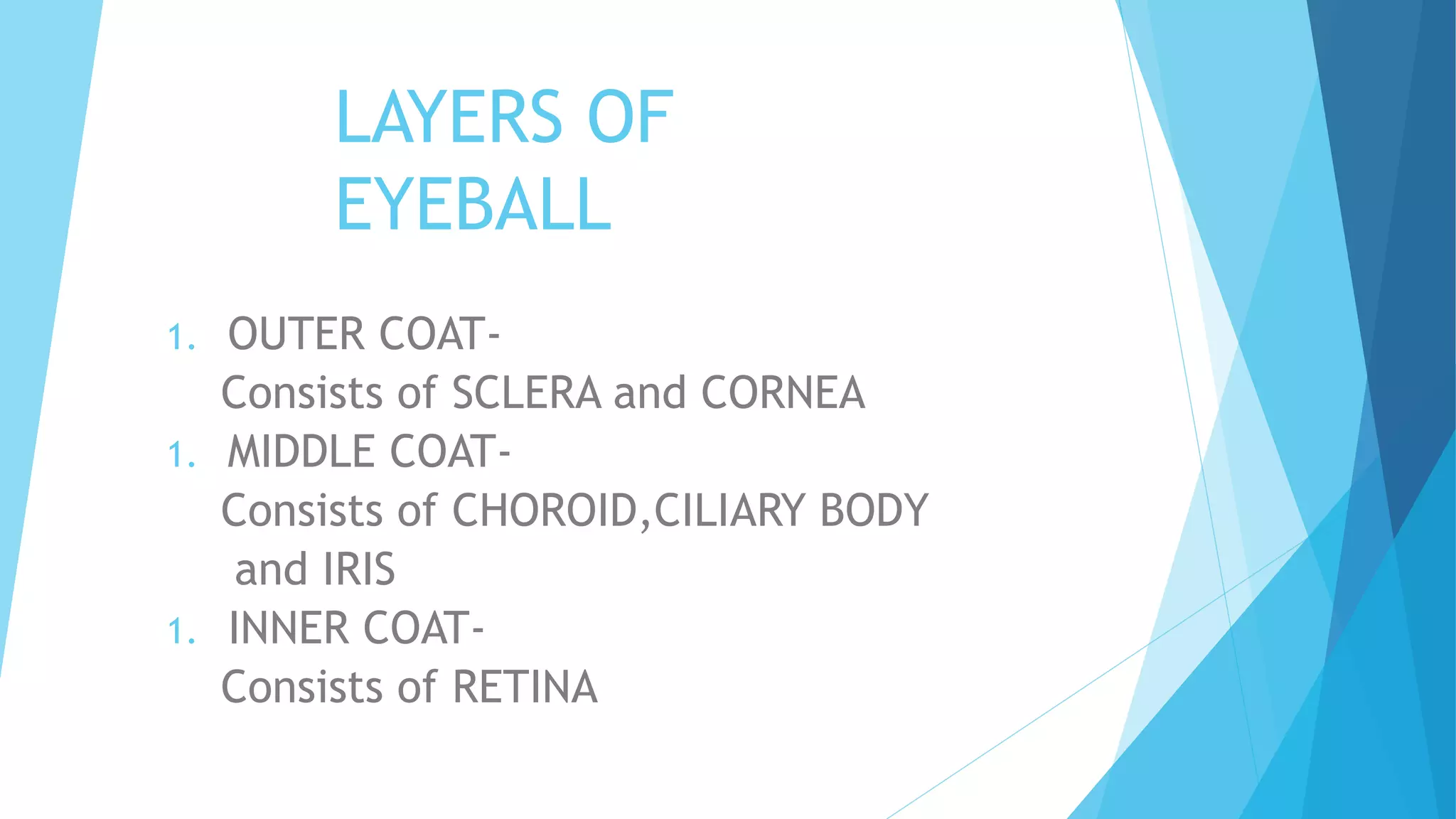
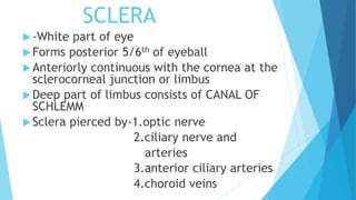
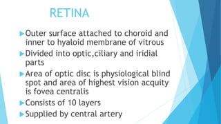

The eyeball consists of three coats or layers: 1. The outer coat includes the sclera and cornea. The sclera forms the white posterior part of the eyeball while the cornea covers the anterior part. 2. The middle coat contains the choroid, ciliary body, and iris. The choroid lies behind the sclera and separates it from the retina. The ciliary body suspends the lens and helps with accommodation. The iris forms the colored circular curtain with the pupil in the center. 3. The inner coat is the retina, which is attached to the choroid on its outer surface and the hyaloid membrane of the vitreous on its inner surface.


