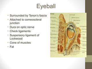
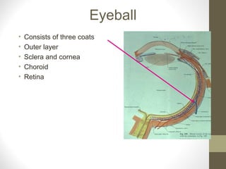
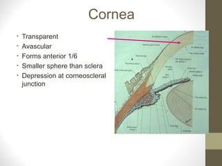
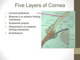
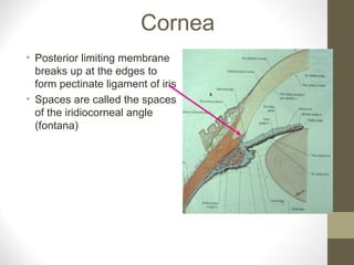
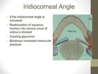
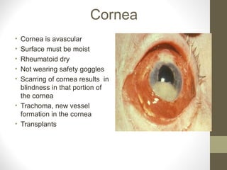
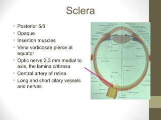
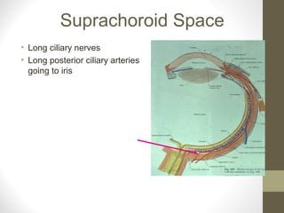
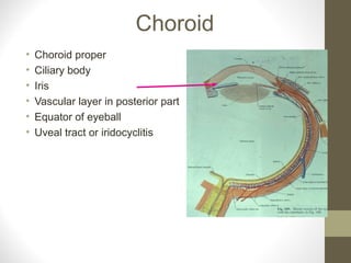
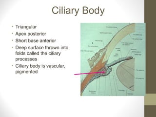
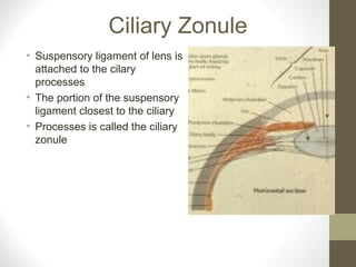
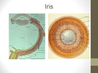
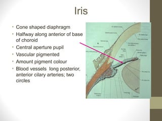
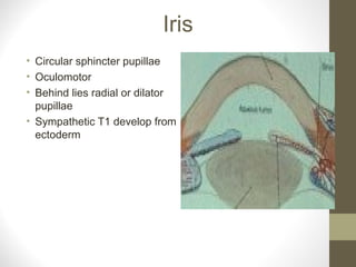
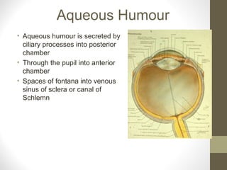
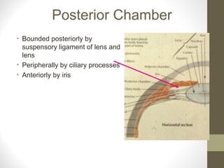
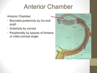
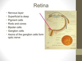
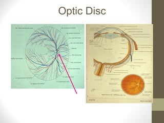
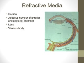
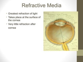
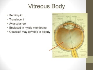
By- Dr. Armaan Singh
Complete Eyeball Info ppt.
Eyeball
• Surrounded by Tenon’s fascia
• Attached to corneoscleral
junction
• Dura on optic nerve
• Check ligaments
• Suspensory ligament of
Lockwood
• Cone of muscles
• Fat
Eyeball
• Consists of three coats
• Outer layer
• Sclera and cornea
• Choroid
• Retina
Cornea
• Transparent
• Avascular
• Forms anterior 1/6
• Smaller sphere than sclera
• Depression at corneoscleral
junction
Five Layers of Cornea
1. Corneal epithelium
2. Bowman’s or anterior limiting
membrane
3. Substantia propria
4. Descement’s or posterior
limiting membrane
5. Endothelium
Cornea
• Posterior limiting membrane
breaks up at the edges to
form pectinate ligament of iris
• Spaces are called the spaces
of the iridiocorneal angle
(fontana)
Iridiocorneal Angle
• If the iridiocorneal angle is
narrowed
• Reabsorption of aqueous
humour into venous sinus of
sclera is blocked
• Causing glaucoma
• Blindness increased intraocular
pressure
Cornea
• Cornea is avascular
• Surface must be moist
• Rheumatoid dry
• Not wearing safety goggles
• Scarring of cornea results in
blindness in that portion of
the cornea
• Trachoma, new vessel
formation in the cornea
• Transplants
Sclera
• Posterior 5/6
• Opaque
• Insertion muscles
• Vena vorticosae pierce at
equator
• Optic nerve 2.3 mm medial to
axis, the lamina cribrosa
• Central artery of retina
• Long and short cilary vessels
and nerves
Suprachoroid Space
• Long ciliary nerves
• Long posterior ciliary arteries
going to iris
Choroid
• Choroid proper
• Ciliary body
• Iris
• Vascular layer in posterior part
• Equator of eyeball
• Uveal tract or iridocyclitis
Choroid
• Outer limiting membrane
• Veins
• Arteries
• Capillaries
• Inner limiting membrane of
bruch
• Attached to pigmented layer
of retina
Ciliary Body
• Triangular
• Apex posterior
• Short base anterior
• Deep surface thrown into
folds called the ciliary
processes
• Ciliary body is vascular,
pigmented
Ciliary Zonule
• Suspensory ligament of lens is
attached to the cilary
processes
• The portion of the suspensory
ligament closest to the ciliary
• Processes is called the ciliary
zonule
Ciliary Muscles
• Smooth muscle
• Radial is attached to scleral
spur
• Circular within anterior part
of radial muscle
• Parasympathetic oculomotor
3rd
nerve
• Relays in ciliary ganglion
Accommodation Reflex
• Contraction of ciliary
muscles
• Ciliary processes pulled
anteriorly
• Relaxes suspensory
ligament of lens
• Elasticity of lens
• It becomes more convex
• Pupils constrict
• Medial rectus contracts
Iris
Iris
• Cone shaped diaphragm
• Halfway along anterior of base
of choroid
• Central aperture pupil
• Vascular pigmented
• Amount pigment colour
• Blood vessels long posterior,
anterior cilary arteries; two
circles
Iris
• Circular sphincter pupillae
• Oculomotor
• Behind lies radial or dilator
pupillae
• Sympathetic T1 develop from
ectoderm
Aqueous Humour
• Aqueous humour is secreted by
ciliary processes into posterior
chamber
• Through the pupil into anterior
chamber
• Spaces of fontana into venous
sinus of sclera or canal of
Schlemn
Posterior Chamber
• Bounded posteriorly by
suspensory ligament of lens and
lens
• Peripherally by ciliary processes
• Anteriorly by iris
Anterior Chamber
Anterior Chamber
• Bounded posteriorly by iris and
pupil
• Anteriorly by cornea
• Peripherally by spaces of fontana
or iridio-corneal angle
Retina
• Nervous layer
• Superficial to deep
• Pigment cells
• Rods and cones
• Bipolar cells
• Ganglion cells
• Axons of the ganglion cells form
optic nerve
Rods
• Rods are found at peripheral
portion of the retina
• 70 rods connected to one
bipolar cell
• Peripheral vision
• Visual purple sees back and
white
Cones
• Cones see colour
• Photoreceptors
• One cone to one bipolar cell
• Found in fovea or macula
lutea
• Directly behind axis
• No nerve fibres or vessels in
front
Blood Supply of Retina
• Rods and cones supplied by
capillaries of choroid by
diffusion
• Bipolar and ganglion cells
supplied by central artery of
the retina
• Enters through optic disc,
blind spot
Optic Disc
Diabetic Hypertensive
Detached Retina
• Outer layer of optic cup
• Pigmented layer
• Inner layer
rods and cones
• Removed from their blood
supply
Refractive Media
• Cornea
• Aqueous humour of anterior
and posterior chamber
• Lens
• Vitreous body
Refractive Media
• Greatest refraction of light
• Takes place at the surface of
the cornea
• Very little refraction after
cornea
Lens
• Crystalline
• Translucent
• Avascular structure
• Lies in hyloid fossa
• Posterior surface is highly
convex
• Does not alter its shape
Lens
• Suspensory ligament is
attached to periphery of the
lens
• Cataract opaque lens
• Nutrition from aqueous humour
Lens
• Supplied by hyloid
• Artery during development
• Hyloid artery regresses remains
as the hyloid canal
• May persist, result a form of
blindness
Vitreous Body
• Semiliquid
• Translucent
• Avascular gel
• Enclosed in hyloid membrane
• Opacities may develop in elderly
Subconjunctival Haemorrhage

Eyeball

  • 1.
    By- Dr. ArmaanSingh Complete Eyeball Info ppt.
  • 2.
    Eyeball • Surrounded byTenon’s fascia • Attached to corneoscleral junction • Dura on optic nerve • Check ligaments • Suspensory ligament of Lockwood • Cone of muscles • Fat
  • 3.
    Eyeball • Consists ofthree coats • Outer layer • Sclera and cornea • Choroid • Retina
  • 4.
    Cornea • Transparent • Avascular •Forms anterior 1/6 • Smaller sphere than sclera • Depression at corneoscleral junction
  • 5.
    Five Layers ofCornea 1. Corneal epithelium 2. Bowman’s or anterior limiting membrane 3. Substantia propria 4. Descement’s or posterior limiting membrane 5. Endothelium
  • 6.
    Cornea • Posterior limitingmembrane breaks up at the edges to form pectinate ligament of iris • Spaces are called the spaces of the iridiocorneal angle (fontana)
  • 7.
    Iridiocorneal Angle • Ifthe iridiocorneal angle is narrowed • Reabsorption of aqueous humour into venous sinus of sclera is blocked • Causing glaucoma • Blindness increased intraocular pressure
  • 8.
    Cornea • Cornea isavascular • Surface must be moist • Rheumatoid dry • Not wearing safety goggles • Scarring of cornea results in blindness in that portion of the cornea • Trachoma, new vessel formation in the cornea • Transplants
  • 9.
    Sclera • Posterior 5/6 •Opaque • Insertion muscles • Vena vorticosae pierce at equator • Optic nerve 2.3 mm medial to axis, the lamina cribrosa • Central artery of retina • Long and short cilary vessels and nerves
  • 10.
    Suprachoroid Space • Longciliary nerves • Long posterior ciliary arteries going to iris
  • 11.
    Choroid • Choroid proper •Ciliary body • Iris • Vascular layer in posterior part • Equator of eyeball • Uveal tract or iridocyclitis
  • 12.
    Choroid • Outer limitingmembrane • Veins • Arteries • Capillaries • Inner limiting membrane of bruch • Attached to pigmented layer of retina
  • 13.
    Ciliary Body • Triangular •Apex posterior • Short base anterior • Deep surface thrown into folds called the ciliary processes • Ciliary body is vascular, pigmented
  • 14.
    Ciliary Zonule • Suspensoryligament of lens is attached to the cilary processes • The portion of the suspensory ligament closest to the ciliary • Processes is called the ciliary zonule
  • 15.
    Ciliary Muscles • Smoothmuscle • Radial is attached to scleral spur • Circular within anterior part of radial muscle • Parasympathetic oculomotor 3rd nerve • Relays in ciliary ganglion
  • 16.
    Accommodation Reflex • Contractionof ciliary muscles • Ciliary processes pulled anteriorly • Relaxes suspensory ligament of lens • Elasticity of lens • It becomes more convex • Pupils constrict • Medial rectus contracts
  • 17.
  • 18.
    Iris • Cone shapeddiaphragm • Halfway along anterior of base of choroid • Central aperture pupil • Vascular pigmented • Amount pigment colour • Blood vessels long posterior, anterior cilary arteries; two circles
  • 19.
    Iris • Circular sphincterpupillae • Oculomotor • Behind lies radial or dilator pupillae • Sympathetic T1 develop from ectoderm
  • 20.
    Aqueous Humour • Aqueoushumour is secreted by ciliary processes into posterior chamber • Through the pupil into anterior chamber • Spaces of fontana into venous sinus of sclera or canal of Schlemn
  • 21.
    Posterior Chamber • Boundedposteriorly by suspensory ligament of lens and lens • Peripherally by ciliary processes • Anteriorly by iris
  • 22.
    Anterior Chamber Anterior Chamber •Bounded posteriorly by iris and pupil • Anteriorly by cornea • Peripherally by spaces of fontana or iridio-corneal angle
  • 23.
    Retina • Nervous layer •Superficial to deep • Pigment cells • Rods and cones • Bipolar cells • Ganglion cells • Axons of the ganglion cells form optic nerve
  • 24.
    Rods • Rods arefound at peripheral portion of the retina • 70 rods connected to one bipolar cell • Peripheral vision • Visual purple sees back and white
  • 25.
    Cones • Cones seecolour • Photoreceptors • One cone to one bipolar cell • Found in fovea or macula lutea • Directly behind axis • No nerve fibres or vessels in front
  • 26.
    Blood Supply ofRetina • Rods and cones supplied by capillaries of choroid by diffusion • Bipolar and ganglion cells supplied by central artery of the retina • Enters through optic disc, blind spot
  • 27.
  • 28.
  • 29.
    Detached Retina • Outerlayer of optic cup • Pigmented layer • Inner layer rods and cones • Removed from their blood supply
  • 30.
    Refractive Media • Cornea •Aqueous humour of anterior and posterior chamber • Lens • Vitreous body
  • 31.
    Refractive Media • Greatestrefraction of light • Takes place at the surface of the cornea • Very little refraction after cornea
  • 32.
    Lens • Crystalline • Translucent •Avascular structure • Lies in hyloid fossa • Posterior surface is highly convex • Does not alter its shape
  • 33.
    Lens • Suspensory ligamentis attached to periphery of the lens • Cataract opaque lens • Nutrition from aqueous humour
  • 34.
    Lens • Supplied byhyloid • Artery during development • Hyloid artery regresses remains as the hyloid canal • May persist, result a form of blindness
  • 35.
    Vitreous Body • Semiliquid •Translucent • Avascular gel • Enclosed in hyloid membrane • Opacities may develop in elderly
  • 36.