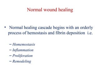
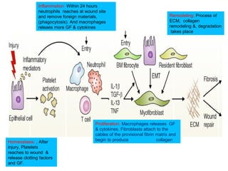
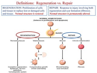
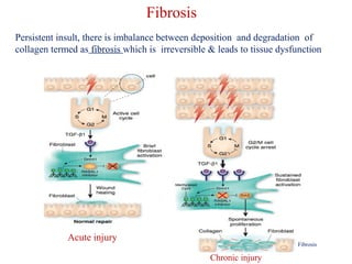
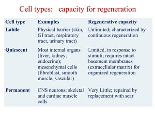
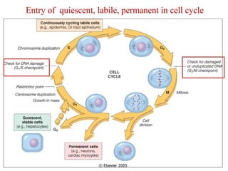
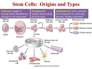
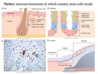
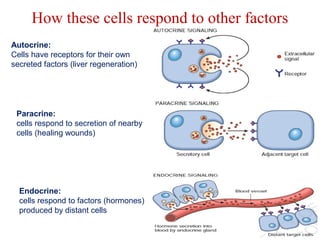
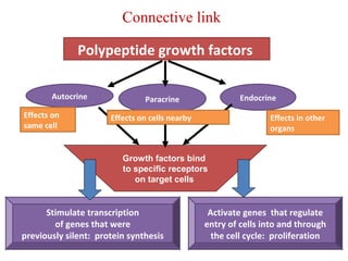
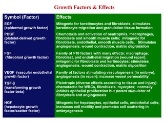
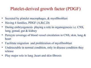
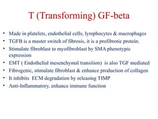
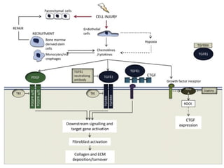
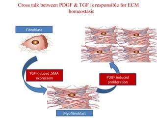
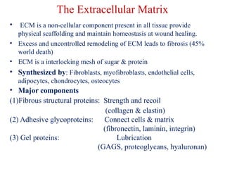
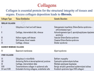
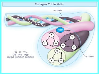
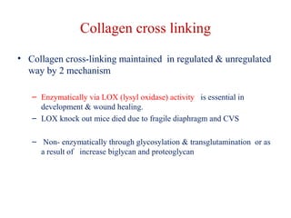
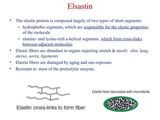
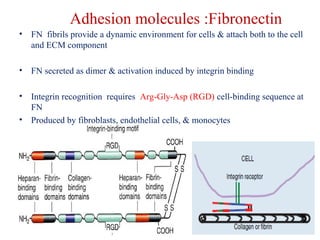
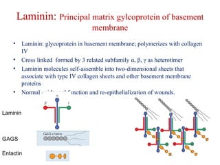
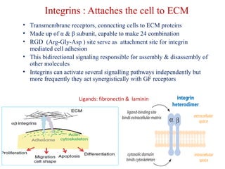
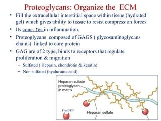
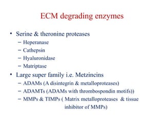
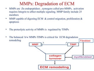
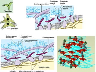
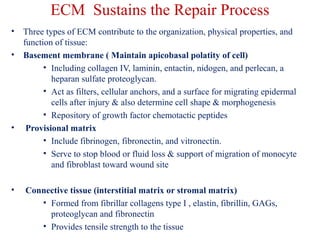
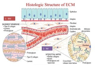
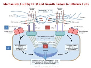
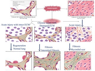
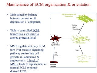
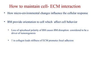
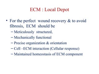
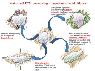
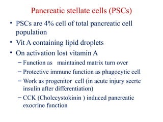
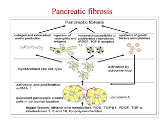
Tissue regeneration & Fibrosis 
Pooja Goswami
Point to be covered 
• Normal Wound healing 
• Regeneration and repair 
• Stem cells: biology & therapeutic applications 
• Cell cycle and regulation of cell replication 
• Growth factors and signaling mechanisms 
• Extracellular Matrix and Cell-Matrix Interactions 
• Summary
Normal wound healing 
• Normal healing cascade begins with an orderly 
process of hemostasis and fibrin deposition i.e. 
– Homemostasis 
– Inflammation 
– Proliferation 
– Remodeling
Inflammation: Within 24 hours 
neutrophils reaches at wound site 
and remove foreign materials, 
(phagocytosis). And macrophages 
releaes more GF & cytokines 
Homeostasis: , After 
injury, Platelets 
reaches to wound & 
release clotting factors 
and GF. 
Remodeling: Process of 
ECM, collagen 
remodeling &, degradation 
takes place 
Proliferation: Macrophages releases GF 
& cytokines. Fibroblasts attach to the 
cables of the provisional fibrin matrix and 
begin to produce collagen
Definitions: Regeneration vs. Repair 
REPAIR: Response to injury involving both 
regeneration and scar formation (fibrosis). 
Normal structure is permanently altered. 
REGENERATION: Proliferation of cells 
and tissues to replace lost or damaged cells 
and tissues. Normal structure is restored.
Fibrosis 
Persistent insult, there is imbalance between deposition and degradation of 
collagen termed as fibrosis which is irreversible & leads to tissue dysfunction 
Chronic injury 
Acute injury 
Fibrosis
Cell types: capacity for regeneration 
Cell type Examples Regenerative capacity 
Labile Physical barrier (skin, 
GI tract, respiratory 
tract, urinary tract) 
Unlimited; characterized by 
continuous regeneration 
Quiescent Most internal organs 
(liver, kidney, 
endocrine); 
mesenchymal cells 
(fibroblast, smooth 
muscle, vascular) 
Limited, in response to 
stimuli; requires intact 
basement membranes 
(extracellular matrix) for 
organized regeneration 
Permanent CNS neurons; skeletal 
and cardiac muscle 
cells 
Very Little; repaired by 
replacement with scar
Entry of quiescent, labile, permanent in cell cycle
Stem Cells: Origins and Types 
Totipotent capable of 
forming almost 200 different 
cell types in the adult body” 
Pluripotent SC: 
capable of generating 
all tissue types 
Multipotent SC: more restricted 
than embryonic SC; eventually 
become “lineage committed”
Niches: microenvironments in which somatic stem cells reside
How these cells respond to other factors 
Autocrine: 
Cells have receptors for their own 
secreted factors (liver regeneration) 
Paracrine: 
cells respond to secretion of nearby 
cells (healing wounds) 
Endocrine: 
cells respond to factors (hormones) 
produced by distant cells
Connective link 
Polypeptide growth factors 
Autocrine Paracrine Endocrine 
Growth factors bind 
to specific receptors 
on target cells 
Stimulate transcription 
of genes that were 
previously silent: protein synthesis 
Activate genes that regulate 
entry of cells into and through 
the cell cycle: proliferation 
Effects on 
same cell 
Effects on cells nearby Effects in other 
organs
Growth Factors & Effects 
Symbol (Factor) Effects 
EGF 
(epidermal growth factor) 
Mitogenic for keratinocytes and fibroblasts, stimulates 
keratinocyte migration and granulation tissue formation 
PDGF 
(platelet-derived growth 
factor) 
Chemotaxis and activation of neutrophils, macrophages, 
fibroblasts and smooth muscle cells; mitogenic for 
fibroblasts, endothelial, smooth muscle cells. Stimulates 
angiogenesis, wound contraction, matrix degradation 
FGF 
(fibroblast growth factor) 
Family of >10 factors with many effects: macrophage, 
fibroblast, and endothelial migration (wound repair), 
mitogenic for fibroblasts and kertinocytes; stimulates 
angiogenesis, wound contraction, matrix deposition 
VEGF (vascular endothelial 
growth factor) 
Family of factors stimulating vasculogenesis (in embryo), 
angiogenesis (in repair); increase vessel permeability 
TGF-b 
(transforming growth 
factor-beta) 
Pleiotropic (diverse effects according to tissue and injury): 
chemotactic for WBCs, fibroblasts, myocytes; normally 
inhibits epithelial proliferation but potent stimulator of 
fibroplasia and angiogenesis 
HGF 
(hepatocyte growth 
factor/scatter factor) 
Mitogenic for hepatocytes, epithelial cells, endothelial cells; 
increases cell motility and promotes cell scattering in 
embryogenesis
Platelet-derived growth factor (PDGF) 
• Secreted by platelet macrophages, & myofibroblast 
• Having 4 families, PDGF (A,B,C,D) 
• During embryogenesis playing a role in organogenesis i.e. CNS, 
lung, gonad, gut & kidney 
• Pericyte coverage of blood vessel circulation in CNS, skin, lung & 
heart 
• Facilitate migration and proliferation of myofibroblast 
• Undetectable in normal condition, only in disease condition they 
release 
• Play major role in lung ,heart and skin fibrosis
T (Transforming) GF-beta 
• Made in platelets, endothelial cells, lymphocytes & macrophages 
• TGFB is a master switch of fibrosis, it is a profibrotic protein. 
• Stimulate fibroblast to myofibroblast by SMA phenotypic 
expression 
• EMT ( Endothelial mesenchymal transition) is also TGF mediated 
• Fibrogenic, stimulate fibroblast & enhance production of collagen 
• It inhibits ECM degradation by releasing TIMP 
• Anti-Inflammatory, enhance immune function
Cross talk between PDGF & TGF is responsible for ECM 
homeostasis 
Fibroblast 
Myofibroblast 
TGF induced ,SMA 
expression PDGF induced 
proliferation
Signal Transduction Pathways 
• Systems which detect extracellular signals through binding of 
ligands to specific receptors, initiating an intracellular cascade of 
events that change gene expression, thus generating a cellular 
response. 
• Pathways usually involve sequential activation of protein kinases 
• Important signal transduction pathways regulating cell growth: 
– Mitogen Activated Protein-kinase (MAP-kinase) 
– Phosphatidylinositol 3-kinase (PI3-kinase) 
– Inositol-triphosphate (IP3) 
– Cyclic adenosine monophosphate (cAMP) 
– JAK/STAT (Janus Kinase/Signal Transducers and Activators of 
Transcription)
Signal Transduction Systems that Require Surface Receptors 
Chemokines, histamine, serotonin, hormones, many drugs 
Steroid hormone 
receptors: in nucleus
Fibroblast to Myofibroblast 
• Granulation tissue, which allows the replacement of the injured tissue, is 
mainly due to fibroblast proliferation, angiogenesis & ECM deposition. 
Fibroblasts acquire smooth muscle (SM) features characterizing the 
myofibroblast which is TGF-β induced 
• Myofibroblasts are also responsible for the synthesis of enzymes involved 
in matrix degradation.
The Extracellular Matrix 
• ECM is a non-cellular component present in all tissue provide 
physical scaffolding and maintain homeostasis at wound healing. 
• Excess and uncontrolled remodeling of ECM leads to fibrosis (45% 
world death) 
• ECM is a interlocking mesh of sugar & protein 
• Synthesized by: Fibroblasts, myofibroblasts, endothelial cells, 
adipocytes, chondrocytes, osteocytes 
• Major components 
(1)Fibrous structural proteins: Strength and recoil 
(collagen & elastin) 
(2) Adhesive glycoproteins: Connect cells & matrix 
(fibronectin, laminin, integrin) 
(3) Gel proteins: Lubrication 
(GAGS, proteoglycans, hyaluronan)
Collagens 
Collagen is essential protein for the structural integrity of tissues and 
organs. Excess collagen deposition leads to fibrosis.
The collagens 
• They are secreted by connective tissue cells, as well 
as by a variety of other cell types 
• Collagens are extremely rich in proline and glycine 
• It is composed mainly of glycine (33%), proline 
(13%), 4-hydroxyproline (9%) 
• Proline stabilizes the helical conformation in each a 
chain 
• Glycine allows the three helical a chains to pack 
tightly together to form the final collagen superhelix 
• Part of the toughness of collagen is accounted by the 
cross-linking of chains via lysine residues
Collagen cross linking 
• Collagen cross-linking maintained in regulated & unregulated 
way by 2 mechanism 
– Enzymatically via LOX (lysyl oxidase) activity is essential in 
development & wound healing. 
– LOX knock out mice died due to fragile diaphragm and CVS 
– Non- enzymatically through glycosylation & transglutamination or as 
a result of increase biglycan and proteoglycan
Elsastin 
• The elastin protein is composed largely of two types of short segments 
– hydrophobic segments, which are responsible for the elastic properties 
of the molecule 
– alanine- and lysine-rich a-helical segments, which form cross-links 
between adjacent molecules 
• Elastic fibers are abundant in organs requiring stretch & recoil: skin, lung, 
uterus, aorta, ligaments 
• Elastin fibers are damaged by aging and sun exposure 
• Resistant to most of the proteolytic enzyme.
Adhesion molecules :Fibronectin 
• FN fibrils provide a dynamic environment for cells & attach both to the cell 
and ECM component 
• FN secreted as dimer & activation induced by integrin binding 
• Integrin recognition requires Arg-Gly-Asp (RGD) cell-binding sequence at 
FN 
• Produced by fibroblasts, endothelial cells, & monocytes
Laminin: Principal matrix gylcoprotein of basement 
membrane 
• Laminin: glycoprotein in basement membrane; polymerizes with collagen 
IV 
• Cross linked formed by 3 related subfamily α, β, γ as heterotimer 
• Laminin molecules self-assemble into two-dimensional sheets that 
associate with type IV collagen sheets and other basement membrane 
proteins 
• Normal epidermal function and re-epithelialization of wounds. 
Laminin 
GAGS 
Entactin
Integrins : Attaches the cell to ECM 
• Transmembrane receptors, connecting cells to ECM proteins 
• Made up of α & β subunit, capable to make 24 combination 
• RGD (Arg-Gly-Asp ) site serve as attachment site for integrin 
mediated cell adhesion 
• This bidirectional signaling responsible for assembly & disassembly of 
other molecules 
• Integrins can activate several signalling pathways independently but 
more frequently they act synergistically with GF receptors 
Ligands: fibronectin & laminin
Proteoglycans: Organize the ECM 
• Fill the extracellular interstitial space within tissue (hydrated 
gel) which gives ability to tissue to resist compression forces 
• Its conc. ↑es in inflammation. 
• Proteoglycans composed of GAGS ( glycosaminoglycans 
chains) linked to core protein 
• GAG are of 2 type, binds to receptors that regulate 
proliferation & migration 
– Sulfated ( Heparin, chondrotin & keratin) 
– Non sulfated (hyaluronic acid) 
Free FGF
ECM degrading enzymes 
• Serine & theronine proteases 
– Heperanase 
– Cathepsin 
– Hyaluronidase 
– Matriptase 
• Large super family i.e. Metzincins 
– ADAMs (A disintegrin & metalloproteases) 
– ADAMTs (ADAMs with thrombospondin motifs)) 
– MMPs & TIMPs ( Matrix metalloproteases & tissue 
inhibitor of MMPs)
MMPs: Degradation of ECM 
• MMPs are Zn endopeptidase , zymogens called pro-MMPs, activation 
requires Integrin to affect multiple signaling. MMP family include 25 
members 
• MMP capable of digesting ECM & control migration, proliferation & 
apoptosis 
• The proteolytic activity of MMPs is regulated by TIMPs 
• The balanced b/w MMPs TIMPs is critical for ECM degradation 
remodeling Fibroblast
ECM Sustains the Repair Process 
• Three types of ECM contribute to the organization, physical properties, and 
function of tissue: 
• Basement membrane ( Maintain apicobasal polatity of cell) 
• Including collagen IV, laminin, entactin, nidogen, and perlecan, a 
heparan sulfate proteoglycan. 
• Act as filters, cellular anchors, and a surface for migrating epidermal 
cells after injury & also determine cell shape & morphogenesis 
• Repository of growth factor chemotactic peptides 
• Provisional matrix 
• Include fibrinogen, fibronectin, and vitronectin. 
• Serve to stop blood or fluid loss & support of migration of monocyte 
and fibroblast toward wound site 
• Connective tissue (interstitial matrix or stromal matrix) 
• Formed from fibrillar collagens type I , elastin, fibrillin, GAGs, 
proteoglycan and fibronectin 
• Provides tensile strength to the tissue
Histologic Structure of ECM 
1: BM 
2: IM
Mechanisms Used by ECM and Growth Factors to Influence Cells 
3 1 
2 
4
Fibrosis 
Interstitial Fibrosis 
Regeneration 
Normal lung 
Fibrosis 
Mycordial scar 
Acute injury with intact ECM 
Chronic injury with 
Damaged ECM Acute injury
How to maintain ECM integrity 
Controlled by specific composition & conc. of matrix and post translational 
modification i.e. glycosylation, transglutamination & cross linking 
• Composition & conc. of matrix affect cell behavior 
• Cross linking of collagen affect mechanical function via 2 ways 
1. Regulated Enzymatically via LOX (lysyl oxidase) activity 
2. Non- enzymatically through glycosylation & transglutamination
Regulated Enzymatically cross-linking of 
Collagen via LOX activity 
• LOX family member 
catalyzes the cross-linking 
of collagen 
through deamidation of 
lysine residues 
• ↑ LOX activity causes 
ECM stiffness 
• Marker of many tumors
Non- enzymatically collagen cross-linking 
• Through glycosylation & transglutamination or as a result of ↑ 
biglycans and proteoglycans 
• ↑ GAGS leads to stiffen the ECM , Age related disease, degenerative 
eye, pulmonary fibrosis, arterial stiffening 
• ↑ Tissue stiffness is specific and cue for tissue dysfunction & 
tumorogenesis
Maintenance of ECM organization & orientation 
• Maintained by balance 
between deposition & 
degradation of component 
• Tightly controlled ECM 
homeostasis sensitive to 
altered protease level 
• MMP regulate not only ECM 
turn over but also signalling 
pathway controlling cell 
growth, inflammation & 
angiogenesis. ↑ level of 
MMPs leads to replacement of 
normal ECM by tumor 
derived ECM.
How to maintain cell- ECM interaction 
• How micro-environmental changes influence the cellular response 
• BM provide orientation to cell which affect cell behavior 
• Loss of apicobasal polarity of BM causes BM disruption considered to be a 
driver of tumorogenesis 
• ↑ in collagen leads stiffness of ECM promotes focal adhesion
ECM : Local Depot 
• For the perfect wound recovery & to avoid 
fibrosis, ECM should be 
– Meticulously structured, 
– Mechanically functional 
– Precise organization & orientation 
– Cell –ECM interaction (Cellular response) 
– Maintained homeostasis of ECM component
Maintained ECM remodeling is important to avoid Fibrosis 
Mechanically signalling 
:matrix elasticity: Intrinsic 
elasticity ( Stiffness) 
regulate cell function and 
modulate cell response to 
micro-environmant 
Mechanically signalling: 
ECM sequester 
Growth factor 
Matricelluler signalling: 
Signals through Adhesive 
molecule ( collegen, elastin, 
Integrins & GAGS) 
ECM interaction 
signalling: ECM transmit 
external forces to the cell 
(Cell –
Pancreatic stellate cells (PSCs) 
• PSCs are 4% cell of total pancreatic cell 
population 
• Vit A containing lipid droplets 
• On activation lost vitamin A 
– Function as maintained matrix turn over 
– Protective immune function as phagocytic cell 
– Work as progenitor cell (in acute injury secrte 
insulin after differentiation) 
– CCK (Cholecystokinin ) induced pancreatic 
exocrine function
Pancreatic fibrosis 
Lost vitamin A
•Cancer cell induced PSCs 
•PSCs stimulate cancer cell proliferation increased survival of cancer 
cell by inhibiting apoptosis 
•ERK 1/2 is the common signalling pathway regulating cancer cell 
induced PSC proliferation

Final 27 aug seminr

  • 1.
    Tissue regeneration &Fibrosis Pooja Goswami
  • 2.
    Point to becovered • Normal Wound healing • Regeneration and repair • Stem cells: biology & therapeutic applications • Cell cycle and regulation of cell replication • Growth factors and signaling mechanisms • Extracellular Matrix and Cell-Matrix Interactions • Summary
  • 3.
    Normal wound healing • Normal healing cascade begins with an orderly process of hemostasis and fibrin deposition i.e. – Homemostasis – Inflammation – Proliferation – Remodeling
  • 4.
    Inflammation: Within 24hours neutrophils reaches at wound site and remove foreign materials, (phagocytosis). And macrophages releaes more GF & cytokines Homeostasis: , After injury, Platelets reaches to wound & release clotting factors and GF. Remodeling: Process of ECM, collagen remodeling &, degradation takes place Proliferation: Macrophages releases GF & cytokines. Fibroblasts attach to the cables of the provisional fibrin matrix and begin to produce collagen
  • 5.
    Definitions: Regeneration vs.Repair REPAIR: Response to injury involving both regeneration and scar formation (fibrosis). Normal structure is permanently altered. REGENERATION: Proliferation of cells and tissues to replace lost or damaged cells and tissues. Normal structure is restored.
  • 6.
    Fibrosis Persistent insult,there is imbalance between deposition and degradation of collagen termed as fibrosis which is irreversible & leads to tissue dysfunction Chronic injury Acute injury Fibrosis
  • 7.
    Cell types: capacityfor regeneration Cell type Examples Regenerative capacity Labile Physical barrier (skin, GI tract, respiratory tract, urinary tract) Unlimited; characterized by continuous regeneration Quiescent Most internal organs (liver, kidney, endocrine); mesenchymal cells (fibroblast, smooth muscle, vascular) Limited, in response to stimuli; requires intact basement membranes (extracellular matrix) for organized regeneration Permanent CNS neurons; skeletal and cardiac muscle cells Very Little; repaired by replacement with scar
  • 8.
    Entry of quiescent,labile, permanent in cell cycle
  • 9.
    Stem Cells: Originsand Types Totipotent capable of forming almost 200 different cell types in the adult body” Pluripotent SC: capable of generating all tissue types Multipotent SC: more restricted than embryonic SC; eventually become “lineage committed”
  • 10.
    Niches: microenvironments inwhich somatic stem cells reside
  • 11.
    How these cellsrespond to other factors Autocrine: Cells have receptors for their own secreted factors (liver regeneration) Paracrine: cells respond to secretion of nearby cells (healing wounds) Endocrine: cells respond to factors (hormones) produced by distant cells
  • 12.
    Connective link Polypeptidegrowth factors Autocrine Paracrine Endocrine Growth factors bind to specific receptors on target cells Stimulate transcription of genes that were previously silent: protein synthesis Activate genes that regulate entry of cells into and through the cell cycle: proliferation Effects on same cell Effects on cells nearby Effects in other organs
  • 13.
    Growth Factors &Effects Symbol (Factor) Effects EGF (epidermal growth factor) Mitogenic for keratinocytes and fibroblasts, stimulates keratinocyte migration and granulation tissue formation PDGF (platelet-derived growth factor) Chemotaxis and activation of neutrophils, macrophages, fibroblasts and smooth muscle cells; mitogenic for fibroblasts, endothelial, smooth muscle cells. Stimulates angiogenesis, wound contraction, matrix degradation FGF (fibroblast growth factor) Family of >10 factors with many effects: macrophage, fibroblast, and endothelial migration (wound repair), mitogenic for fibroblasts and kertinocytes; stimulates angiogenesis, wound contraction, matrix deposition VEGF (vascular endothelial growth factor) Family of factors stimulating vasculogenesis (in embryo), angiogenesis (in repair); increase vessel permeability TGF-b (transforming growth factor-beta) Pleiotropic (diverse effects according to tissue and injury): chemotactic for WBCs, fibroblasts, myocytes; normally inhibits epithelial proliferation but potent stimulator of fibroplasia and angiogenesis HGF (hepatocyte growth factor/scatter factor) Mitogenic for hepatocytes, epithelial cells, endothelial cells; increases cell motility and promotes cell scattering in embryogenesis
  • 14.
    Platelet-derived growth factor(PDGF) • Secreted by platelet macrophages, & myofibroblast • Having 4 families, PDGF (A,B,C,D) • During embryogenesis playing a role in organogenesis i.e. CNS, lung, gonad, gut & kidney • Pericyte coverage of blood vessel circulation in CNS, skin, lung & heart • Facilitate migration and proliferation of myofibroblast • Undetectable in normal condition, only in disease condition they release • Play major role in lung ,heart and skin fibrosis
  • 15.
    T (Transforming) GF-beta • Made in platelets, endothelial cells, lymphocytes & macrophages • TGFB is a master switch of fibrosis, it is a profibrotic protein. • Stimulate fibroblast to myofibroblast by SMA phenotypic expression • EMT ( Endothelial mesenchymal transition) is also TGF mediated • Fibrogenic, stimulate fibroblast & enhance production of collagen • It inhibits ECM degradation by releasing TIMP • Anti-Inflammatory, enhance immune function
  • 17.
    Cross talk betweenPDGF & TGF is responsible for ECM homeostasis Fibroblast Myofibroblast TGF induced ,SMA expression PDGF induced proliferation
  • 18.
    Signal Transduction Pathways • Systems which detect extracellular signals through binding of ligands to specific receptors, initiating an intracellular cascade of events that change gene expression, thus generating a cellular response. • Pathways usually involve sequential activation of protein kinases • Important signal transduction pathways regulating cell growth: – Mitogen Activated Protein-kinase (MAP-kinase) – Phosphatidylinositol 3-kinase (PI3-kinase) – Inositol-triphosphate (IP3) – Cyclic adenosine monophosphate (cAMP) – JAK/STAT (Janus Kinase/Signal Transducers and Activators of Transcription)
  • 19.
    Signal Transduction Systemsthat Require Surface Receptors Chemokines, histamine, serotonin, hormones, many drugs Steroid hormone receptors: in nucleus
  • 20.
    Fibroblast to Myofibroblast • Granulation tissue, which allows the replacement of the injured tissue, is mainly due to fibroblast proliferation, angiogenesis & ECM deposition. Fibroblasts acquire smooth muscle (SM) features characterizing the myofibroblast which is TGF-β induced • Myofibroblasts are also responsible for the synthesis of enzymes involved in matrix degradation.
  • 21.
    The Extracellular Matrix • ECM is a non-cellular component present in all tissue provide physical scaffolding and maintain homeostasis at wound healing. • Excess and uncontrolled remodeling of ECM leads to fibrosis (45% world death) • ECM is a interlocking mesh of sugar & protein • Synthesized by: Fibroblasts, myofibroblasts, endothelial cells, adipocytes, chondrocytes, osteocytes • Major components (1)Fibrous structural proteins: Strength and recoil (collagen & elastin) (2) Adhesive glycoproteins: Connect cells & matrix (fibronectin, laminin, integrin) (3) Gel proteins: Lubrication (GAGS, proteoglycans, hyaluronan)
  • 22.
    Collagens Collagen isessential protein for the structural integrity of tissues and organs. Excess collagen deposition leads to fibrosis.
  • 23.
    The collagens •They are secreted by connective tissue cells, as well as by a variety of other cell types • Collagens are extremely rich in proline and glycine • It is composed mainly of glycine (33%), proline (13%), 4-hydroxyproline (9%) • Proline stabilizes the helical conformation in each a chain • Glycine allows the three helical a chains to pack tightly together to form the final collagen superhelix • Part of the toughness of collagen is accounted by the cross-linking of chains via lysine residues
  • 24.
    Collagen cross linking • Collagen cross-linking maintained in regulated & unregulated way by 2 mechanism – Enzymatically via LOX (lysyl oxidase) activity is essential in development & wound healing. – LOX knock out mice died due to fragile diaphragm and CVS – Non- enzymatically through glycosylation & transglutamination or as a result of increase biglycan and proteoglycan
  • 25.
    Elsastin • Theelastin protein is composed largely of two types of short segments – hydrophobic segments, which are responsible for the elastic properties of the molecule – alanine- and lysine-rich a-helical segments, which form cross-links between adjacent molecules • Elastic fibers are abundant in organs requiring stretch & recoil: skin, lung, uterus, aorta, ligaments • Elastin fibers are damaged by aging and sun exposure • Resistant to most of the proteolytic enzyme.
  • 26.
    Adhesion molecules :Fibronectin • FN fibrils provide a dynamic environment for cells & attach both to the cell and ECM component • FN secreted as dimer & activation induced by integrin binding • Integrin recognition requires Arg-Gly-Asp (RGD) cell-binding sequence at FN • Produced by fibroblasts, endothelial cells, & monocytes
  • 27.
    Laminin: Principal matrixgylcoprotein of basement membrane • Laminin: glycoprotein in basement membrane; polymerizes with collagen IV • Cross linked formed by 3 related subfamily α, β, γ as heterotimer • Laminin molecules self-assemble into two-dimensional sheets that associate with type IV collagen sheets and other basement membrane proteins • Normal epidermal function and re-epithelialization of wounds. Laminin GAGS Entactin
  • 28.
    Integrins : Attachesthe cell to ECM • Transmembrane receptors, connecting cells to ECM proteins • Made up of α & β subunit, capable to make 24 combination • RGD (Arg-Gly-Asp ) site serve as attachment site for integrin mediated cell adhesion • This bidirectional signaling responsible for assembly & disassembly of other molecules • Integrins can activate several signalling pathways independently but more frequently they act synergistically with GF receptors Ligands: fibronectin & laminin
  • 29.
    Proteoglycans: Organize theECM • Fill the extracellular interstitial space within tissue (hydrated gel) which gives ability to tissue to resist compression forces • Its conc. ↑es in inflammation. • Proteoglycans composed of GAGS ( glycosaminoglycans chains) linked to core protein • GAG are of 2 type, binds to receptors that regulate proliferation & migration – Sulfated ( Heparin, chondrotin & keratin) – Non sulfated (hyaluronic acid) Free FGF
  • 30.
    ECM degrading enzymes • Serine & theronine proteases – Heperanase – Cathepsin – Hyaluronidase – Matriptase • Large super family i.e. Metzincins – ADAMs (A disintegrin & metalloproteases) – ADAMTs (ADAMs with thrombospondin motifs)) – MMPs & TIMPs ( Matrix metalloproteases & tissue inhibitor of MMPs)
  • 31.
    MMPs: Degradation ofECM • MMPs are Zn endopeptidase , zymogens called pro-MMPs, activation requires Integrin to affect multiple signaling. MMP family include 25 members • MMP capable of digesting ECM & control migration, proliferation & apoptosis • The proteolytic activity of MMPs is regulated by TIMPs • The balanced b/w MMPs TIMPs is critical for ECM degradation remodeling Fibroblast
  • 33.
    ECM Sustains theRepair Process • Three types of ECM contribute to the organization, physical properties, and function of tissue: • Basement membrane ( Maintain apicobasal polatity of cell) • Including collagen IV, laminin, entactin, nidogen, and perlecan, a heparan sulfate proteoglycan. • Act as filters, cellular anchors, and a surface for migrating epidermal cells after injury & also determine cell shape & morphogenesis • Repository of growth factor chemotactic peptides • Provisional matrix • Include fibrinogen, fibronectin, and vitronectin. • Serve to stop blood or fluid loss & support of migration of monocyte and fibroblast toward wound site • Connective tissue (interstitial matrix or stromal matrix) • Formed from fibrillar collagens type I , elastin, fibrillin, GAGs, proteoglycan and fibronectin • Provides tensile strength to the tissue
  • 34.
    Histologic Structure ofECM 1: BM 2: IM
  • 35.
    Mechanisms Used byECM and Growth Factors to Influence Cells 3 1 2 4
  • 36.
    Fibrosis Interstitial Fibrosis Regeneration Normal lung Fibrosis Mycordial scar Acute injury with intact ECM Chronic injury with Damaged ECM Acute injury
  • 37.
    How to maintainECM integrity Controlled by specific composition & conc. of matrix and post translational modification i.e. glycosylation, transglutamination & cross linking • Composition & conc. of matrix affect cell behavior • Cross linking of collagen affect mechanical function via 2 ways 1. Regulated Enzymatically via LOX (lysyl oxidase) activity 2. Non- enzymatically through glycosylation & transglutamination
  • 38.
    Regulated Enzymatically cross-linkingof Collagen via LOX activity • LOX family member catalyzes the cross-linking of collagen through deamidation of lysine residues • ↑ LOX activity causes ECM stiffness • Marker of many tumors
  • 39.
    Non- enzymatically collagencross-linking • Through glycosylation & transglutamination or as a result of ↑ biglycans and proteoglycans • ↑ GAGS leads to stiffen the ECM , Age related disease, degenerative eye, pulmonary fibrosis, arterial stiffening • ↑ Tissue stiffness is specific and cue for tissue dysfunction & tumorogenesis
  • 40.
    Maintenance of ECMorganization & orientation • Maintained by balance between deposition & degradation of component • Tightly controlled ECM homeostasis sensitive to altered protease level • MMP regulate not only ECM turn over but also signalling pathway controlling cell growth, inflammation & angiogenesis. ↑ level of MMPs leads to replacement of normal ECM by tumor derived ECM.
  • 41.
    How to maintaincell- ECM interaction • How micro-environmental changes influence the cellular response • BM provide orientation to cell which affect cell behavior • Loss of apicobasal polarity of BM causes BM disruption considered to be a driver of tumorogenesis • ↑ in collagen leads stiffness of ECM promotes focal adhesion
  • 42.
    ECM : LocalDepot • For the perfect wound recovery & to avoid fibrosis, ECM should be – Meticulously structured, – Mechanically functional – Precise organization & orientation – Cell –ECM interaction (Cellular response) – Maintained homeostasis of ECM component
  • 43.
    Maintained ECM remodelingis important to avoid Fibrosis Mechanically signalling :matrix elasticity: Intrinsic elasticity ( Stiffness) regulate cell function and modulate cell response to micro-environmant Mechanically signalling: ECM sequester Growth factor Matricelluler signalling: Signals through Adhesive molecule ( collegen, elastin, Integrins & GAGS) ECM interaction signalling: ECM transmit external forces to the cell (Cell –
  • 44.
    Pancreatic stellate cells(PSCs) • PSCs are 4% cell of total pancreatic cell population • Vit A containing lipid droplets • On activation lost vitamin A – Function as maintained matrix turn over – Protective immune function as phagocytic cell – Work as progenitor cell (in acute injury secrte insulin after differentiation) – CCK (Cholecystokinin ) induced pancreatic exocrine function
  • 45.
  • 46.
    •Cancer cell inducedPSCs •PSCs stimulate cancer cell proliferation increased survival of cancer cell by inhibiting apoptosis •ERK 1/2 is the common signalling pathway regulating cancer cell induced PSC proliferation

Editor's Notes

  • #16 This particular GF looks like it has many inhibitory functions, rather than stimulatory ones, i.e., control! Might you think of TGF-beta as having many opposite effects of TGF-alpha? Yes!