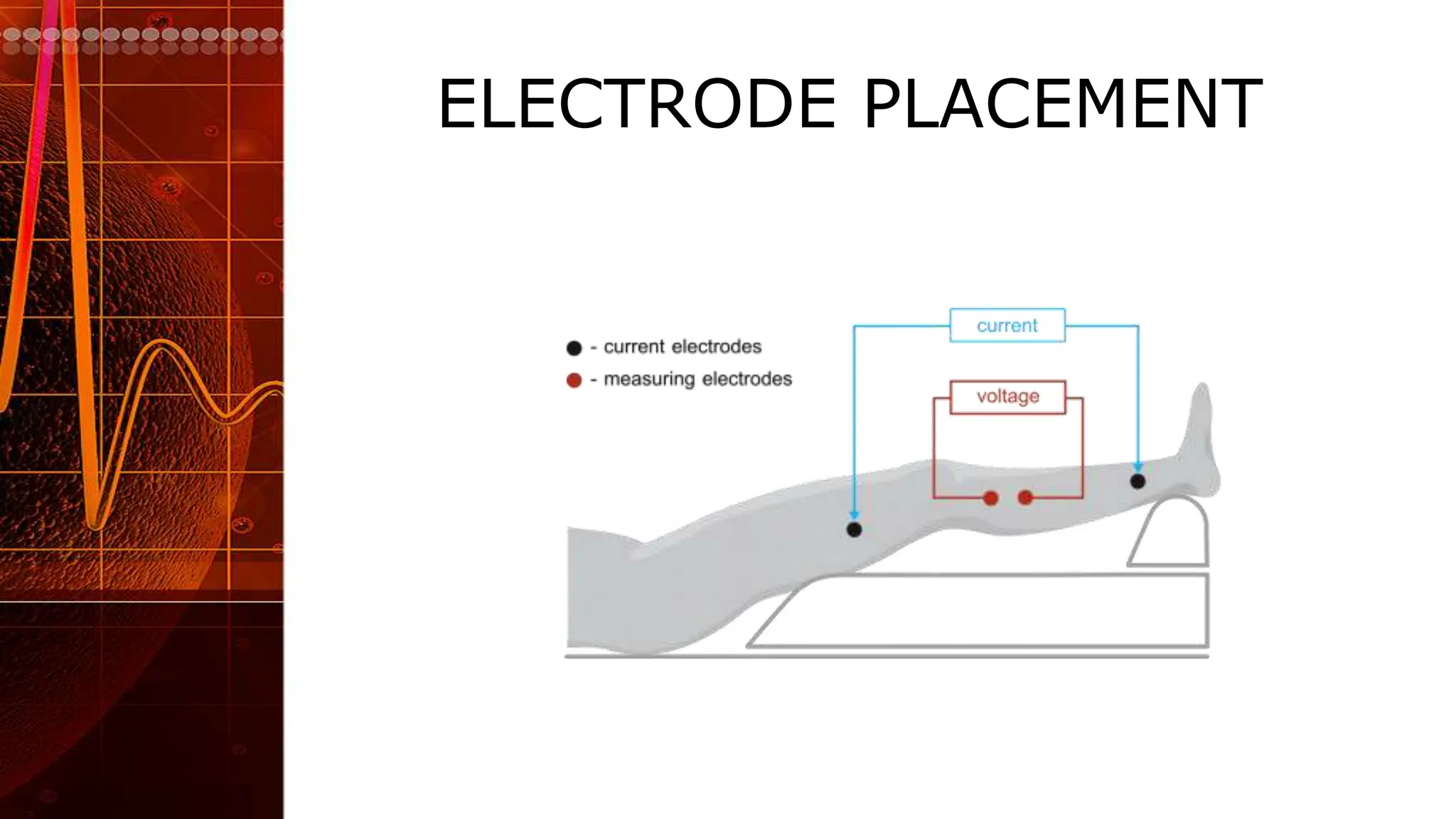
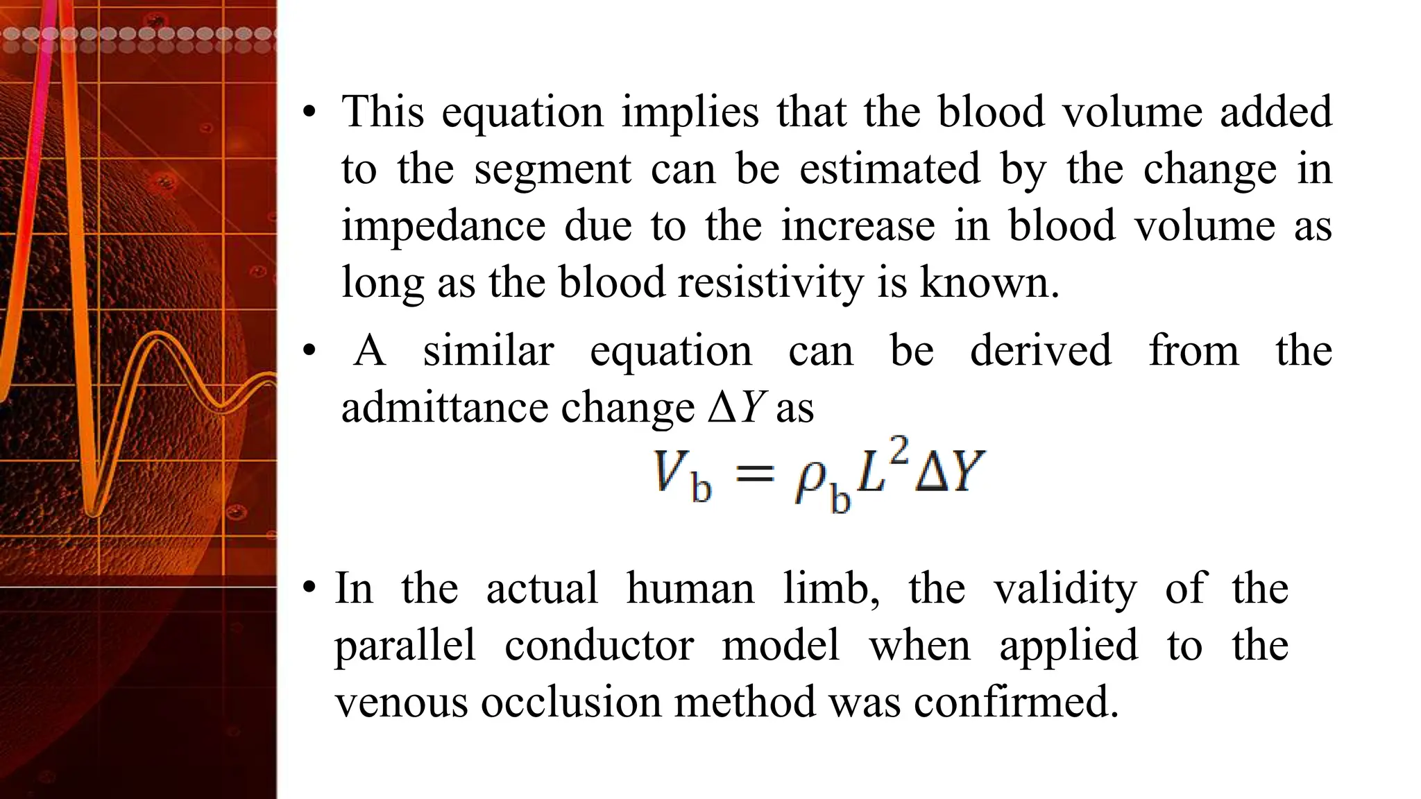
Impedance plethysmography
Impedance
Plethysmography(IPG)
• This method also named Rheography .
• It allows to measure arterial and venous blood
volume changes in nearly each body segment
(arms, legs, head, …)
• It works non-invasively and continuously and is
suitable to be used for arterial and venous vascular
diagnosis.
• The IPG is based on the measurement of the
electrical impedance (resistance) of a selected body
segment.
INTRODUCTION
• The IPG is based on the measurement of the
electrical impedance (resistance) of a selected
body segment.
• In comparison to other tissue, such as,
muscle or bone, blood has a much lower
impedance.
• Therefore, blood volume variations in a body
segment correspond with measurable changes
of the electrical impedance whereby an
increase of the blood volume results in a lower
impedance.
MEASUREMENT PRINCIPLE
• IPG relies on the fact that blood vessels expand
and contract with each heartbeat, leading to
changes in blood volume.
• These changes in blood volume cause alterations
in the electrical impedance of the arteries.
• By applying a weak electrical current and
measuring the resulting voltage through electrodes
attached to the skin, IPG captures these impedance
variations.
BLOCK DIAGRAM
ELECTRODES
• For the measurement of the electrical impedance usually 4 electrodes are
applied to the body surface approximately in a line.
• The 2 outer electrodes (usually called current electrodes) are used to
pass a very low and constant alternating current (1.5 mA, 86 kHz)
through the body segment.
• The 2 inner electrodes (usually called measuring electrodes) are placed
between the 2 current electrodes and measure the voltage which is caused
when the current flows through the body segment.
• This voltage corresponds with the impedance of the body segment which
changes depending on venous and arterial blood volume variations.
ELECTRODE PLACEMENT
EXPLANATION
• BLOCK DIAGRAM shows a schematic outline of
blood flow measurement using the electrical
impedance in a segment of a limb.
• A constant AC current of 0.1–10 mA and 20–
200 kHz is supplied to the outer electrodes, and the
voltage is measured at the inner electrodes.
• The impedance, Z, of the limb segment is defined as
the ratio of the voltage across the segment and the
supplied current.
• The reciprocal of Z is the admittance, Y.
Measurement of blood volume change in the limb by
electrical impedance is based on the parallel
conductor model
PARALLEL CONDUCTOR MODEL
• Consider a limb segment of length L, volume V0, and
tissue resistivity ρ0.
• The cross-sectional area, A0, is assumed to be constant
along the segment axis such that A0 = V0/L.
• When additional blood of volume Vb and
resistivity ρb is added to this segment, the parallel
conductor model postulates that the blood is distributed
uniformly in the segment forming a parallel conductor
of length L, cross-sectional area Ab = Vb/L, and
resistivity ρb.
DERIVATION AND EXPRESSION
and the impedance, Z, composed of parallel
conductors is expressed by
• This equation implies that the blood volume added
to the segment can be estimated by the change in
impedance due to the increase in blood volume as
long as the blood resistivity is known.
• A similar equation can be derived from the
admittance change ΔY as
• In the actual human limb, the validity of the
parallel conductor model when applied to the
venous occlusion method was confirmed.
BLOOD FLOW IN ARTERIES
• For Arteries, blood flow in any area like thigh, calf, arm or
forearm can be measured by Impedance Arteriography.
• Pulsatile flow of arteries will give characteristic Electrical
Resistance that can be recorded as a Graph.
• Along with simultaneous recording of ECG, parameters
like PAT, PTT can be measured.
Impedance Arteriography
pulse transit time (PTT) -represents the time between
two pulses measured at two locations
Pulse Arrival Time(PAT) is the time it takes for the
pulse to travel from the heart to a peripheral artery
BLOOD VOLUME IN VENOUS
• For leg veins, the test measures blood volume in the lower leg due to temporary venous
obstruction.
• This is done by inflating a cuff around the thigh to sufficient pressure to cut off venous flow
but not arterial flow, causing the venous blood pressure to rise.
• When the cuff is released there is a rapid venous return and a prompt return to the resting
blood volume.
• Cuff Inflation & Deflation will alter the Electrical Resistance of respective region, that will give
a characteristic Graph.
• Delayed emptying of veins in any venous pathology will change the normal response.
IMPEDANCE VENOGRAPHY
IPG
ADVANTAGES
• Noninvasiveness: IPG is a noninvasive technique,
meaning it doesn’t require any surgical incisions or
invasive procedures.
• Deep Artery Measurement: Unlike some other
methods, IPG allows the observation of a pulse in
every artery, regardless of its depth.
• Miniaturization Potential: This simplicity enables
the development of miniaturized devices, which can
be portable and convenient for various applications
ADVANTAGES
DRAWBACK
• The drawback is found to be in the motion
artefacts caused by the slight movements of the
hand, thus decreasing accuracy and also that a
regulator/potentiometer is required for signal
adjustment.
CLINICAL APPLICATIONS
• IPG is commonly used for peripheral vascular studies,
assessing blood flow and volume changes.
• It provides valuable information about venous return,
arterial pulsations, and overall vascular health.
• Researchers and clinicians utilize EIP to study
conditions such as deep vein thrombosis, venous
insufficiency, and peripheral artery disease.
APPLICATIONS

6. Impedance plethysmography .pptx

  • 1.
  • 2.
    Impedance Plethysmography(IPG) • This methodalso named Rheography . • It allows to measure arterial and venous blood volume changes in nearly each body segment (arms, legs, head, …) • It works non-invasively and continuously and is suitable to be used for arterial and venous vascular diagnosis. • The IPG is based on the measurement of the electrical impedance (resistance) of a selected body segment.
  • 3.
    INTRODUCTION • The IPGis based on the measurement of the electrical impedance (resistance) of a selected body segment. • In comparison to other tissue, such as, muscle or bone, blood has a much lower impedance. • Therefore, blood volume variations in a body segment correspond with measurable changes of the electrical impedance whereby an increase of the blood volume results in a lower impedance.
  • 4.
    MEASUREMENT PRINCIPLE • IPGrelies on the fact that blood vessels expand and contract with each heartbeat, leading to changes in blood volume. • These changes in blood volume cause alterations in the electrical impedance of the arteries. • By applying a weak electrical current and measuring the resulting voltage through electrodes attached to the skin, IPG captures these impedance variations.
  • 5.
  • 6.
    ELECTRODES • For themeasurement of the electrical impedance usually 4 electrodes are applied to the body surface approximately in a line. • The 2 outer electrodes (usually called current electrodes) are used to pass a very low and constant alternating current (1.5 mA, 86 kHz) through the body segment. • The 2 inner electrodes (usually called measuring electrodes) are placed between the 2 current electrodes and measure the voltage which is caused when the current flows through the body segment. • This voltage corresponds with the impedance of the body segment which changes depending on venous and arterial blood volume variations.
  • 7.
  • 8.
    EXPLANATION • BLOCK DIAGRAMshows a schematic outline of blood flow measurement using the electrical impedance in a segment of a limb. • A constant AC current of 0.1–10 mA and 20– 200 kHz is supplied to the outer electrodes, and the voltage is measured at the inner electrodes. • The impedance, Z, of the limb segment is defined as the ratio of the voltage across the segment and the supplied current.
  • 9.
    • The reciprocalof Z is the admittance, Y. Measurement of blood volume change in the limb by electrical impedance is based on the parallel conductor model
  • 10.
  • 11.
    • Consider alimb segment of length L, volume V0, and tissue resistivity ρ0. • The cross-sectional area, A0, is assumed to be constant along the segment axis such that A0 = V0/L. • When additional blood of volume Vb and resistivity ρb is added to this segment, the parallel conductor model postulates that the blood is distributed uniformly in the segment forming a parallel conductor of length L, cross-sectional area Ab = Vb/L, and resistivity ρb. DERIVATION AND EXPRESSION
  • 12.
    and the impedance,Z, composed of parallel conductors is expressed by
  • 13.
    • This equationimplies that the blood volume added to the segment can be estimated by the change in impedance due to the increase in blood volume as long as the blood resistivity is known. • A similar equation can be derived from the admittance change ΔY as • In the actual human limb, the validity of the parallel conductor model when applied to the venous occlusion method was confirmed.
  • 14.
    BLOOD FLOW INARTERIES • For Arteries, blood flow in any area like thigh, calf, arm or forearm can be measured by Impedance Arteriography. • Pulsatile flow of arteries will give characteristic Electrical Resistance that can be recorded as a Graph. • Along with simultaneous recording of ECG, parameters like PAT, PTT can be measured.
  • 15.
    Impedance Arteriography pulse transittime (PTT) -represents the time between two pulses measured at two locations Pulse Arrival Time(PAT) is the time it takes for the pulse to travel from the heart to a peripheral artery
  • 16.
    BLOOD VOLUME INVENOUS • For leg veins, the test measures blood volume in the lower leg due to temporary venous obstruction. • This is done by inflating a cuff around the thigh to sufficient pressure to cut off venous flow but not arterial flow, causing the venous blood pressure to rise. • When the cuff is released there is a rapid venous return and a prompt return to the resting blood volume. • Cuff Inflation & Deflation will alter the Electrical Resistance of respective region, that will give a characteristic Graph. • Delayed emptying of veins in any venous pathology will change the normal response.
  • 17.
  • 18.
  • 19.
    ADVANTAGES • Noninvasiveness: IPGis a noninvasive technique, meaning it doesn’t require any surgical incisions or invasive procedures. • Deep Artery Measurement: Unlike some other methods, IPG allows the observation of a pulse in every artery, regardless of its depth. • Miniaturization Potential: This simplicity enables the development of miniaturized devices, which can be portable and convenient for various applications
  • 20.
  • 21.
    DRAWBACK • The drawbackis found to be in the motion artefacts caused by the slight movements of the hand, thus decreasing accuracy and also that a regulator/potentiometer is required for signal adjustment.
  • 22.
    CLINICAL APPLICATIONS • IPGis commonly used for peripheral vascular studies, assessing blood flow and volume changes. • It provides valuable information about venous return, arterial pulsations, and overall vascular health. • Researchers and clinicians utilize EIP to study conditions such as deep vein thrombosis, venous insufficiency, and peripheral artery disease.
  • 23.