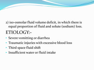
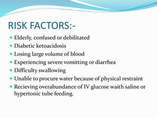
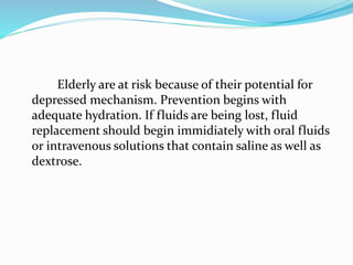
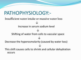
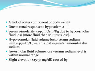
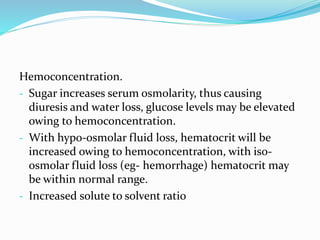
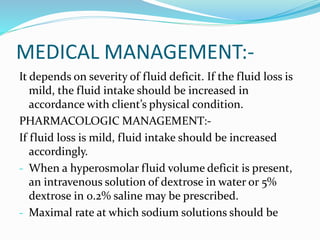
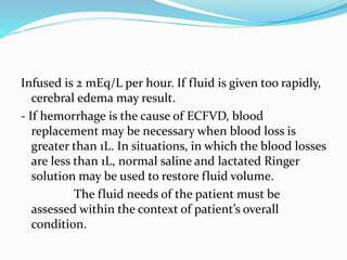
PRESENTED BY:-KHUSHNASIB
ACN
 The five types of fluid imbalances that may occur are
extracellular fluid volume deficit (ECFVD),
extracellular fluid volume excess (ECFVE),
extracellular fluid volume shift, extracellular fluid
volume deficit.
 (Sodium is the major ion that influences fluid balance
and imbalance)
ECFVD:-
EXTRACELLULAR
FLUID VOLUME
DEFICIT:-
 An ECFVD is a decrease in intravascular and
interstitial fluids. ECFVD is a common and serious
fluid imbalance that results in vascular fluid volume
loss (hypovolemia). ECFVD can lead to cellular fluid
loss (hypovolemia) owing to fluid shifting from the
cells to the vascular fluid to restore fluid balance.
There are major two types of extracellular
fluid volume deficits
1) Hyperosmolar fluid volume deficit, in which fluid loss
is greater than the solute (sodium) loss.
2) iso-osmolar fluid volume deficit, in which there is
equal proportion of fluid and solute (sodium) loss.
ETIOLOGY:-
• Severe vomitting or diarrhea
• Traumatic injuries with excessive blood loss
• Third space fluid shift
• Insufficient water or fluid intake
RISK FACTORS:-
 Elderly, confused or debilitated
 Diabetic ketoacidosis
 Losing large volume of blood
 Experiencing severe vomitting or diarrhea
 Difficulty swallowing
 Unable to procure water because of physical restraint
 Recieving overabundance of IV glucose waith saline or
hypertonic tube feeding.
Elderly are at risk because of their potential for
depressed mechanism. Prevention begins with
adequate hydration. If fluids are being lost, fluid
replacement should begin immidiately with oral fluids
or intravenous solutions that contain saline as well as
dextrose.
PATHOPHYSIOLOGY:-
Insufficient water intake or massive water loss
↓
Increase in serum sodium level
↓
Shifting of water from cells to vascular space
↓
Decrease the hyperosmolarity (caused by water loss)
↓
This shift causes cells to shrink and cellular dehydration
occurs
↓
Severe fluid volume deficit
↓
Shock or vascular collapse can occur
If ECFVD develops rapidly, symptoms are more severe.
Severe brain shrinkage during water deficit may cause
vascular damage and intracerebral hemorrhage. When
ECFVD occurs slowly, the brain cells adapt to the
increased intracellular osmolarity by producing extra
Intracellular particles (idiogenic osmoles) which prevent
large amount of water from leaving the cell.
CLINICAL MANIFESTATIONS:-
- With mild ECFVD, 1-2 L of water & 2% of body weight
is lost. Moderate ECFVD is evidenced by 3-5 L of water
loss & 5% weight loss.
- In severe ECFVD, the water loss is increased to 5-10 L &
weight loss is increased to 8%, the systolic blood
pressure becomes alarmingly low, as demonstrated by
a reading of less than 70 mm Hg.
 Thirst
 Decreased skin turgor
 Dry mucus memb, dry crackled lips or tongue
 Eyeballs soft or sunken (severe deficit)
 Apprehension & restlessness, coma insevere deficit
 Elevated temperature
 Tachycardia
 Postural systolic blood pressure>15 mmHg and
diastolic fall>10 mm Hg
 Narrowed pulse pressure, decreased central venous
pressure and pulmonary capillary wedge pressure
 Flattened neck veins in supine position
PATHOPHYSIOLOGIC BASIS:-
- Cells shrink, stimulating thirst osmorecceptors in the
hypothalamus, with iso-osmolar fluid loss, thirst
usually does not occur.
- Decreased interstitial fluid causes skin tissue to stick
together.
- Cells of mucus memb and tongue dry out.
 Water tension in eye balls decreased
 Cerebral dehydration
 Less fluid available for evaporation
 Pulse greater than 100 bpm may be due to circulatory
compensation by the heart.
 With iso-osmolar fluid loss, the plasma volume is
inadequate owing to hypovolemia, systolic pressure
begin to fall.
 Decreased venous return.
 A lack of water component of body weight.
 Due to renal response to hypovolemia
 Serum osmolarity> 295 mOsm/Kg due to hypoosmolar
fluid loss (more fluid than solutes is lost).
 Hypo-osmolar fluid volume loss:- serum sodium
level>145mEq/L, water is lost in greater amounts tahn
sodium.
 Iso-osmolar fluid volume loss –serum sodium level is
within normal range.
 Slight elevation (25-35 mg/dl) caused by
Hemoconcentration.
- Sugar increases serum osmolarity, thus causing
diuresis and water loss, glucose levels may be elevated
owing to hemoconcentration.
- With hypo-osmolar fluid loss, hematocrit will be
increased owing to hemoconcentration, with iso-
osmolar fluid loss (eg- hemorrhage) hematocrit may
be within normal range.
- Increased solute to solvent ratio
MEDICAL MANAGEMENT:-
It depends on severity of fluid deficit. If the fluid loss is
mild, the fluid intake should be increased in
accordance with client’s physical condition.
PHARMACOLOGIC MANAGEMENT:-
If fluid loss is mild, fluid intake should be increased
accordingly.
- When a hyperosmolar fluid volume deficit is present,
an intravenous solution of dextrose in water or 5%
dextrose in 0.2% saline may be prescribed.
- Maximal rate at which sodium solutions should be
Infused is 2 mEq/L per hour. If fluid is given too rapidly,
cerebral edema may result.
- If hemorrhage is the cause of ECFVD, blood
replacement may be necessary when blood loss is
greater than 1L. In situations, in which the blood losses
are less than 1L, normal saline and lactated Ringer
solution may be used to restore fluid volume.
The fluid needs of the patient must be
assessed within the context of patient’s overall
condition.
A patient with severe ECFVD and also severe heart or
kidney disease cannot be given large volume of fluids
or sodium.
DIETARY MANAGEMENT:-
Avoid fatty or fried foods and milk products.
NURSING MANAGEMENT:-
1) Fluid volume deficit r/t insufficient fluid intake,
vomotting, diarrhea, hemorrhage or third space fluid
loss (ascites, burns).
 Expected outcomes:- The patient’s fluid balance will
be restored, as evidenced by vital signs within normal
range, return to baseline body weight, absence of
causative factors of ECFVD, equal intake and output,
urine output greater than 600 ml/day.
 Implementation-
- Check vital signs 2-4 hrly
- Check positional BP to determine degree of
orthostasis. If systolic BP falls 10-15 mm Hg or more,
the results should be reported and patient protected
from injury while ambulating.
- Assess urine output hrly to indicate renal insufficiency
because of decreased renal perfusion. (Absence of
adequate renal perfusion for several hrs may result in
permanent renal damage)
- Measure sp. gravity of urine, to determine osmolality.
- Monitor daily weight. Weight loss of approx. 3.1 Kg is
indicative of fluid volume deficit.
- Apply lotion on skin to maintain skin integrity.
- Provide oral care 2 hrly with non alcohol based
solution.
- Serum sodium, BUN, glucose, hematocrit level should
be monitored closely.
- C/M of fluid volume overload- dyspnea, crackles and
jugular vein engorgement.
Thank you

Extracellular fluid volum deficit

  • 1.
  • 2.
     The fivetypes of fluid imbalances that may occur are extracellular fluid volume deficit (ECFVD), extracellular fluid volume excess (ECFVE), extracellular fluid volume shift, extracellular fluid volume deficit.  (Sodium is the major ion that influences fluid balance and imbalance)
  • 3.
  • 4.
     An ECFVDis a decrease in intravascular and interstitial fluids. ECFVD is a common and serious fluid imbalance that results in vascular fluid volume loss (hypovolemia). ECFVD can lead to cellular fluid loss (hypovolemia) owing to fluid shifting from the cells to the vascular fluid to restore fluid balance. There are major two types of extracellular fluid volume deficits 1) Hyperosmolar fluid volume deficit, in which fluid loss is greater than the solute (sodium) loss.
  • 5.
    2) iso-osmolar fluidvolume deficit, in which there is equal proportion of fluid and solute (sodium) loss. ETIOLOGY:- • Severe vomitting or diarrhea • Traumatic injuries with excessive blood loss • Third space fluid shift • Insufficient water or fluid intake
  • 6.
    RISK FACTORS:-  Elderly,confused or debilitated  Diabetic ketoacidosis  Losing large volume of blood  Experiencing severe vomitting or diarrhea  Difficulty swallowing  Unable to procure water because of physical restraint  Recieving overabundance of IV glucose waith saline or hypertonic tube feeding.
  • 7.
    Elderly are atrisk because of their potential for depressed mechanism. Prevention begins with adequate hydration. If fluids are being lost, fluid replacement should begin immidiately with oral fluids or intravenous solutions that contain saline as well as dextrose.
  • 8.
    PATHOPHYSIOLOGY:- Insufficient water intakeor massive water loss ↓ Increase in serum sodium level ↓ Shifting of water from cells to vascular space ↓ Decrease the hyperosmolarity (caused by water loss) ↓ This shift causes cells to shrink and cellular dehydration occurs
  • 9.
    ↓ Severe fluid volumedeficit ↓ Shock or vascular collapse can occur If ECFVD develops rapidly, symptoms are more severe. Severe brain shrinkage during water deficit may cause vascular damage and intracerebral hemorrhage. When ECFVD occurs slowly, the brain cells adapt to the increased intracellular osmolarity by producing extra
  • 10.
    Intracellular particles (idiogenicosmoles) which prevent large amount of water from leaving the cell. CLINICAL MANIFESTATIONS:- - With mild ECFVD, 1-2 L of water & 2% of body weight is lost. Moderate ECFVD is evidenced by 3-5 L of water loss & 5% weight loss. - In severe ECFVD, the water loss is increased to 5-10 L & weight loss is increased to 8%, the systolic blood pressure becomes alarmingly low, as demonstrated by a reading of less than 70 mm Hg.
  • 11.
     Thirst  Decreasedskin turgor  Dry mucus memb, dry crackled lips or tongue  Eyeballs soft or sunken (severe deficit)  Apprehension & restlessness, coma insevere deficit  Elevated temperature  Tachycardia  Postural systolic blood pressure>15 mmHg and diastolic fall>10 mm Hg
  • 12.
     Narrowed pulsepressure, decreased central venous pressure and pulmonary capillary wedge pressure  Flattened neck veins in supine position PATHOPHYSIOLOGIC BASIS:- - Cells shrink, stimulating thirst osmorecceptors in the hypothalamus, with iso-osmolar fluid loss, thirst usually does not occur. - Decreased interstitial fluid causes skin tissue to stick together. - Cells of mucus memb and tongue dry out.
  • 13.
     Water tensionin eye balls decreased  Cerebral dehydration  Less fluid available for evaporation  Pulse greater than 100 bpm may be due to circulatory compensation by the heart.  With iso-osmolar fluid loss, the plasma volume is inadequate owing to hypovolemia, systolic pressure begin to fall.  Decreased venous return.
  • 14.
     A lackof water component of body weight.  Due to renal response to hypovolemia  Serum osmolarity> 295 mOsm/Kg due to hypoosmolar fluid loss (more fluid than solutes is lost).  Hypo-osmolar fluid volume loss:- serum sodium level>145mEq/L, water is lost in greater amounts tahn sodium.  Iso-osmolar fluid volume loss –serum sodium level is within normal range.  Slight elevation (25-35 mg/dl) caused by
  • 15.
    Hemoconcentration. - Sugar increasesserum osmolarity, thus causing diuresis and water loss, glucose levels may be elevated owing to hemoconcentration. - With hypo-osmolar fluid loss, hematocrit will be increased owing to hemoconcentration, with iso- osmolar fluid loss (eg- hemorrhage) hematocrit may be within normal range. - Increased solute to solvent ratio
  • 16.
    MEDICAL MANAGEMENT:- It dependson severity of fluid deficit. If the fluid loss is mild, the fluid intake should be increased in accordance with client’s physical condition. PHARMACOLOGIC MANAGEMENT:- If fluid loss is mild, fluid intake should be increased accordingly. - When a hyperosmolar fluid volume deficit is present, an intravenous solution of dextrose in water or 5% dextrose in 0.2% saline may be prescribed. - Maximal rate at which sodium solutions should be
  • 17.
    Infused is 2mEq/L per hour. If fluid is given too rapidly, cerebral edema may result. - If hemorrhage is the cause of ECFVD, blood replacement may be necessary when blood loss is greater than 1L. In situations, in which the blood losses are less than 1L, normal saline and lactated Ringer solution may be used to restore fluid volume. The fluid needs of the patient must be assessed within the context of patient’s overall condition.
  • 18.
    A patient withsevere ECFVD and also severe heart or kidney disease cannot be given large volume of fluids or sodium. DIETARY MANAGEMENT:- Avoid fatty or fried foods and milk products. NURSING MANAGEMENT:- 1) Fluid volume deficit r/t insufficient fluid intake, vomotting, diarrhea, hemorrhage or third space fluid loss (ascites, burns).
  • 19.
     Expected outcomes:-The patient’s fluid balance will be restored, as evidenced by vital signs within normal range, return to baseline body weight, absence of causative factors of ECFVD, equal intake and output, urine output greater than 600 ml/day.  Implementation- - Check vital signs 2-4 hrly - Check positional BP to determine degree of orthostasis. If systolic BP falls 10-15 mm Hg or more, the results should be reported and patient protected
  • 20.
    from injury whileambulating. - Assess urine output hrly to indicate renal insufficiency because of decreased renal perfusion. (Absence of adequate renal perfusion for several hrs may result in permanent renal damage) - Measure sp. gravity of urine, to determine osmolality. - Monitor daily weight. Weight loss of approx. 3.1 Kg is indicative of fluid volume deficit. - Apply lotion on skin to maintain skin integrity.
  • 21.
    - Provide oralcare 2 hrly with non alcohol based solution. - Serum sodium, BUN, glucose, hematocrit level should be monitored closely. - C/M of fluid volume overload- dyspnea, crackles and jugular vein engorgement.
  • 22.