Downloaded 1,658 times
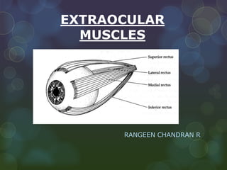


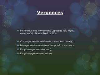
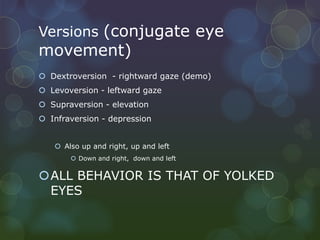


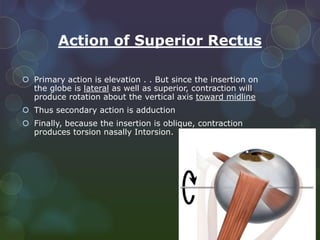


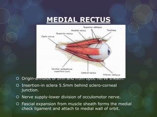


The extraocular muscles include 6 rectus muscles and 2 oblique muscles that control movement of the eyeball. The rectus muscles are the superior, inferior, medial, and lateral rectus and act to move the eye in specific directions. The oblique muscles are the superior and inferior oblique and produce torsional movements as their primary action with secondary actions of moving the eye. All the muscles are supplied by specific cranial nerves with some variations in their blood supply.


