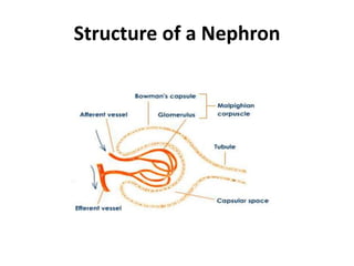
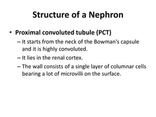
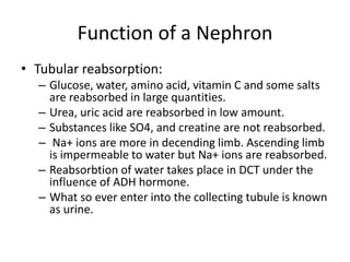
HAFIZ
MUHAMM
AD
WASEEM
Physiology of Excretion
Excretory System
• Back ground:
– The process of removal of nitrogenous wastes
along with excess water and pigments is called
excretion.
– Elimination of metabolic waste products
accompanying digestion and respiration and/ or
maintenance of water and electrolytes is known
as excretion.
– It maintains the homeostasis of the body.
Excretory System
• Back ground:
– Maintain proper internal pH by removing excess acids and
bases.
– Eliminate byproducts of nitrogen metabolism.
– Due to catabolism of proteins amino (-NH2) group is released.
– The accumulation of -NH2 group is very toxic.
– So it must be either used in the body or must be expelled out.
– Similarly, water is also formed due to oxidative phosphorylation
and pigments are formed by the metabolism of haemoglobin of
dead RBCs.
– Excessive of water is to be expelled to prevent the dilution of
fluids.
Excretory System
• Important Nitrogenous wastes
– Different animals expel different nitrogenous
wastes like:
• Ammonia
• Urea
• Uric acid
Important Nitrogenous wastes
• Ammonia:
– Generally it is formed in aquatic animals.
– Soluble in water and highly toxic.
– Easily diffusible in water media.
– Animals which produce ammonia are known as ammonotelic.
– It is formed by the deamination of proteins.
– One gram of ammonia needs about 300-500ml of water to be
expelled out of the body.
– So this process involves loss of considerable amount of water.
– That is the reason why ammonotelic excretion is found in
aquatic animals.
• Examples: Hydra,Teleosts
Important Nitrogenous wastes
• Urea:
– It is characteristic of terrestrial animals.
– In higher animals it is produced in liver by the
deamination process.
– It is known as ornithine cycle.
– Animals which produce urea are known as ureotelic.
– One gram of urea needs about 50ml of water to be
expelled out.
– So it is suitable for terrestrial mode of life with
tendency to conserve water.
• Example. Man, Elasmobranch fish.
Important Nitrogenous wastes
• Uric acid:
– It is characteristic of animals where conservation of water is needed.
– Uric acid is formed in liver.
– It is formed due to absence of arginase enzyme.
– Animals which excrete uric acid are known as uricotelic.
– It is found in birds, insects and desert animals.
– It is formed from ammonia and purines (adenine and guanine) in liver
and kidney.
– In human beings uric acid is formed by the catabolism of purines.
– It is less soluble in water. One gram of uric acid needs 10 ml of water
to be expelled out.
– Besides it is less toxic and can remain in the tissues for long period.
– So excretion of uric acid is of greater advantage to land animals.
Excretory system of Man
• It is formed of
– One pair of kidneys
– One pair of ureters
– A urinary bladder
– A urethra
Excretory system of Man
• Kidneys
– They are the major excretory organs of man and other
vertebrates.
– They are paired, large sized, bean shaped, dark red and present
in upper part of the abdominal cavity one on either side of the
vertebral column just below the diaphragm.
– Located between the last thoracic vertebra and the third lumbar
vertebra.
– They are protected by the last 2 ribs.
– The kidneys are covered by the peritoneum in the front and on
the side and they rest against the abdominal muscles.
– Their position is slightly asymmetrical, the left kidney being a
little higher than the right.
Excretory system of Man
• Kidneys
– The outer surface is convex and the inner surface is
concave.
– The inner surface has a deep notch called hilus.
– The kidney is divided into 2 regions, an outer region called
renal cortex and the inner region termed renal medulla.
– The medulla is subdivided into conical masses, the renal
pyramids, each having a broad base towards the cortex
and a narrow end called renal papilla towards the pelvis.
– The renal papilla projects into the wide funnel - like
structure called the pelvis.
Excretory system of Man
• Kidneys
– The pelvis leads into the ureter between the
pyramids.
– The cortex extends into the medulla as renal column
of Bertin.
– Internally each kidney is formed of about one million
uriniferous or renal tubules or nephrons.
– They are embedded in renal fat called renal facia.
– It forms a shock - absorbing cushion and the renal
fascia fixes the kidney to the abdominal walls.
– They help in urine formation.
Excretory system of Man
• Ureters
– They are paired, long, narrow, muscular and
tubular structures.
– Each ureter arises from the hilus of the kidney.
– They run backwards and open into urinary
bladder.
– These are lined by transitional epithelium.
– They conduct the urine from kidneys to urinary
bladder.
Excretory system of Man
• Urinary bladder:
– It is large, thin walled, distensible, pear shaped sac present in
the pelvic region of the abdominal cavity.
– It is lined by smooth muscles and transitional epithelium.
– It temporarily stores the urine.
• Urethra:
– It is muscular and tubular.
– In female it is about 4cm long and opens directly outside
through vaginal orifice.
– In man, it joins the ejaculatory duct to form urino gential canal
which passes through the penis.
– It is about 20 cm long and common passages for both urine and
semen.
Structure of a Nephron
• Uriniferous tubule/ Nephron
– The basic structural and functional unit of the kidney.
– Its chief function is to regulate the concentration of
water.
– A nephron consists of a twisted tubule closed at one
end, open at the other with a network of associated
blood vessels.
– Each kidney of man is formed of about one million
nephrons.
– Each nephron has a length of about 3 cm.
Structure of a Nephron
• It is differentiated into 4 regions having
different anatomical features and different
physiological roles:
– Bowman's capsule
– Proximal convoluted tubule (PCT)
– Loop of Henle
– Distal convoluted tubule (DCT)
Structure of a Nephron
• Bowman’s Capsule
– These were first reported by M.Malphigi (1966).
– It is a large double walled cup.
– It lies in the renal cortex.
– It contains a tuft of capillaries called glomerulus.
– The space between the two walls of the Bowman's
capsule is continuous with the lumen of the next part
of the nephron.
– The Bowman’s capsule and the glomerulus together
constitute the renal corpuscle or Malpighian body.
Structure of a Nephron
• Proximal convoluted tubule (PCT)
– It starts from the neck of the Bowman's capsule
and it is highly convoluted.
– It lies in the renal cortex.
– The wall consists of a single layer of columnar cells
bearing a lot of microvilli on the surface.
Structure of a Nephron
• Loop of Henle
– It is a V/U shaped segment of the nephron.
– It is located in the renal medulla.
– It consists of two straight parallel limbs known as a
descending limb and an ascending limb.
– Descending limb is a continuation of the PCT and
enters into the renal medulla and an ascending limb
which reenters the renal cortex and joins the DCT.
– It is short or absent in other vertebrates like reptiles.
Structure of a Nephron
• Distal convoluted tubule (DCT)
– It is greatly twisted like the PCT and lies in the renal cortex.
– The terminal relatively short part of the DCT is called the
collecting tubule.
– It opens into the collecting duct.
– It is lined by cuboidal epithelium.
– The collecting ducts receive the collecting tubules of several
nephrons.
– They pass into the renal medulla and join each other forming
large ducts of Bellini which pass through renal pyramids, open
into minor calices which open in larger major calices (4-5 in
number).
– Major calices finally open in a funnel shaped pelvis.
– Pelvis opens into ureter.
Structure of a Nephron
Function of a Nephron
• Urine formation is the main function of
neuron.
• It occurs in three steps:
– Pressure filtration/ultrafiltration
– Tubular reabsorption
– Tubular secretion
Function of a Nephron
• Pressure filtration/ Ultrafiltration
– The process through which fluid is filtered and enters
Bowman’s capsule and is than termed as filtrate or
ultrafiltrate .
– This process of ultrafiltration occurs when the hydrostatic
pressure outside of tubule is higher than inside of tubule
lumen .
– This filtrate only consist of water and solutes that pass
with water.
– The blood plasma is source of water and solutes .
– The blood pressure produced by heart is the pressure that
drives ultrafiltration.
Function of a Nephron
• Pressure filtration/ Ultrafiltration
– GHP= glomerular hydrostatic pressure is about 70mm
Hg. It is due to wider afferent and narrow efferent
arteriole. But it is resisted by osmotic pressure of
blood (BCOP) and cells of Bowman’s capsule (CHP).
– BCOP= Blood Colloidal Osmotic Pressure is due to the
presence of plasma proteins (albumin). It is about 30
mm Hg.
– CHP= Capsular Hydrostatic pressure. It is due to the
pre existing fluid in Bowman’s capsule. It is about 20
mmHg.
Function of a Nephron
• Pressure filtration/ Ultrafiltration
– So the Effective filtration Pressure:
– EFP= GHP-(BCOP+CHP) = 70- (30+20) =20mm Hg.
It is also known as Net filtration rate (NFR).
– What so ever filtered in Bowman’s capsule is
known as nephric filtrate or glomerulus filtrate.
– It contains both useful and non useful substances.
– Nephric filtrate =Blood- (Blood cells+proteins).
Function of a Nephron
• Regulation of Glomerular Filtration:
– Intrinsic Mechanism: It includes renal
autoregulation. In this kidney itself adjust the
changes by constriction or dilation of afferent
arterioles.
– Extrinsic mechanism: This include neural and
hormonal control e:g, renin-angiotensin-
aldosterone system.
Function of a Nephron- regulation
Function of a Nephron
• Tubular reabsorption:
– Nephric filtrate is moved in the nephron towards collecting
tubule.
– During this passage all useful and selective substances are
reabsorbed. So, it is called as selective reabsorbtion.
– It takes place in PCT+ Loop of Henle+ DCT.
– All glucose and amino acids reabsorb.
– Most of the electrolyte reabsorbed.
– 96% of water reabsorbed.
– Na+, glucose, amino acid are reabsorbed by active
transport.
– Water and Cl- are reabsorbed by passive transport.
Function of a Nephron
• Tubular reabsorption:
– Glucose, water, amino acid, vitamin C and some salts
are reabsorbed in large quantities.
– Urea, uric acid are reabsorbed in low amount.
– Substances like SO4, and creatine are not reabsorbed.
– Na+ ions are more in decending limb. Ascending limb
is impermeable to water but Na+ ions are reabsorbed.
– Reabsorbtion of water takes place in DCT under the
influence of ADH hormone.
– What so ever enter into the collecting tubule is known
as urine.
Function of a Nephron
• Tubular secretion:
– It is supporting and supplementary mechanism.
– It is mainly aided by the glandular cells of DCT.
– Cells of tubule remove non thresh hold substances
and foreign materials
– from the blood.
– Most of the K+ is eliminated in exchange of Na+.
– As a result of these three processes the waste are
changed into urine.
Function of a Nephron
• Composition of urine
– It is pale yellow in color due presence of a pigment
called urochrome, formed by the haemoglobin of
dead RBCs.
– It is acidic in nature and has pH 6.1- 6.5.
– It has a faint aromatic odour. The bad smell is due
degradation of urea into ammonia by microbes.
– Daily output of urine in normal person is 1.5 1.8 litres.
– Chemically it consists of 95-96% water, urea 2%, uric
acid 0.2%, ammonia 0.25%, creatine 0.5% and salts
1%.
Function of a Nephron
• Conduction of urine
– The filtrate left after tubular absorption and secretion
is called urine.
– Contains 95% water, nitrogenous wastes, ions,
metabolites and blood cells.
– It is transported through ureter to bladder and is
stored there.
– It leaves body through urethra. When urinary bladder
becomes filled with urine tension increases on its
walls.
– This stimulates the response, and sphincter muscles
are relaxed at entrance of urethra. This response is
called urination.

Excretory system

  • 1.
  • 2.
  • 3.
    Excretory System • Background: – The process of removal of nitrogenous wastes along with excess water and pigments is called excretion. – Elimination of metabolic waste products accompanying digestion and respiration and/ or maintenance of water and electrolytes is known as excretion. – It maintains the homeostasis of the body.
  • 4.
    Excretory System • Background: – Maintain proper internal pH by removing excess acids and bases. – Eliminate byproducts of nitrogen metabolism. – Due to catabolism of proteins amino (-NH2) group is released. – The accumulation of -NH2 group is very toxic. – So it must be either used in the body or must be expelled out. – Similarly, water is also formed due to oxidative phosphorylation and pigments are formed by the metabolism of haemoglobin of dead RBCs. – Excessive of water is to be expelled to prevent the dilution of fluids.
  • 5.
    Excretory System • ImportantNitrogenous wastes – Different animals expel different nitrogenous wastes like: • Ammonia • Urea • Uric acid
  • 6.
    Important Nitrogenous wastes •Ammonia: – Generally it is formed in aquatic animals. – Soluble in water and highly toxic. – Easily diffusible in water media. – Animals which produce ammonia are known as ammonotelic. – It is formed by the deamination of proteins. – One gram of ammonia needs about 300-500ml of water to be expelled out of the body. – So this process involves loss of considerable amount of water. – That is the reason why ammonotelic excretion is found in aquatic animals. • Examples: Hydra,Teleosts
  • 7.
    Important Nitrogenous wastes •Urea: – It is characteristic of terrestrial animals. – In higher animals it is produced in liver by the deamination process. – It is known as ornithine cycle. – Animals which produce urea are known as ureotelic. – One gram of urea needs about 50ml of water to be expelled out. – So it is suitable for terrestrial mode of life with tendency to conserve water. • Example. Man, Elasmobranch fish.
  • 8.
    Important Nitrogenous wastes •Uric acid: – It is characteristic of animals where conservation of water is needed. – Uric acid is formed in liver. – It is formed due to absence of arginase enzyme. – Animals which excrete uric acid are known as uricotelic. – It is found in birds, insects and desert animals. – It is formed from ammonia and purines (adenine and guanine) in liver and kidney. – In human beings uric acid is formed by the catabolism of purines. – It is less soluble in water. One gram of uric acid needs 10 ml of water to be expelled out. – Besides it is less toxic and can remain in the tissues for long period. – So excretion of uric acid is of greater advantage to land animals.
  • 9.
    Excretory system ofMan • It is formed of – One pair of kidneys – One pair of ureters – A urinary bladder – A urethra
  • 10.
    Excretory system ofMan • Kidneys – They are the major excretory organs of man and other vertebrates. – They are paired, large sized, bean shaped, dark red and present in upper part of the abdominal cavity one on either side of the vertebral column just below the diaphragm. – Located between the last thoracic vertebra and the third lumbar vertebra. – They are protected by the last 2 ribs. – The kidneys are covered by the peritoneum in the front and on the side and they rest against the abdominal muscles. – Their position is slightly asymmetrical, the left kidney being a little higher than the right.
  • 11.
    Excretory system ofMan • Kidneys – The outer surface is convex and the inner surface is concave. – The inner surface has a deep notch called hilus. – The kidney is divided into 2 regions, an outer region called renal cortex and the inner region termed renal medulla. – The medulla is subdivided into conical masses, the renal pyramids, each having a broad base towards the cortex and a narrow end called renal papilla towards the pelvis. – The renal papilla projects into the wide funnel - like structure called the pelvis.
  • 12.
    Excretory system ofMan • Kidneys – The pelvis leads into the ureter between the pyramids. – The cortex extends into the medulla as renal column of Bertin. – Internally each kidney is formed of about one million uriniferous or renal tubules or nephrons. – They are embedded in renal fat called renal facia. – It forms a shock - absorbing cushion and the renal fascia fixes the kidney to the abdominal walls. – They help in urine formation.
  • 13.
  • 14.
    Excretory system ofMan • Ureters – They are paired, long, narrow, muscular and tubular structures. – Each ureter arises from the hilus of the kidney. – They run backwards and open into urinary bladder. – These are lined by transitional epithelium. – They conduct the urine from kidneys to urinary bladder.
  • 15.
    Excretory system ofMan • Urinary bladder: – It is large, thin walled, distensible, pear shaped sac present in the pelvic region of the abdominal cavity. – It is lined by smooth muscles and transitional epithelium. – It temporarily stores the urine. • Urethra: – It is muscular and tubular. – In female it is about 4cm long and opens directly outside through vaginal orifice. – In man, it joins the ejaculatory duct to form urino gential canal which passes through the penis. – It is about 20 cm long and common passages for both urine and semen.
  • 16.
    Structure of aNephron • Uriniferous tubule/ Nephron – The basic structural and functional unit of the kidney. – Its chief function is to regulate the concentration of water. – A nephron consists of a twisted tubule closed at one end, open at the other with a network of associated blood vessels. – Each kidney of man is formed of about one million nephrons. – Each nephron has a length of about 3 cm.
  • 17.
    Structure of aNephron • It is differentiated into 4 regions having different anatomical features and different physiological roles: – Bowman's capsule – Proximal convoluted tubule (PCT) – Loop of Henle – Distal convoluted tubule (DCT)
  • 18.
  • 19.
    Structure of aNephron • Bowman’s Capsule – These were first reported by M.Malphigi (1966). – It is a large double walled cup. – It lies in the renal cortex. – It contains a tuft of capillaries called glomerulus. – The space between the two walls of the Bowman's capsule is continuous with the lumen of the next part of the nephron. – The Bowman’s capsule and the glomerulus together constitute the renal corpuscle or Malpighian body.
  • 20.
  • 21.
    Structure of aNephron • Proximal convoluted tubule (PCT) – It starts from the neck of the Bowman's capsule and it is highly convoluted. – It lies in the renal cortex. – The wall consists of a single layer of columnar cells bearing a lot of microvilli on the surface.
  • 22.
    Structure of aNephron • Loop of Henle – It is a V/U shaped segment of the nephron. – It is located in the renal medulla. – It consists of two straight parallel limbs known as a descending limb and an ascending limb. – Descending limb is a continuation of the PCT and enters into the renal medulla and an ascending limb which reenters the renal cortex and joins the DCT. – It is short or absent in other vertebrates like reptiles.
  • 23.
    Structure of aNephron • Distal convoluted tubule (DCT) – It is greatly twisted like the PCT and lies in the renal cortex. – The terminal relatively short part of the DCT is called the collecting tubule. – It opens into the collecting duct. – It is lined by cuboidal epithelium. – The collecting ducts receive the collecting tubules of several nephrons. – They pass into the renal medulla and join each other forming large ducts of Bellini which pass through renal pyramids, open into minor calices which open in larger major calices (4-5 in number). – Major calices finally open in a funnel shaped pelvis. – Pelvis opens into ureter.
  • 24.
  • 25.
    Function of aNephron • Urine formation is the main function of neuron. • It occurs in three steps: – Pressure filtration/ultrafiltration – Tubular reabsorption – Tubular secretion
  • 26.
  • 27.
    Function of aNephron • Pressure filtration/ Ultrafiltration – The process through which fluid is filtered and enters Bowman’s capsule and is than termed as filtrate or ultrafiltrate . – This process of ultrafiltration occurs when the hydrostatic pressure outside of tubule is higher than inside of tubule lumen . – This filtrate only consist of water and solutes that pass with water. – The blood plasma is source of water and solutes . – The blood pressure produced by heart is the pressure that drives ultrafiltration.
  • 28.
    Function of aNephron • Pressure filtration/ Ultrafiltration – GHP= glomerular hydrostatic pressure is about 70mm Hg. It is due to wider afferent and narrow efferent arteriole. But it is resisted by osmotic pressure of blood (BCOP) and cells of Bowman’s capsule (CHP). – BCOP= Blood Colloidal Osmotic Pressure is due to the presence of plasma proteins (albumin). It is about 30 mm Hg. – CHP= Capsular Hydrostatic pressure. It is due to the pre existing fluid in Bowman’s capsule. It is about 20 mmHg.
  • 29.
    Function of aNephron • Pressure filtration/ Ultrafiltration – So the Effective filtration Pressure: – EFP= GHP-(BCOP+CHP) = 70- (30+20) =20mm Hg. It is also known as Net filtration rate (NFR). – What so ever filtered in Bowman’s capsule is known as nephric filtrate or glomerulus filtrate. – It contains both useful and non useful substances. – Nephric filtrate =Blood- (Blood cells+proteins).
  • 30.
  • 31.
    Function of aNephron • Regulation of Glomerular Filtration: – Intrinsic Mechanism: It includes renal autoregulation. In this kidney itself adjust the changes by constriction or dilation of afferent arterioles. – Extrinsic mechanism: This include neural and hormonal control e:g, renin-angiotensin- aldosterone system.
  • 32.
    Function of aNephron- regulation
  • 33.
    Function of aNephron • Tubular reabsorption: – Nephric filtrate is moved in the nephron towards collecting tubule. – During this passage all useful and selective substances are reabsorbed. So, it is called as selective reabsorbtion. – It takes place in PCT+ Loop of Henle+ DCT. – All glucose and amino acids reabsorb. – Most of the electrolyte reabsorbed. – 96% of water reabsorbed. – Na+, glucose, amino acid are reabsorbed by active transport. – Water and Cl- are reabsorbed by passive transport.
  • 34.
    Function of aNephron • Tubular reabsorption: – Glucose, water, amino acid, vitamin C and some salts are reabsorbed in large quantities. – Urea, uric acid are reabsorbed in low amount. – Substances like SO4, and creatine are not reabsorbed. – Na+ ions are more in decending limb. Ascending limb is impermeable to water but Na+ ions are reabsorbed. – Reabsorbtion of water takes place in DCT under the influence of ADH hormone. – What so ever enter into the collecting tubule is known as urine.
  • 35.
    Function of aNephron • Tubular secretion: – It is supporting and supplementary mechanism. – It is mainly aided by the glandular cells of DCT. – Cells of tubule remove non thresh hold substances and foreign materials – from the blood. – Most of the K+ is eliminated in exchange of Na+. – As a result of these three processes the waste are changed into urine.
  • 36.
    Function of aNephron • Composition of urine – It is pale yellow in color due presence of a pigment called urochrome, formed by the haemoglobin of dead RBCs. – It is acidic in nature and has pH 6.1- 6.5. – It has a faint aromatic odour. The bad smell is due degradation of urea into ammonia by microbes. – Daily output of urine in normal person is 1.5 1.8 litres. – Chemically it consists of 95-96% water, urea 2%, uric acid 0.2%, ammonia 0.25%, creatine 0.5% and salts 1%.
  • 37.
  • 38.
    Function of aNephron • Conduction of urine – The filtrate left after tubular absorption and secretion is called urine. – Contains 95% water, nitrogenous wastes, ions, metabolites and blood cells. – It is transported through ureter to bladder and is stored there. – It leaves body through urethra. When urinary bladder becomes filled with urine tension increases on its walls. – This stimulates the response, and sphincter muscles are relaxed at entrance of urethra. This response is called urination.