This document provides an overview of the physical examination of the shoulder, including:
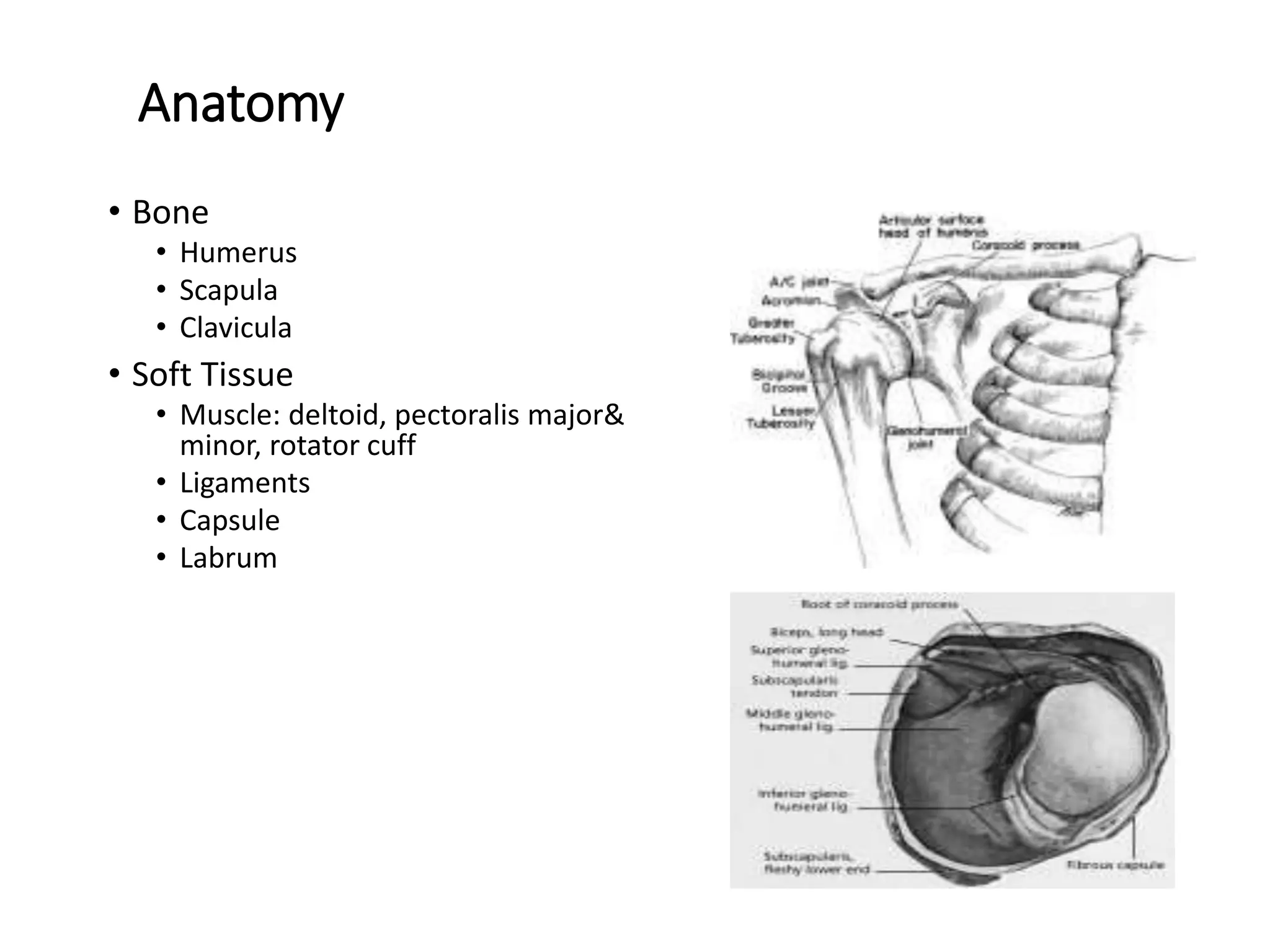
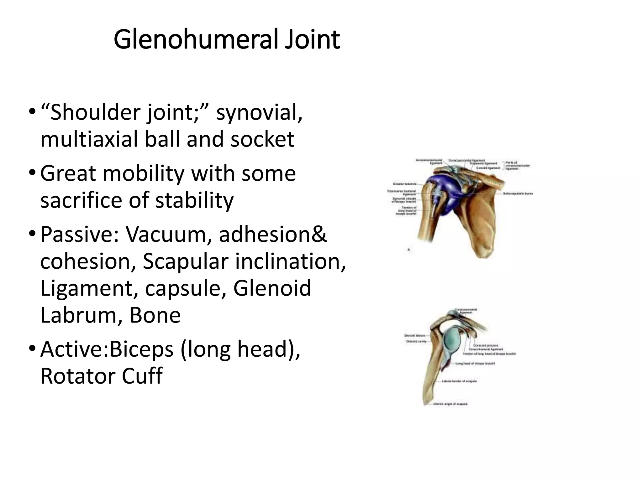
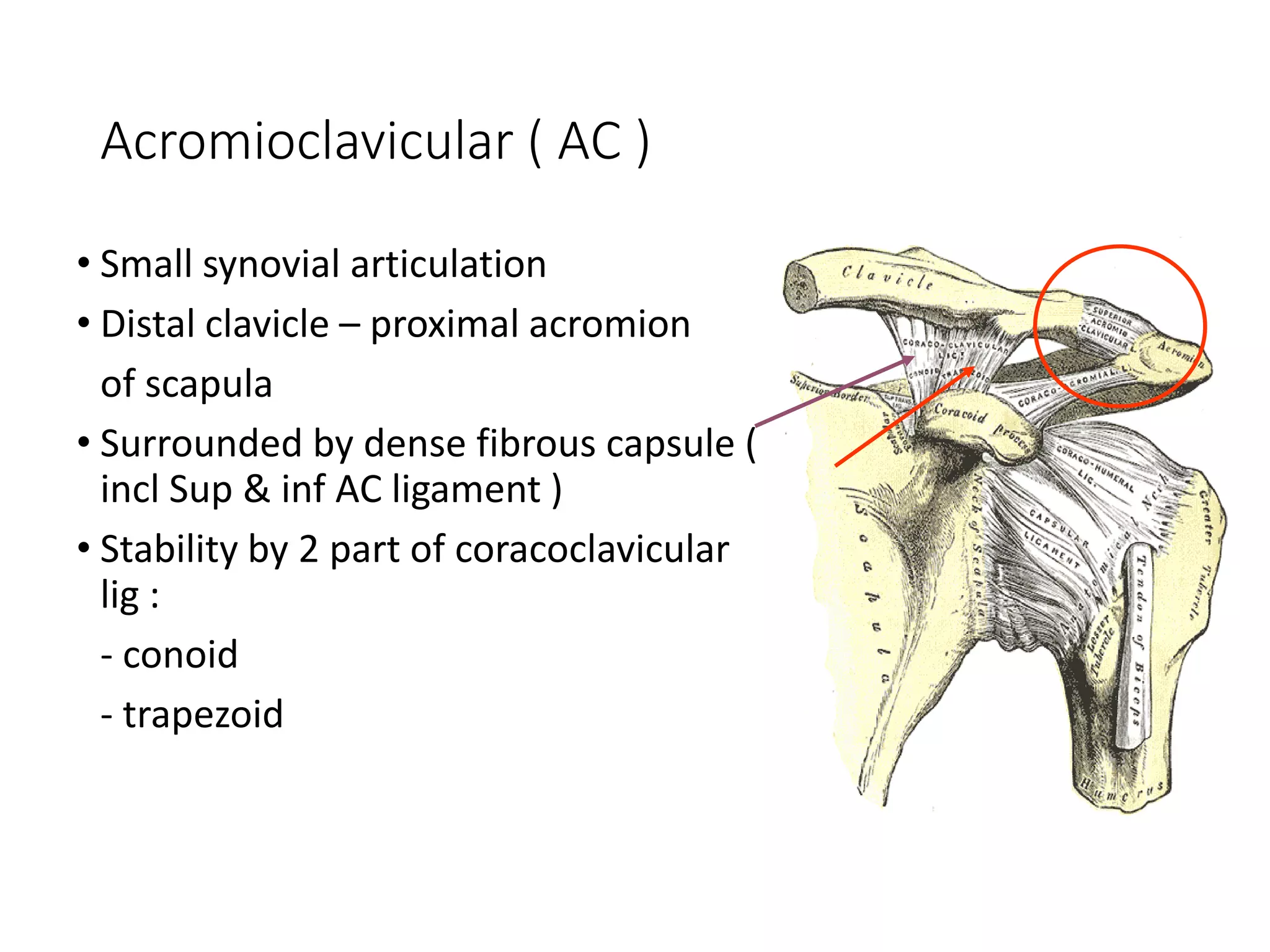
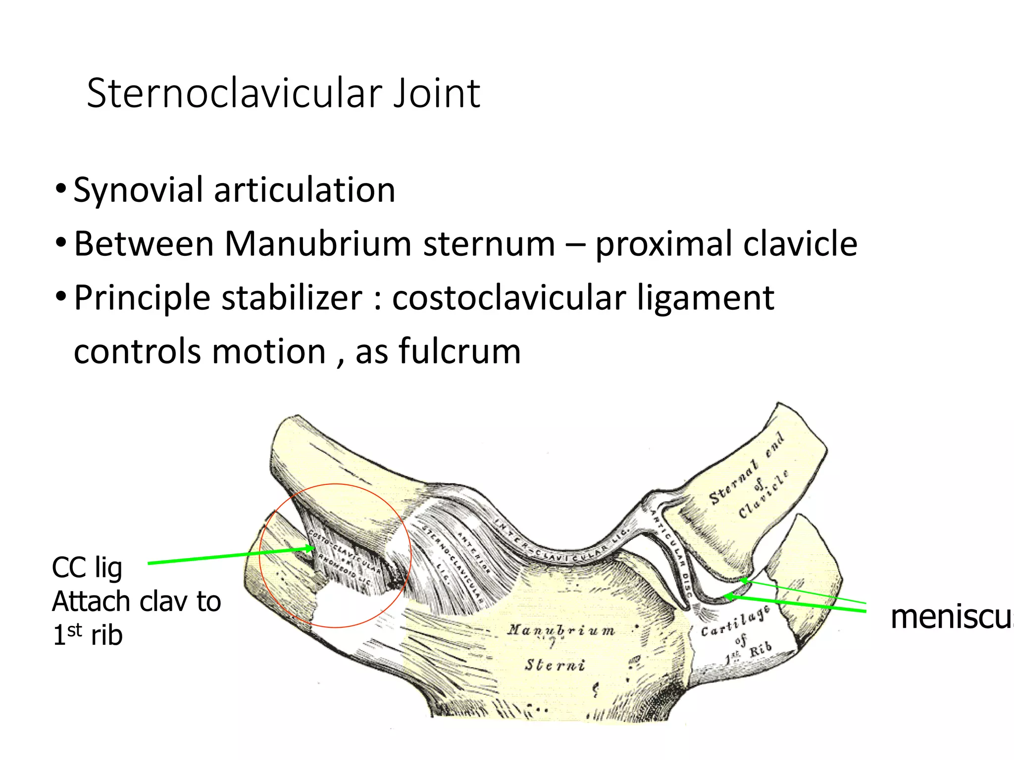
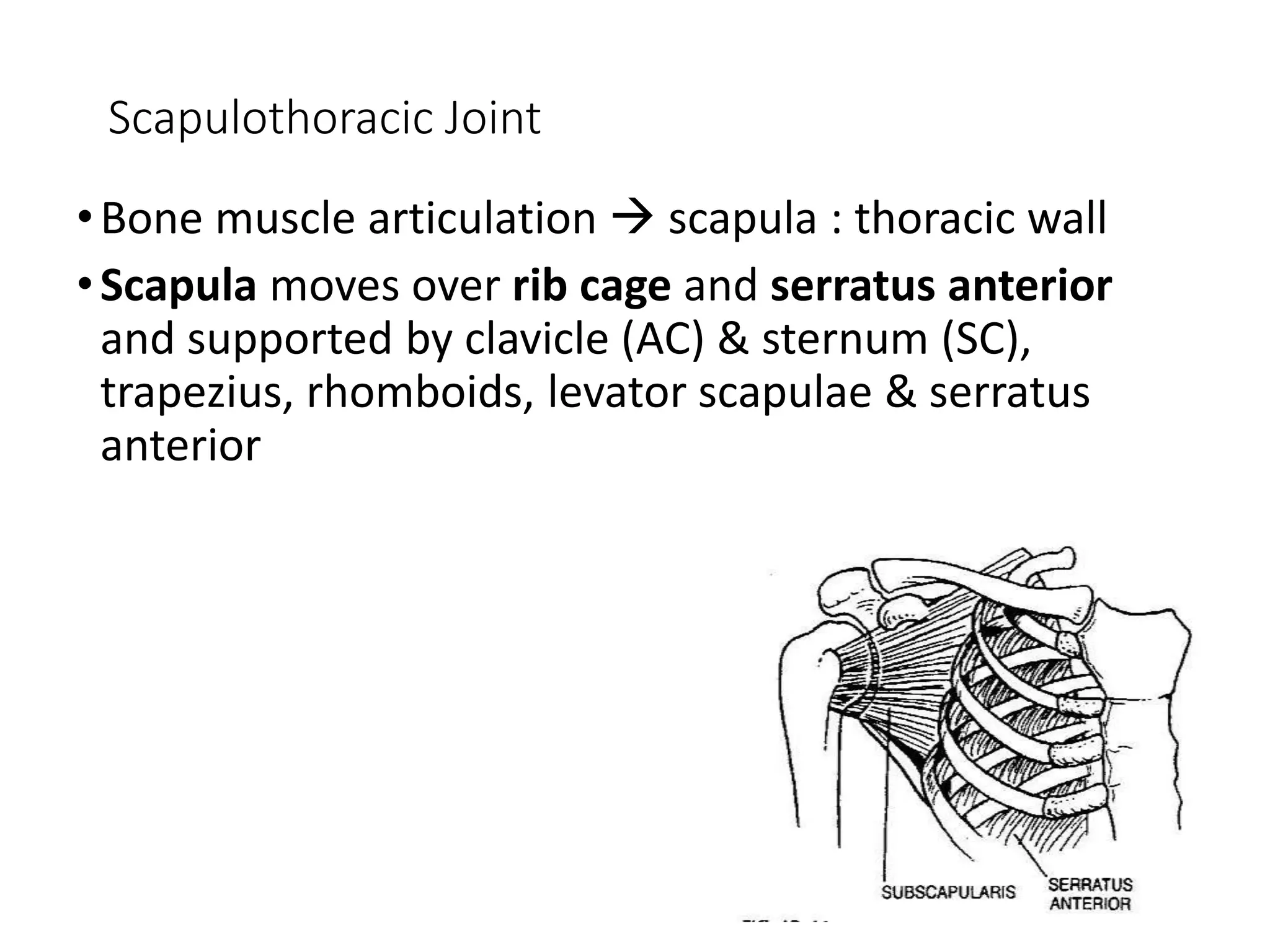
1. The anatomy of the shoulder including bones, muscles, and ligaments.
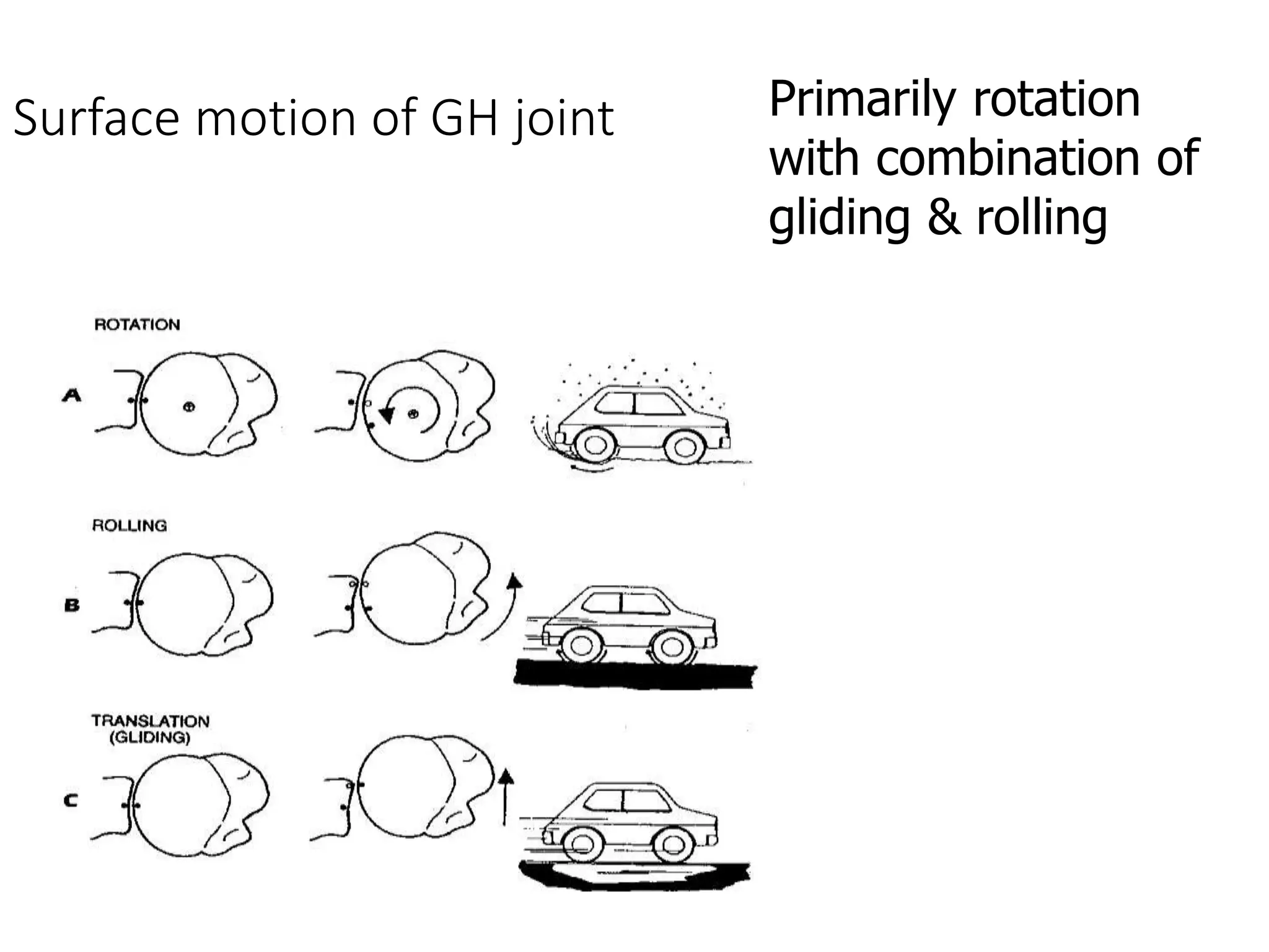
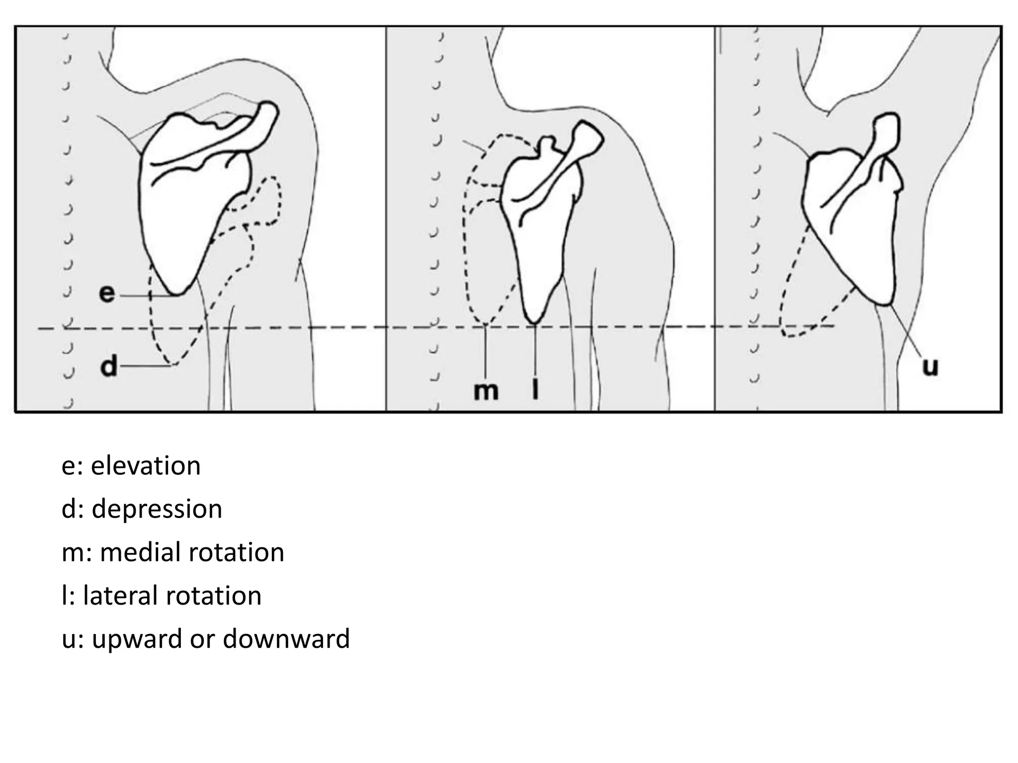
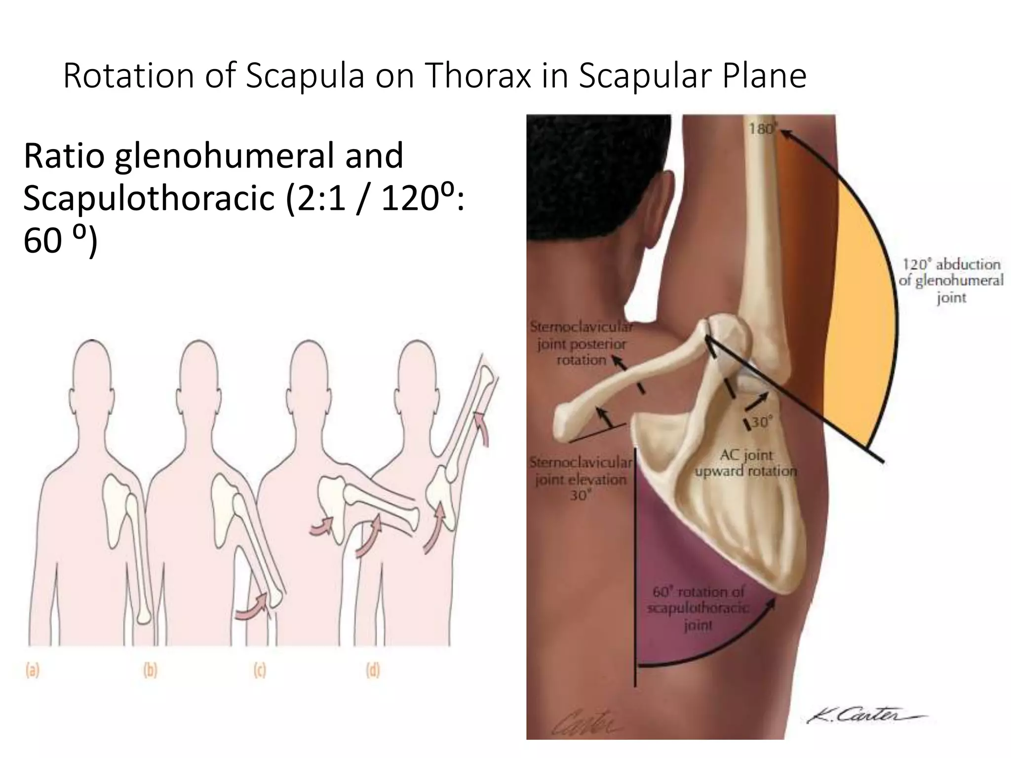
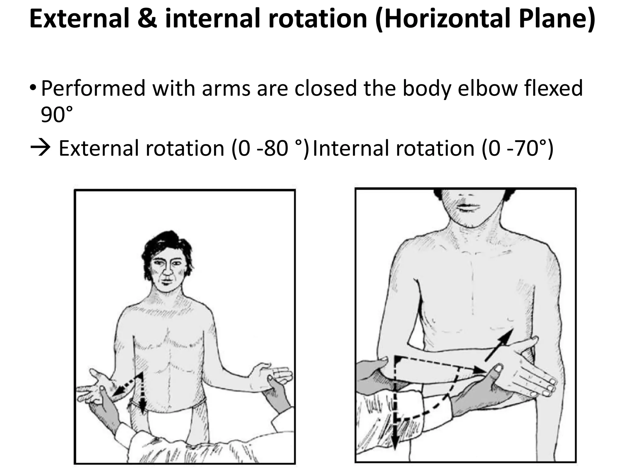
2. Descriptions of various motions of the shoulder like flexion, extension, abduction, and rotation.
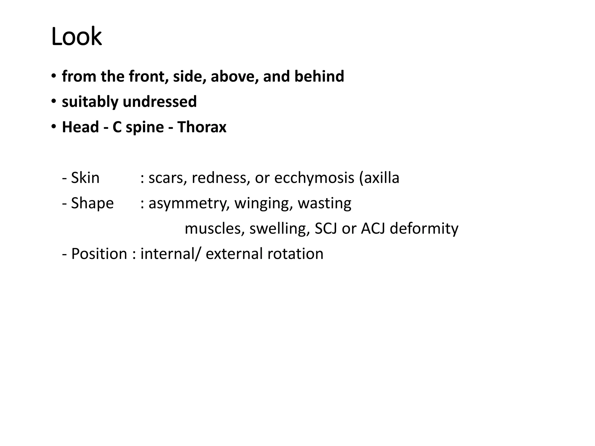
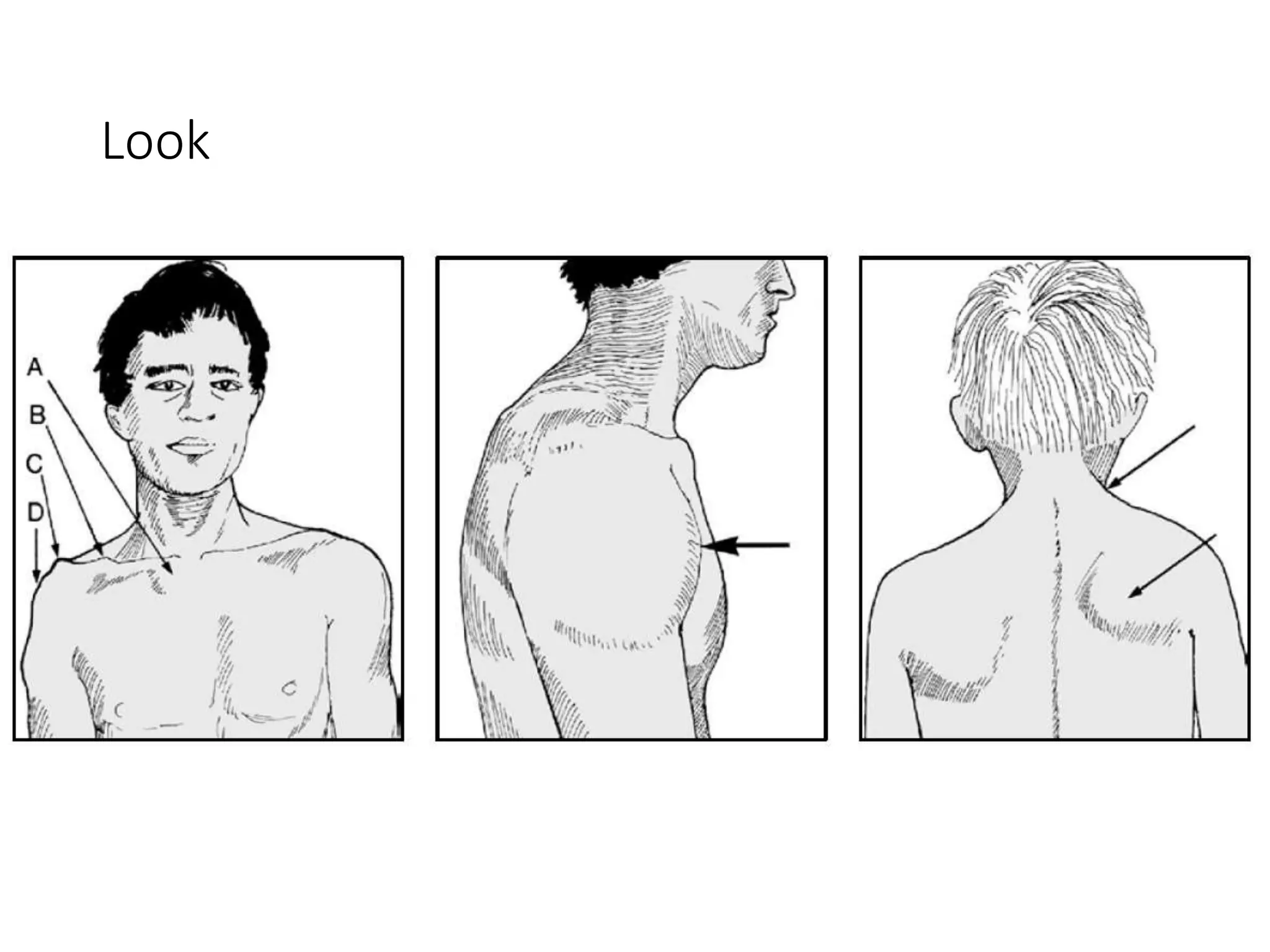
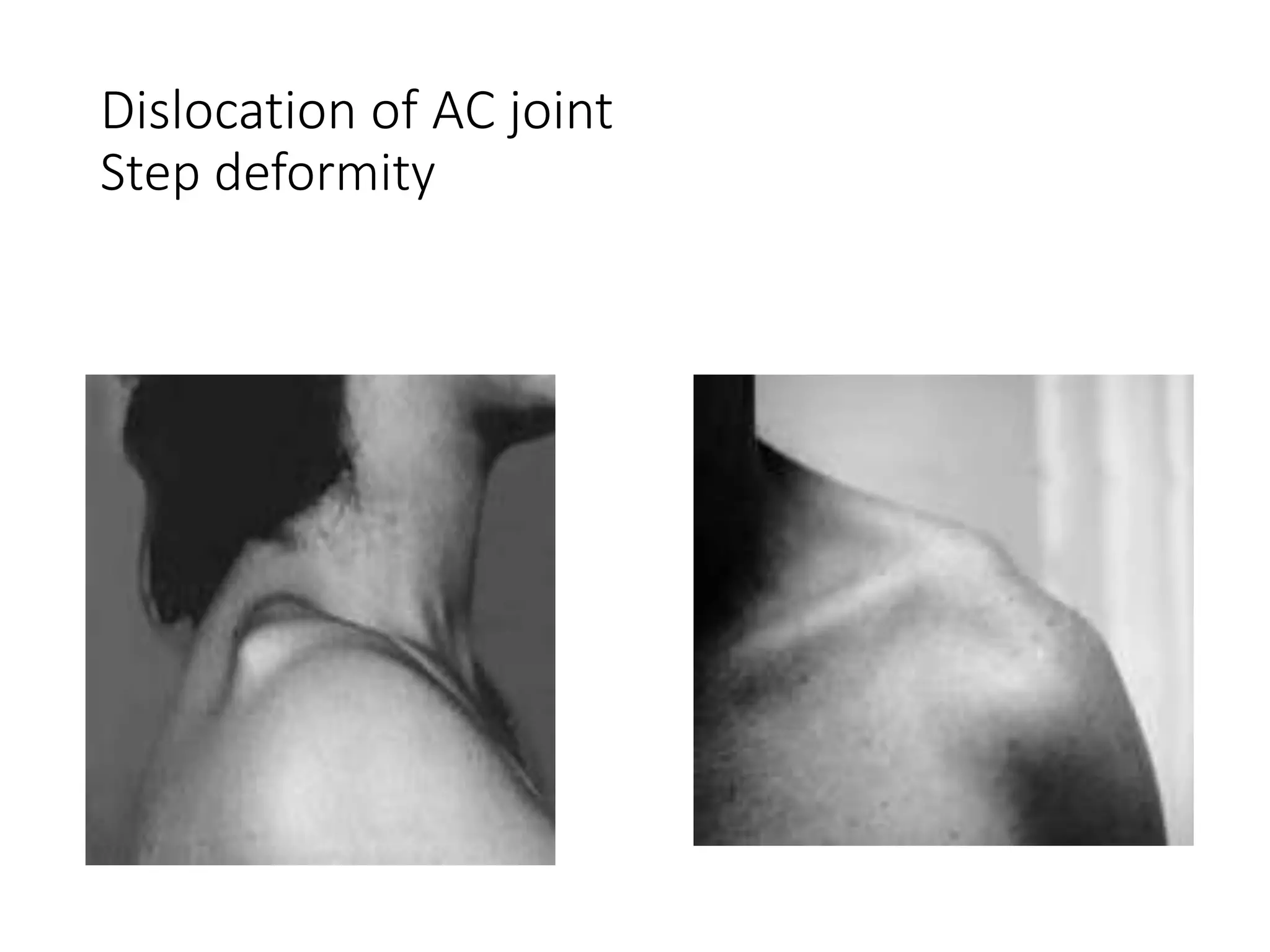
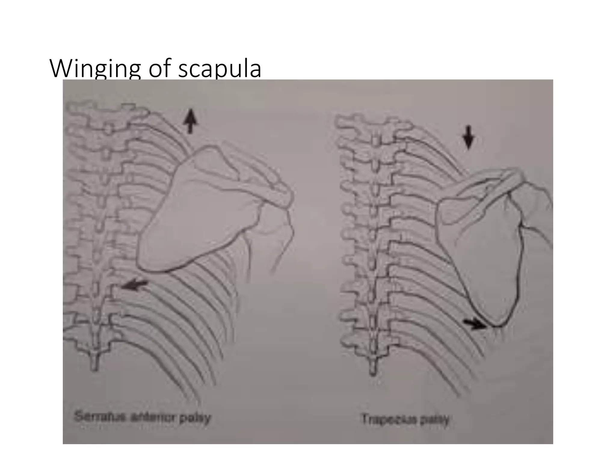
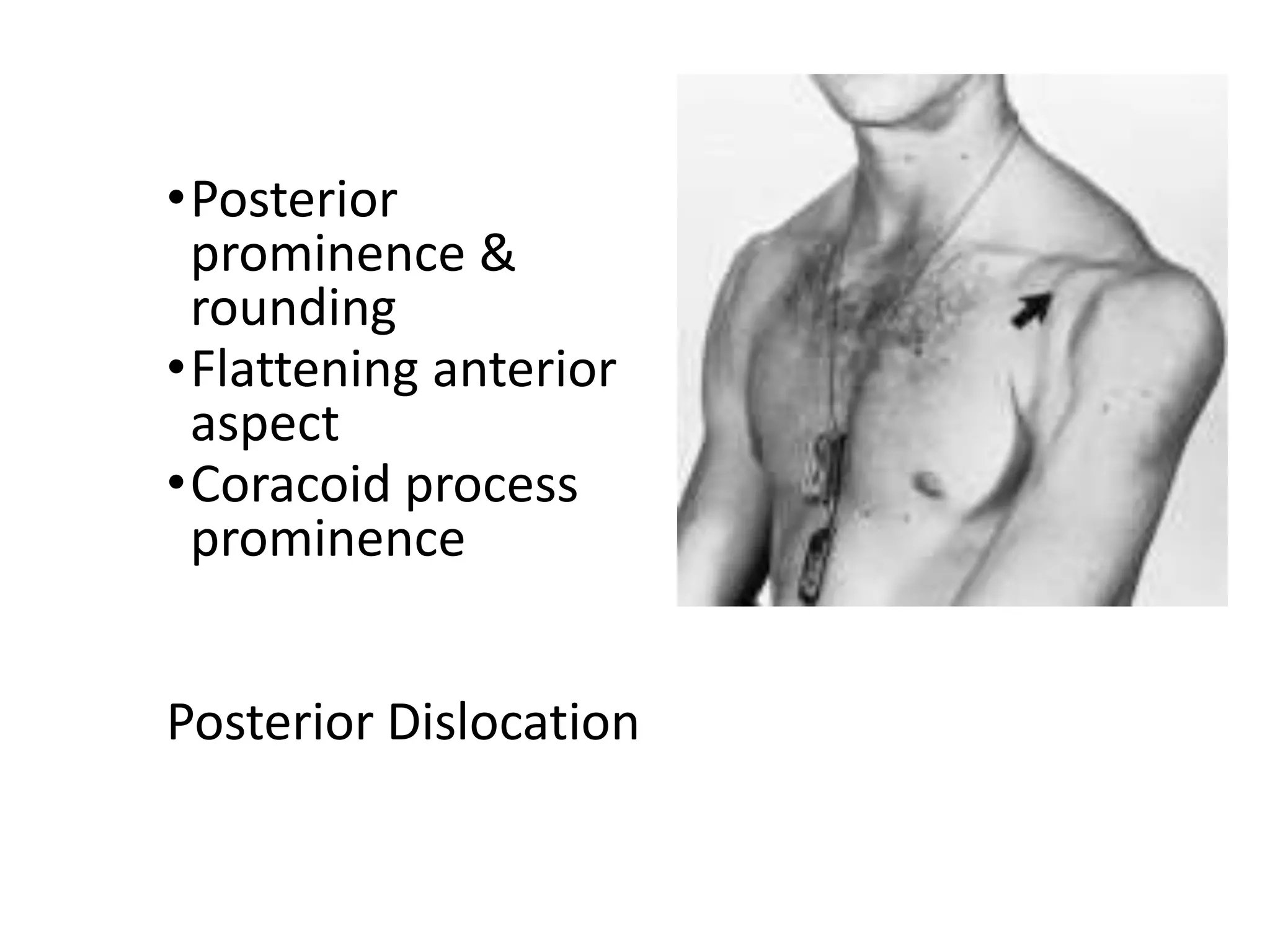
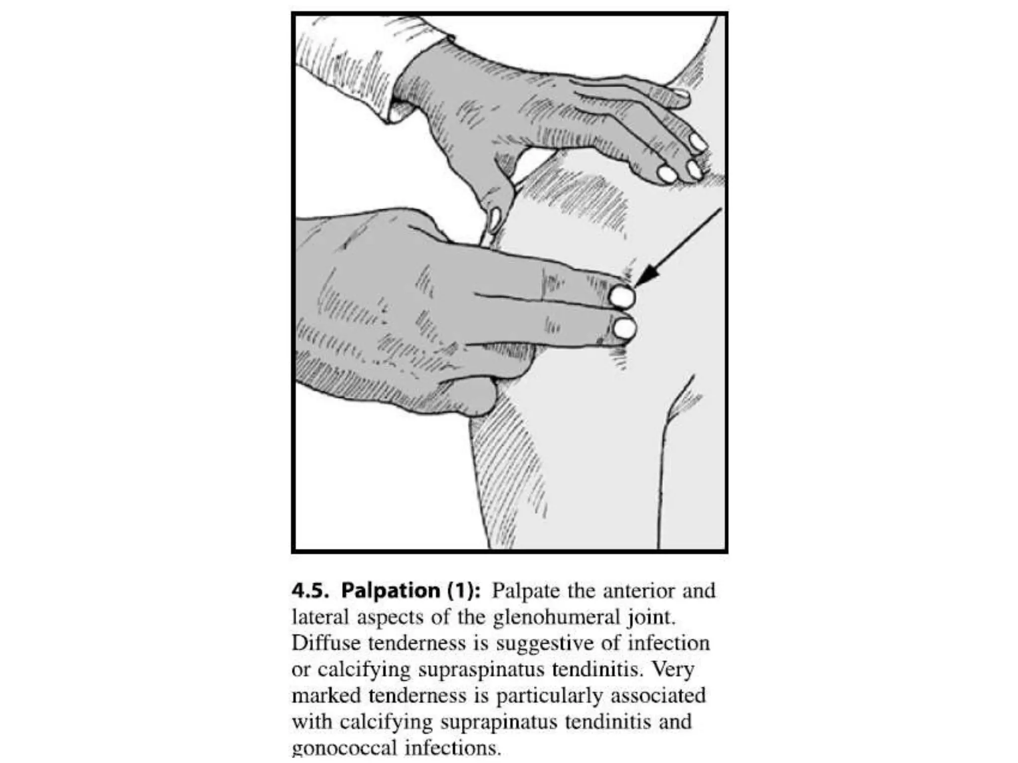
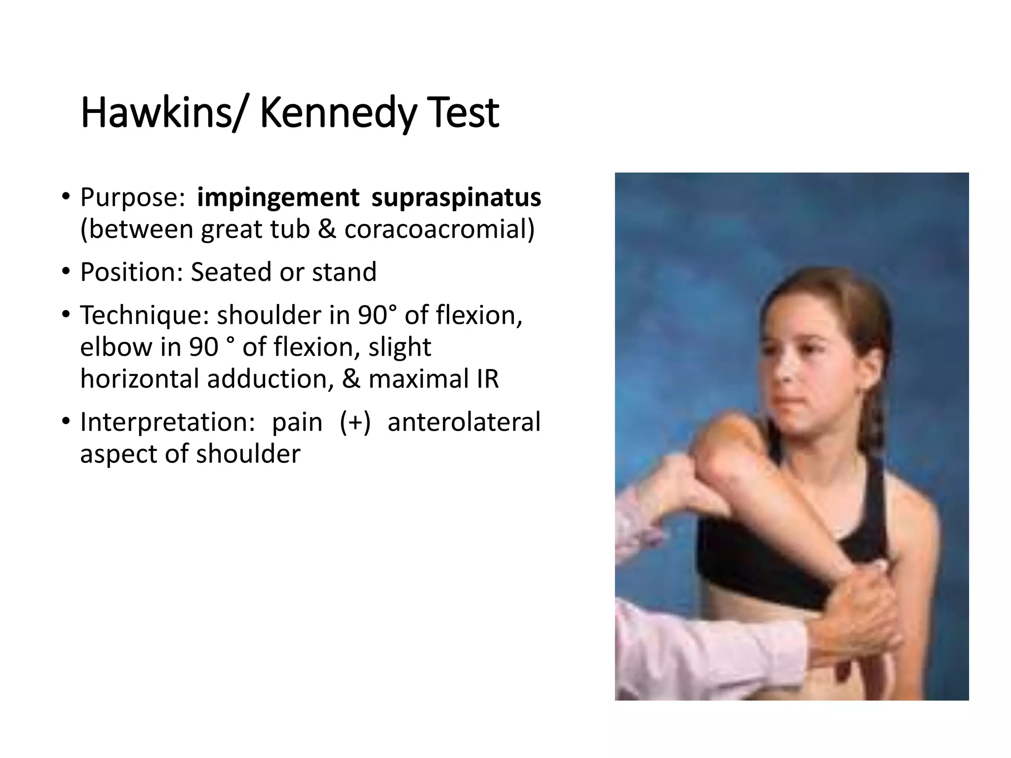
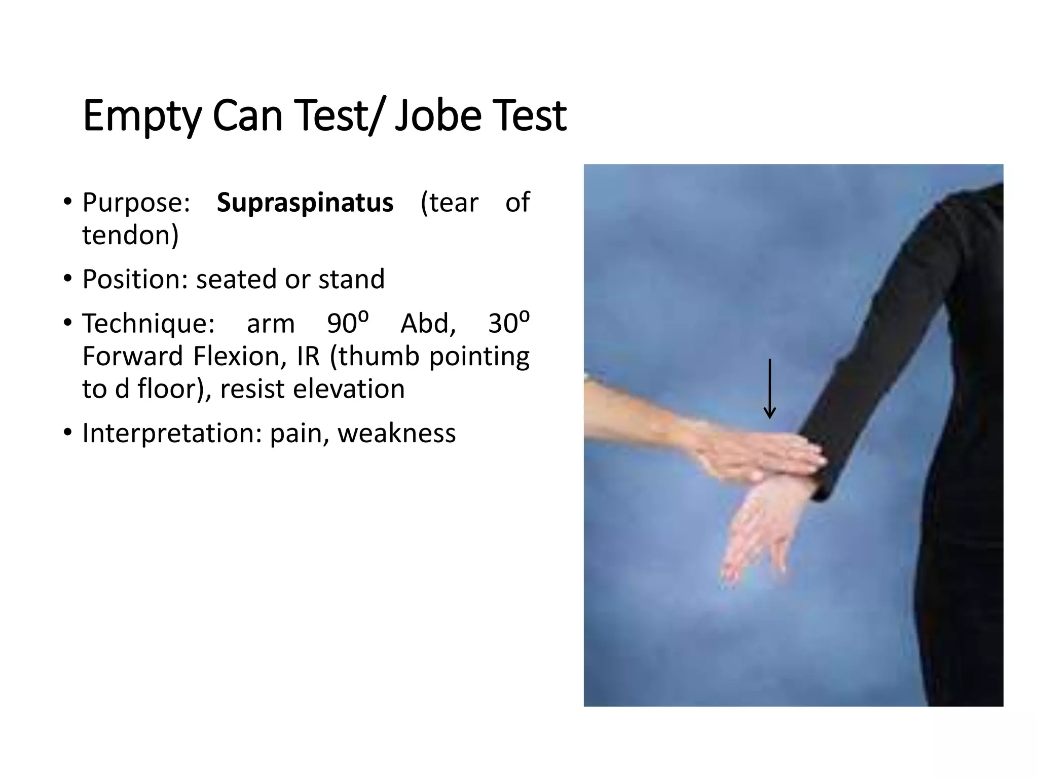
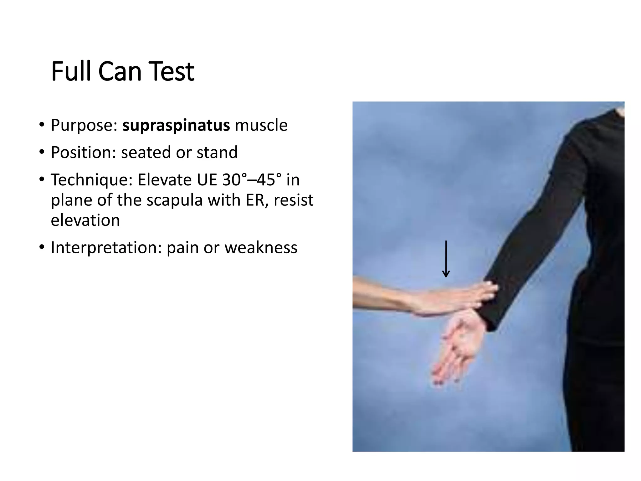
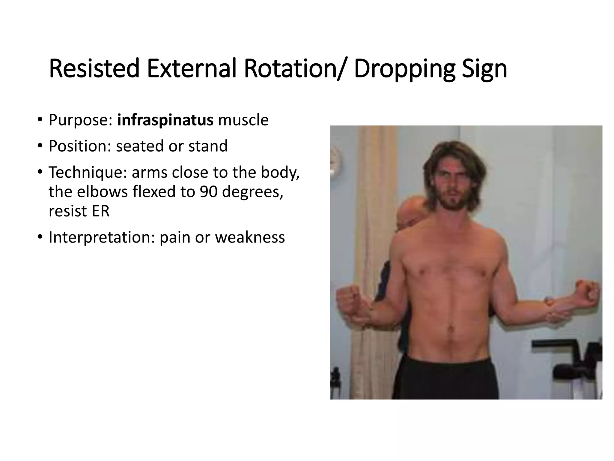
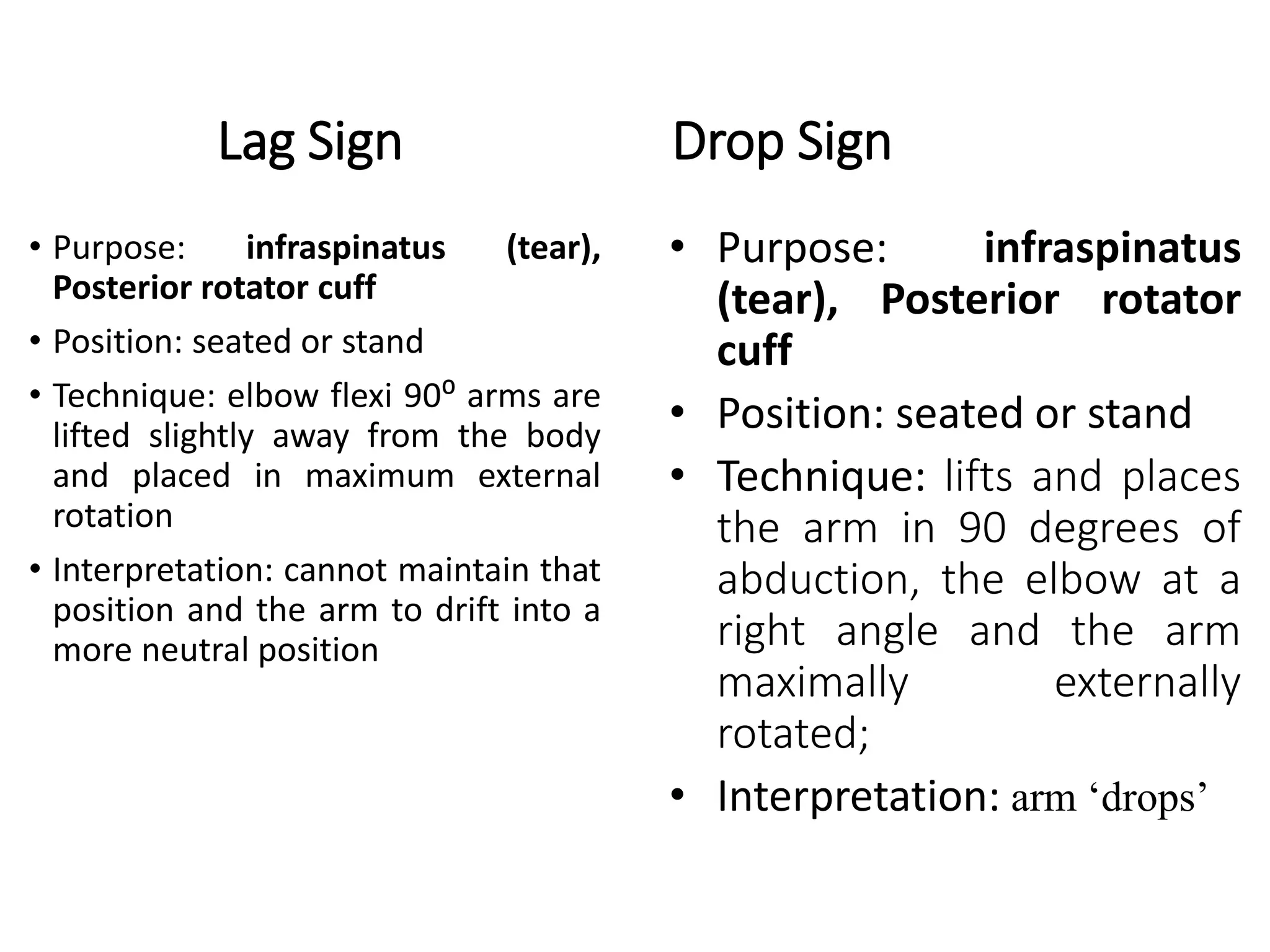
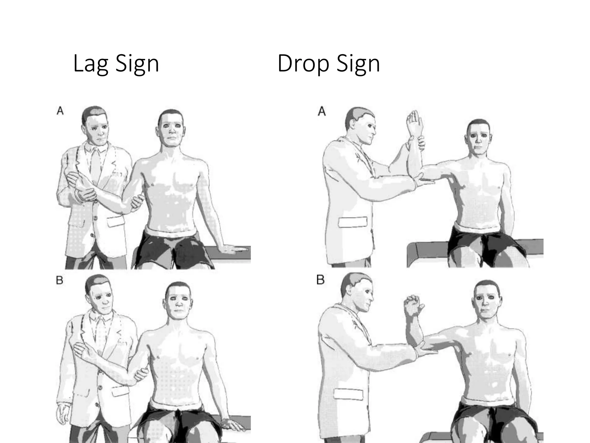
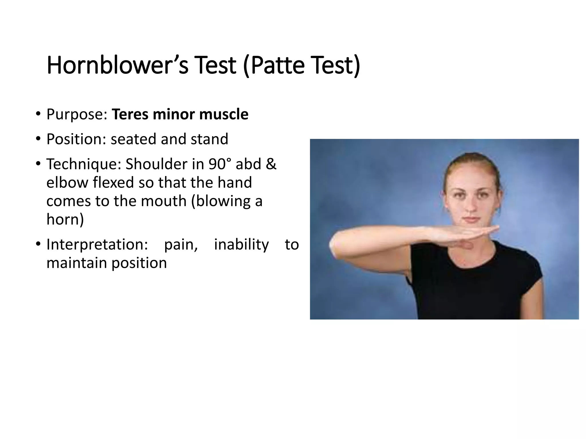
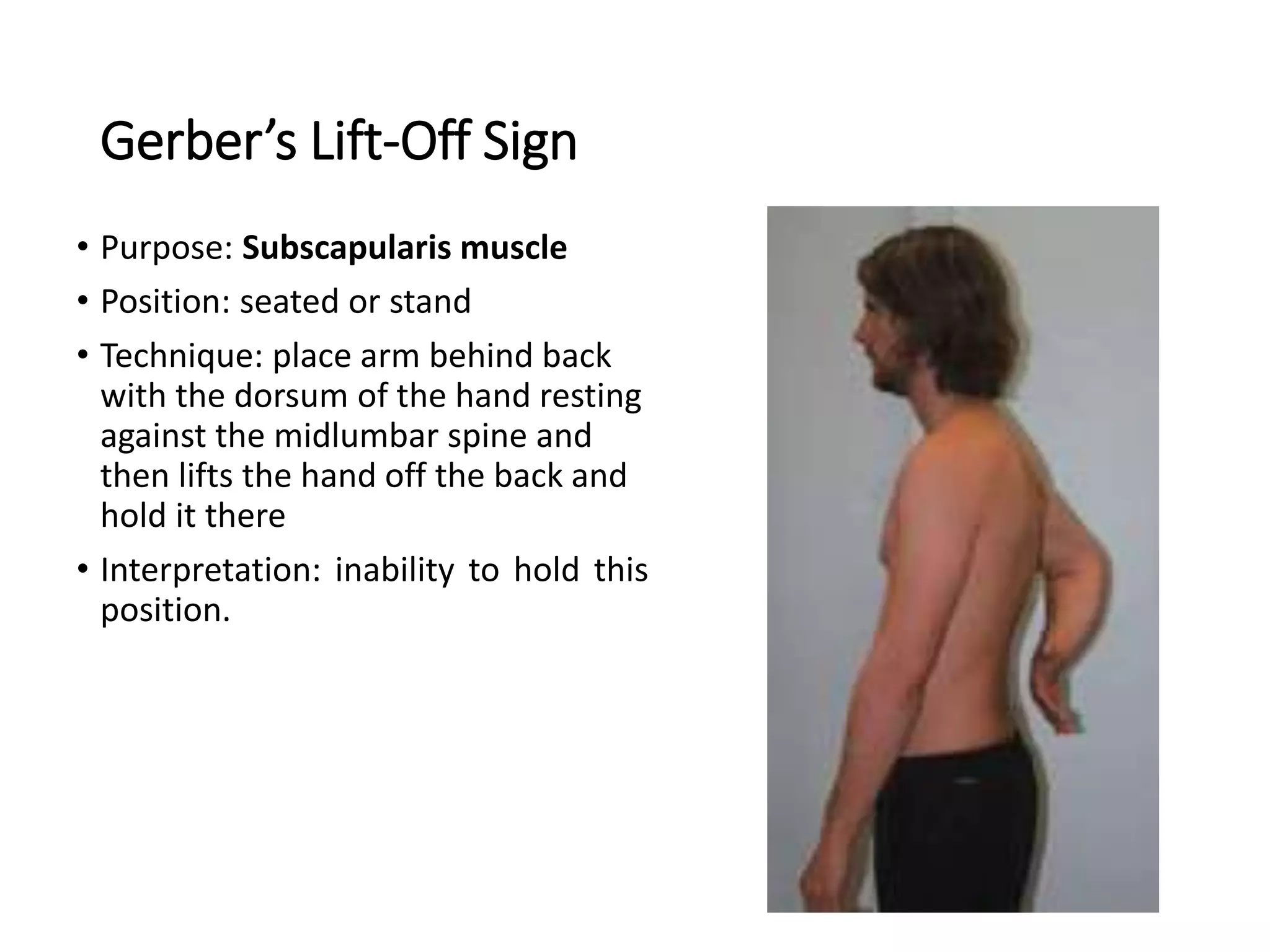
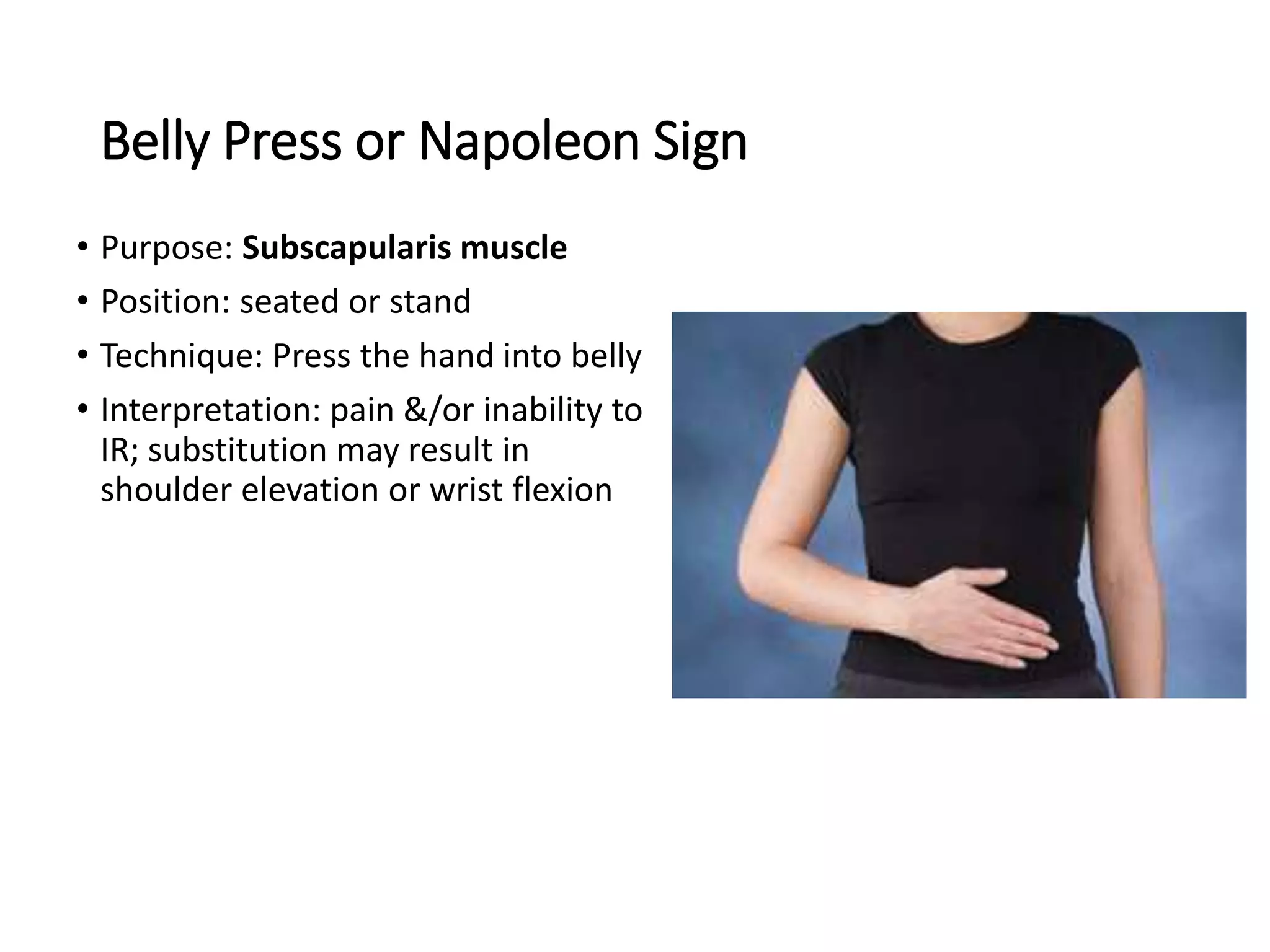
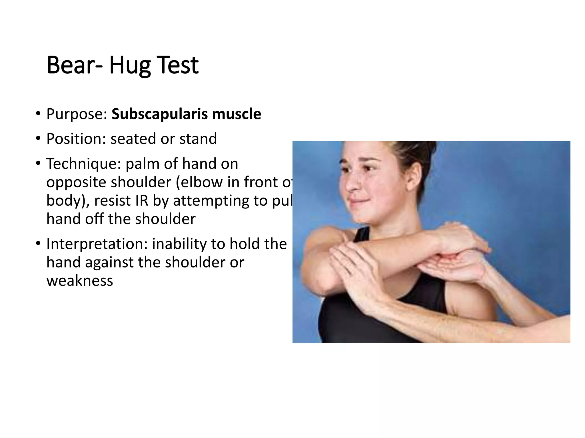
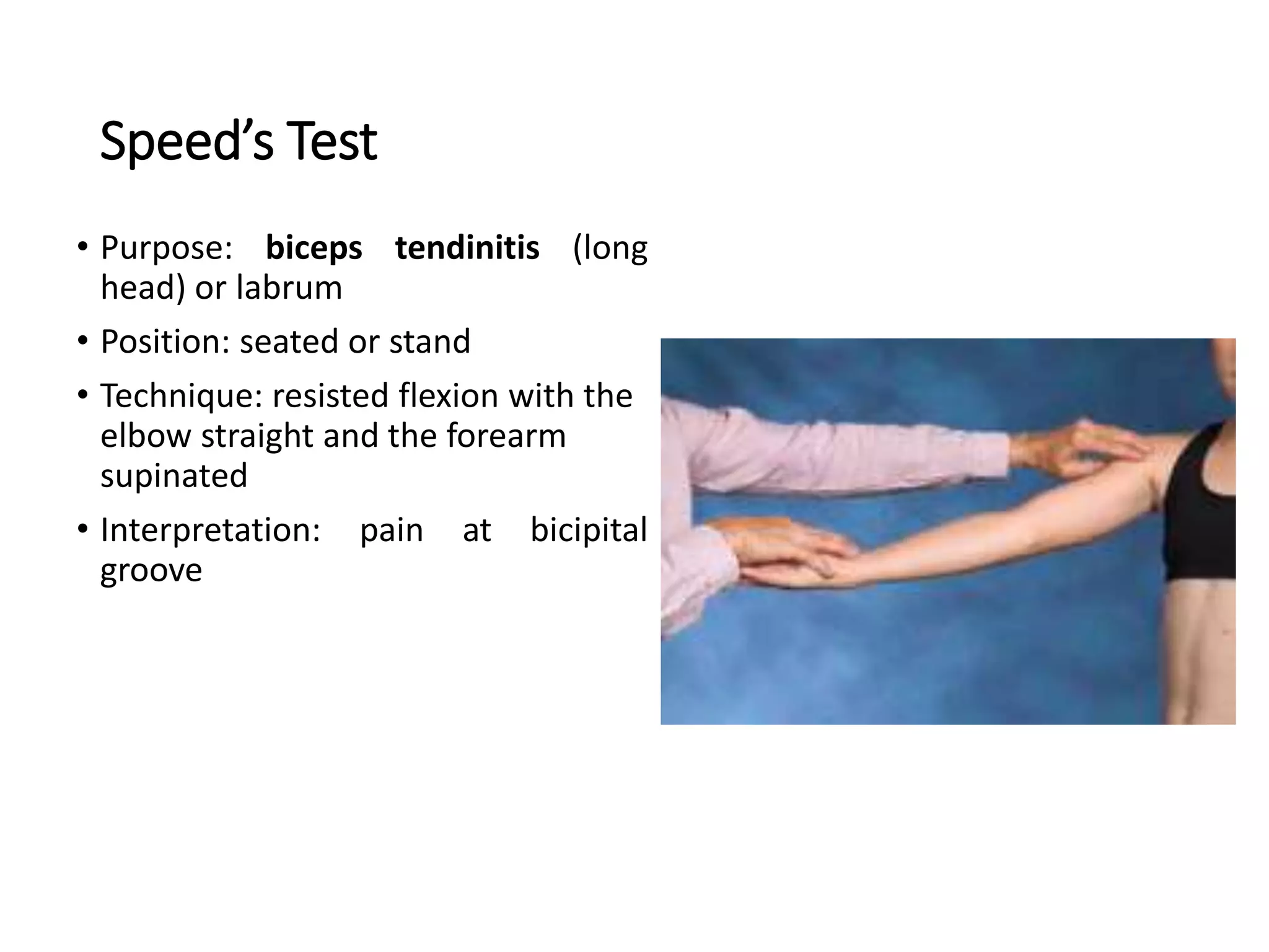
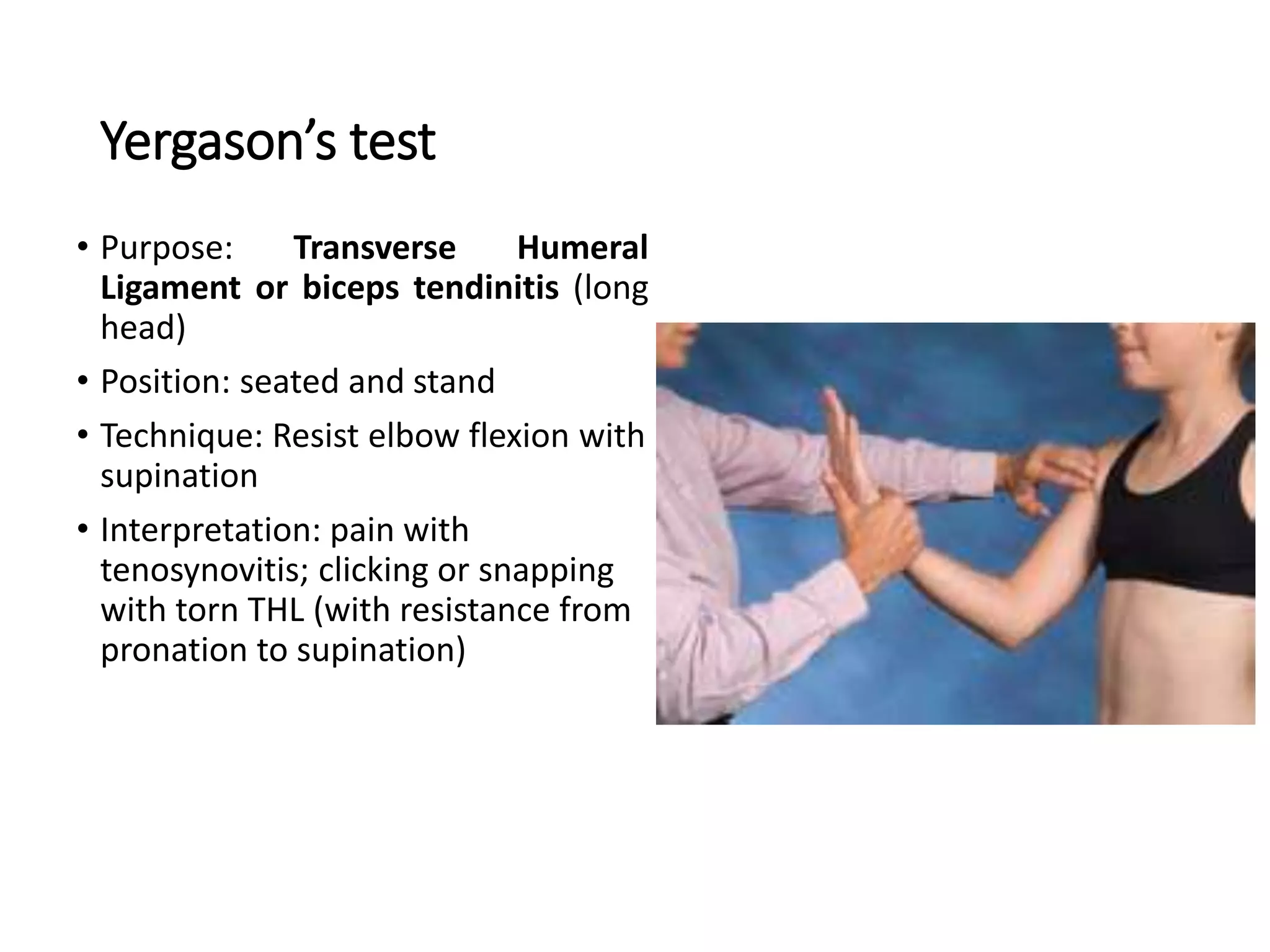
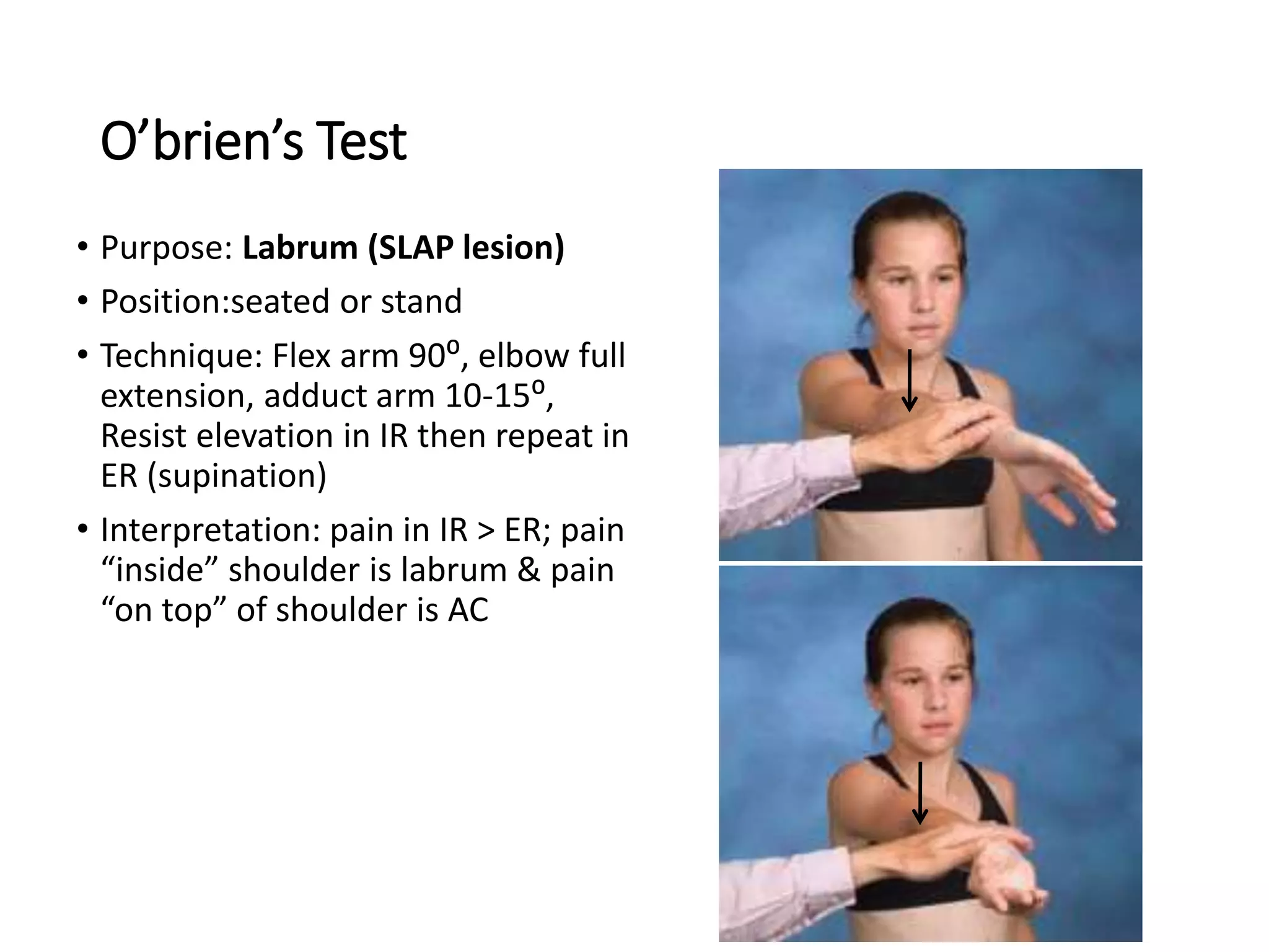
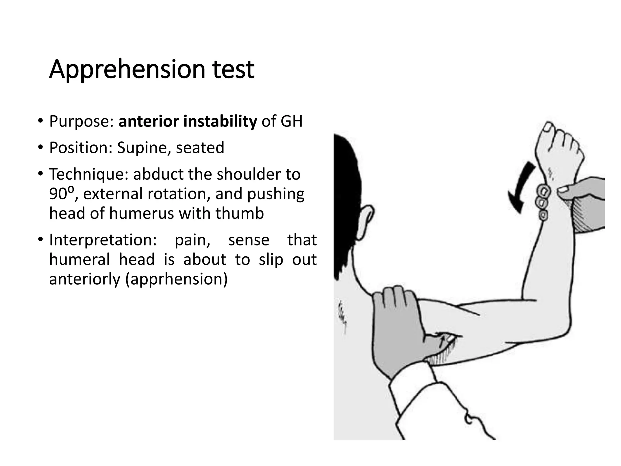
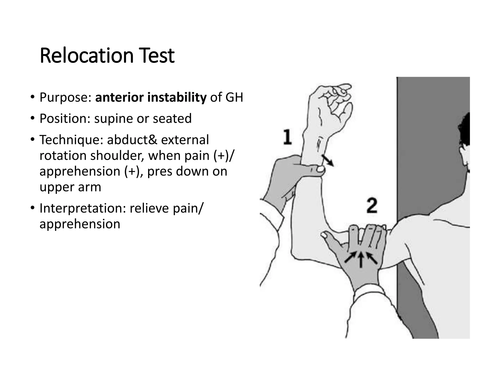
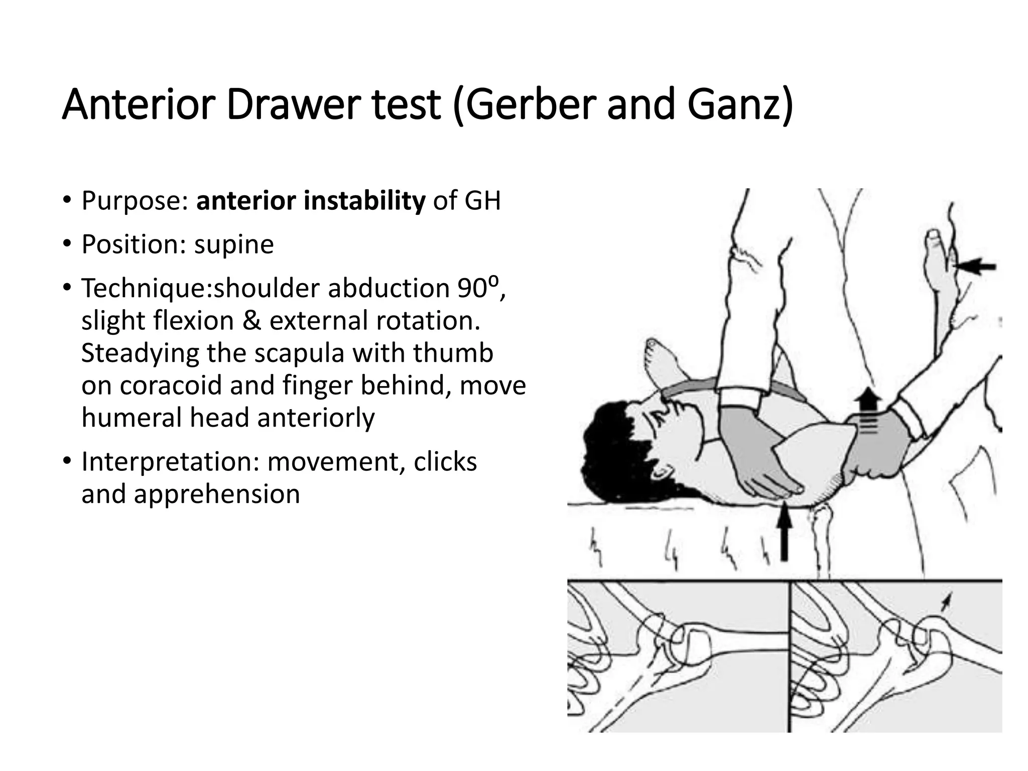
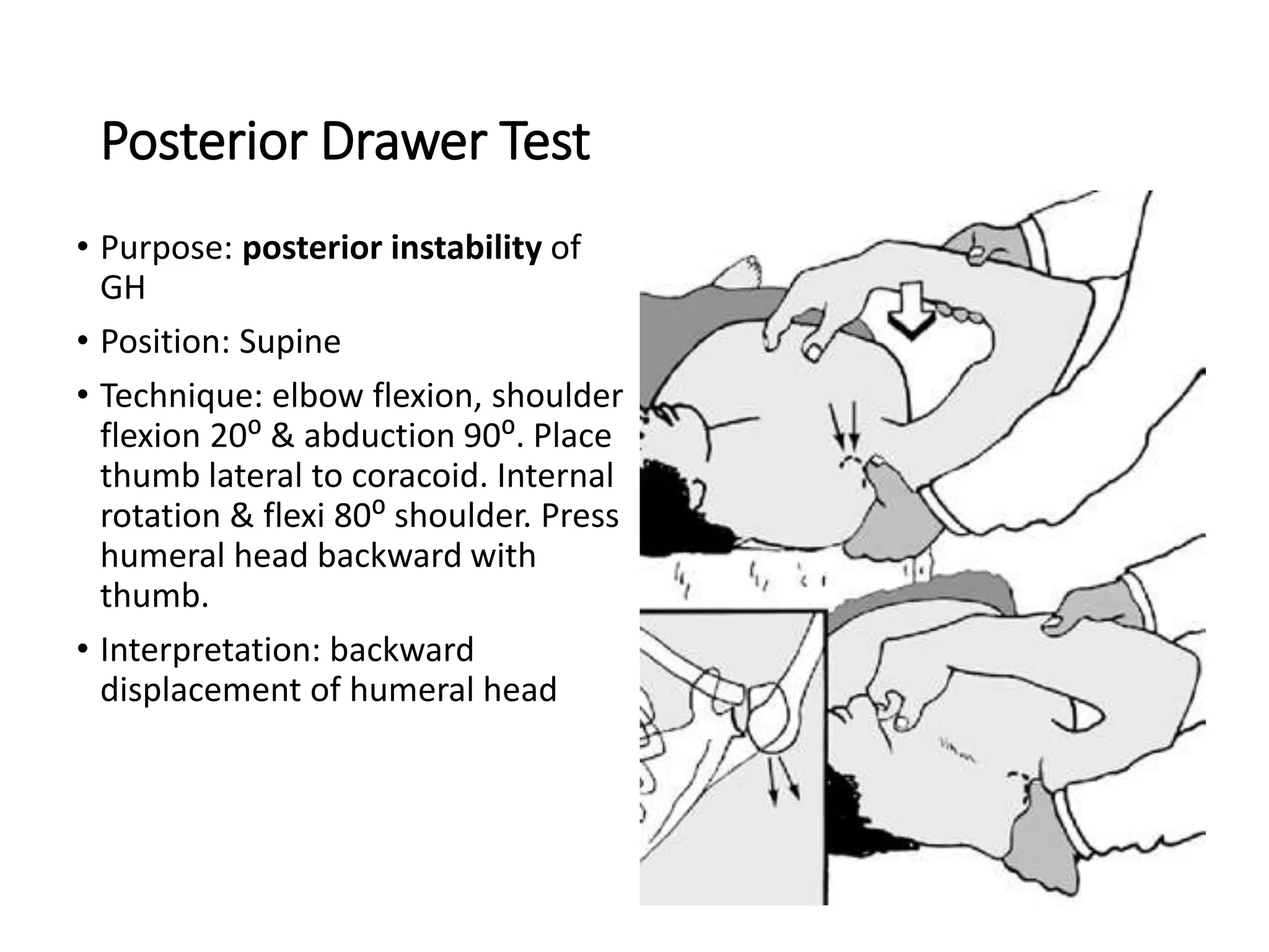
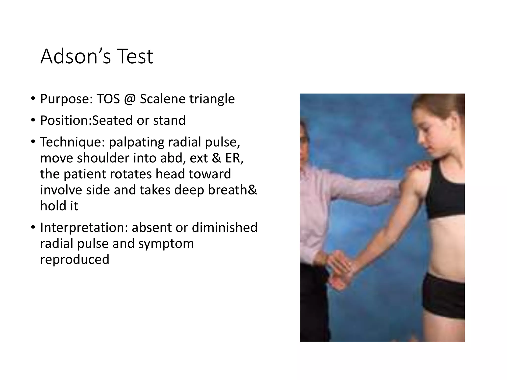
3. Details on clinical assessment including inspection, palpation, and special tests to evaluate the shoulder for issues like impingement, instability, and thoracic outlet syndrome. Special tests evaluate specific muscles, ligaments, and structures of the shoulder.
This document provides guidance on performing an orthopedic examination of the shoulder. It outlines the basic steps as look, feel, move, and special tests. For the shoulder examination, it emphasizes tailoring the exam based on suspected problems such as instability in young patients or impingement/rotator cuff tears in older patients. Key parts of examination include inspection for deformities or wasting, palpation of bony landmarks and soft tissues, and assessing the full range of motion including any painful arcs. Special tests are described to detect impingement, specific rotator cuff injuries, or instability. The document stresses performing the exam with knowledge of anatomy and potential diagnoses to both prove and rule out clinical findings.
This document provides an overview of examining the elbow joint, including:
- The anatomy of the elbow joint as a hinge joint between the humerus, radius, and ulna.
- Details on taking a history, including trauma, pain symptoms, and deformities.
- Instructions on visually inspecting and palpating the elbow joint area, including measurements.
- How to assess the range of motion of flexion, extension, supination, and pronation.
- Special tests to evaluate for conditions like lateral epicondylitis.
- Evaluation of nerves like the ulnar nerve and vascular structures.
The document outlines the process for examining a patient's shoulder, including:
1) Inspecting for deformities, swelling, atrophy, and other abnormalities.
2) Palpating bony landmarks and soft tissues to check for tenderness.
3) Testing the full range of motion both actively and passively while observing for pain.
4) Performing special tests to isolate specific structures like the rotator cuff muscles and labrum.
5) Examining other joints and the neck for full evaluation of the shoulder.
6) Ordering relevant x-rays to assess for fractures or other bone abnormalities.
The document discusses various injuries and conditions that can affect the elbow. It describes the anatomy of the elbow and then discusses fractures of the olecranon and radial head, which usually result from falls onto an outstretched hand. It also covers tendonopathies like lateral epicondylitis ("tennis elbow") and medial epicondylitis ("golfer's elbow"), which cause pain from inflammation and microtears of tendons. Other topics include biceps tears/ruptures, nerve compression like cubital tunnel syndrome, and various treatment options for these conditions ranging from splinting, anti-inflammatory medications, cortisone injections, surgery, and physical therapy.
The document provides an overview of rotator cuff rehabilitation. It discusses that the rotator cuff has different roles as a stabilizer and torque producer depending on the position of the shoulder. Rehabilitation should focus on improving control, proprioception, timing, loading through the range of motion, and endurance. Exercises should incorporate the kinetic chain and provide rotational control and loading through the full range of movement.
Pes cavus and pes planus are foot deformities characterized by high and low arches, respectively. Pes cavus, or a high arched foot, can be congenital or acquired and results in clawing of the toes. Pes planus, or a flat foot, is caused by the collapse of the medial longitudinal arch. Both conditions can cause foot, ankle, and leg pain and abnormal shoe wear. Treatment involves orthotics, physical therapy, and sometimes surgery to correct muscle imbalances and bony deformities.
The document provides information on performing a physical examination of the shoulder, including:
An overview of the anatomy of the shoulder joint and surrounding structures. Descriptions of various tests to assess range of motion, impingement, rotator cuff integrity, labral disorders, and instability. Special tests include Neer's impingement sign, Hawkins' test, relocation test, and others. A thorough shoulder exam evaluates history, inspection, palpation, range of motion, and results of special tests to identify potential pathology.
Biomechanics of Ankle joint- intended to share the powerpoint with first year undergraduate students at Kathmandu University School of Medical Sciences, Nepal.
This document provides an overview of the assessment and special tests used to evaluate the hip joint. It discusses collecting demographic data, medical history, and performing an examination including observation, palpation, range of motion testing, and special tests like the Patrick test and hip scour test. The goal of the assessment is to evaluate the hip for conditions like arthritis, fractures, muscle injuries, and neurological disorders.
The document discusses several electrical modalities used in obstetrics and gynecology, including ultrasound, TENS, shortwave diathermy, and laser therapy. Ultrasound is used during pregnancy to monitor fetal development and assess gestational age. TENS is used for pain relief during labor and postpartum. Shortwave diathermy and laser therapy are used to promote healing after procedures like episiotomy or cesarean section. The modalities are also discussed for treating issues like breast engorgement, mastitis, and pelvic pain conditions during and after pregnancy. Precautions for safe application of the modalities are provided.
Tightness test for lower quadrent muscleRachita Hada
This document discusses tests to assess tightness in the lower body muscles. It provides details on:
- The muscles involved - iliopsoas, rectus femoris, hamstrings, iliotibial band, gastrocnemius, soleus, gluteus medius and minimus.
- Tests for each muscle including positioning, the examiner's actions, and interpretation of tightness. Tests include Thomas, straight leg raise, Ober, and ankle dorsiflexion range of motion tests.
- It also discusses tests for quadratus lumborum and piriformis muscles. Positioning, examiner actions and interpreting tightness are outlined for each test.
This document describes various special tests used to examine the knee joint. It lists the Patellar Grinding Test for the patella, Anterior Drawer Test for the ACL, Posterior Drawer Test for the PCL, Valgus Stress Test for the MCL, and Varus Stress Test for the LCL. It also mentions Apley's Grinding Test and McMurray's Test which are used to examine the meniscus. For each test, it provides a brief description of the procedures and positioning.
The extensor mechanism of the knee involves four quadriceps muscles that connect the femur to the tibia via the patella. The quadriceps muscles include the rectus femoris, vastus lateralis, vastus intermedius, and vastus medialis. They originate on the femur and connect to the patella. The patella then connects to the tibia via the patellar tendon. This mechanism improves the efficiency of knee extension by increasing the lever arm of the quadriceps muscles. It functions via a "screw home mechanism" where the tibia rotates internally at the end of knee extension, maximally stabilizing the knee joint.
basic info to understand forward head posture...if any objection then plz leave a comment and for more details you can send message to harisubedi12@yahoo.com
The patient underwent ACL reconstruction and meniscal repair surgery using a patellar tendon graft. At 10 weeks post-op, she was experiencing pain when ascending/descending stairs or weight bearing for long periods. Physical therapy is focusing on strengthening, proprioception, and functional exercises to address deficits and meet goals of returning to prior activity levels without pain. Progress includes increased strength and range of motion but some pain with stairs remains.
complete Knee joint assessment from physiotherapeutic point of view. Includes observation , palpation , assessment, special test, differential diagnosis of knee joint .
The posterior cruciate ligament (PCL) is one of the four major ligaments of the knee. It connects the posterior intercondylar area of the tibia to the medial condyle of the femur. The PCL prevents the femur from sliding off the anterior edge of the tibia and prevents hyperextension of the knee. Injuries to the PCL typically occur from direct blows to the flexed knee, falling on the knee, or hyperextension injuries. Treatment involves rest, bracing, and physical therapy, with surgery required for complete tears.
Shoulder Impingement Evidence Based Case Study Rumy Petkov
Used evidence based literature to compare laser therapy treatment versus corticosteroid injections, ultrasound, rehab exercises, and Kinesio taping to treat shoulder impingement.
The document discusses rotator cuff injuries, providing information on anatomy, causes, symptoms, diagnosis and treatment. It describes the rotator cuff muscles, how injury can result from impingement or overuse, and common symptoms like shoulder pain. Physical exams like empty can and lift-off tests help identify injuries. Imaging like x-rays and ultrasound can diagnose conditions like tears or calcification. Surgery may be needed for severe, unresponsive tears of the rotator cuff tendons.
rotator cuff injuries, its causes, types,symptoms, special test and its pt management
special test for every injury types which includes rotator cuff tear, tendonitis, impingement syndrome,painfull arc syndrome, frozen shoulder.....special test includes neer's impingent, empty cane, full cane, speed test
This document discusses shoulder instability, including definitions, anatomy, evaluation, and treatment. It defines instability as the inability to maintain the humeral head in the glenoid fossa, ranging from dislocation to laxity. Static stabilizers include the labrum and ligaments, while dynamic stabilizers are the rotator cuff and scapulothoracic muscles. Evaluation involves history, exam, and imaging to classify instability by direction, degree, and etiology. Treatment depends on classification but may include immobilization, rehabilitation, or surgical repair of labral tears or bone defects.
This document discusses the anatomy and clinical assessment of the rotator cuff. It describes the four muscles that make up the rotator cuff, their innervation and attachments. Common rotator cuff injuries like impingement syndrome and ruptures are explained. The physical exam involves assessing range of motion and performing special tests like Neer's, Hawkins-Kennedy, and lift-off to identify injuries.
Rotator cuff tear is a very common orthopedic condition, which causes shoulder pain and stiffness. The slides are on rotator cuff tears and its management by open repair, mini open repair & by arthroscopy
Scapular dyskinesis refers to abnormal or dysfunctional movement of the scapula. It can impair shoulder function and create issues like decreased subacromial space and rotator cuff weakness. Scapular dyskinesis is often associated with shoulder injuries like labral tears, impingement, and rotator cuff injuries. Rehabilitation focuses on strengthening the scapular stabilizing muscles like the serratus anterior and lower trapezius to improve scapular control and positioning during arm movements.
The document discusses the Berg Balance Scale (BBS), which is a 14-item clinical test used to assess static and dynamic balance abilities. It involves tasks of increasing difficulty ranging from sitting to standing to walking. Scores are interpreted on a scale of 0 to 4 for each item, with a maximum total of 56 indicating low fall risk. The BBS has been validated as a reliable tool for measuring balance impairment, especially in elderly patients and those with conditions like stroke. However, it may lack sensitivity for early-stage stroke patients and not adequately challenge very high-functioning individuals.
Calcific tendonitis is characterized by the deposition of calcium hydroxyapatite in tendons, most commonly in the shoulder. The exact pathogenesis is unclear but it involves a cell-mediated process with formative, calcific, resorptive, and reparative phases. Diagnosis is typically made based on x-ray or ultrasound findings of calcific deposits. Initial treatment involves rest, NSAIDs, physiotherapy, and steroid injections. If conservative treatment fails, extracorporeal shockwave therapy or surgical debridement may be considered to remove the calcium deposits.
Special Tests for Lower Leg, Ankle, and FootJulie Jane
This document describes various physical examination tests for the lower leg, ankle, and foot. It provides procedures and implications for tests that assess the neutral position of the talus, leg and foot alignment, tibial torsion, ligamentous instability, and other conditions. Key tests include those for the anterior drawer test of the ankle, tibial torsion in sitting and supine positions, leg-heel alignment, and Feiss line to assess for flat foot. The document contains detailed steps for examiners to accurately perform various physical exams of the lower extremity.
The document discusses the assessment of the shoulder joint, including its components, restraints, and common injuries. It describes the anatomy of the shoulder joint and surrounding structures. Various tests are outlined to assess different conditions of the shoulder like instability, impingement, and nerve injuries.
This document provides an overview of an examination of the shoulder, including:
1) Anatomy of the shoulder bones, joints, and muscles like the rotator cuff.
2) Traditional steps for examining the shoulder including taking a history, inspecting, palpating, assessing range of motion, and performing special tests.
3) Details on examining range of motion, palpating for tenderness, and performing special tests to identify issues like impingement, rotator cuff tears, and instability.
This document summarizes the steps for examining a patient's shoulder, including inspection from multiple angles to identify scars, swelling, alignment and muscle wasting. It describes palpating various parts of the shoulder joint. Range of motion and special tests are outlined to assess the rotator cuff, impingement, AC joint, biceps, deltoid, serratus anterior and instability. Specific tests described include Neer's sign, Hawkin's sign, Jobe's test, belly-press test, apprehension test and relocation test. The examination provides a thorough evaluation of the shoulder to identify any injuries or limitations.
1. The document discusses shoulder pain and provides a differential diagnosis. It covers anatomy of the shoulder joint, muscles, nerves and bursae.
2. Common causes of shoulder pain discussed include degenerative arthritis, rotator cuff tears, shoulder instability, adhesive capsulitis, bursitis and tendonitis.
3. The differential diagnosis section covers the history, examinations, tests and investigations for various shoulder conditions. It describes conditions like rotator cuff tears, shoulder dislocations, impingement syndrome and frozen shoulder.
This document provides an overview of the anatomy of the shoulder, including bones, joints, ligaments, tendons, muscles, nerves and range of motion. It describes the key bones (humerus, scapula, clavicle, ribs, vertebrae), joints (glenohumeral, acromioclavicular, sternoclavicular, scapulothoracic) and muscles (deltoid, rotator cuff, latissimus dorsi, trapezius, serratus anterior, pectoralis) of the shoulder. It also discusses common shoulder injuries like rotator cuff tears, tendonitis and frozen shoulder, and examines physical exam tests and treatment approaches
The document summarizes the glenohumeral joint and rotator cuff. It describes the anatomy of the joint and rotator cuff muscles. It then discusses rotator cuff pathology, symptoms, physical exam maneuvers, and special tests to assess the rotator cuff including the empty can test, lift-off test, and external rotation lag sign.
Exaggerated thoracic kyphosis and compensatory lumbar lordosisMiriam Clavel
This document describes the physical examination findings and treatment plan for a 21-year-old student presenting with thoracic hyperkyphosis and compensatory lumbar lordosis. The examination revealed forward head, hyperextended cervical spine, abducted scapulae, increased thoracic flexion, hyperextended lumbar spine, and anterior pelvic tilt. Range of motion testing showed hypomobility in the upper dorsal and lower dorsal spine. The treatment plan focuses on strengthening spinal extensors and stabilizer muscles, improving posture and proprioception, and increasing joint mobility in the restricted dorsal spine areas.
This document provides guidance on conducting a physical examination of the musculoskeletal system. It outlines general principles such as ensuring privacy and comfort for the patient. The objectives are to apply anatomy and physiology knowledge to differentiate normal from abnormal findings through physical assessment. Assessments should be conducted when examining bone, muscle or joint injuries, disorders, or pain. Common disorders are described. The assessment involves subjective history collection and physical examination. Key areas to examine include joints, muscles, nerves and blood vessels. Common examination techniques like inspection, palpation, and assessing range of motion are outlined for each body area. Common abnormalities that may be found and diagnostic tests are also summarized.
The document provides details on examining the shoulder joint, including history taking and physical examination. It discusses taking a thorough history regarding pain, swelling, range of motion, instability, and related factors. The physical examination section covers inspection, palpation, range of motion testing, and special tests to evaluate the rotator cuff, instability, and other shoulder pathologies. A number of clinical tests are described to isolate injuries or conditions of specific shoulder structures.
The document describes muscle tests of the knee joint. It discusses the hamstring muscles - semitendinosus, semimembranosus, and biceps femoris - and their actions, innervation, and testing positions. It also discusses the knee extensor muscles - rectus femoris, vastus intermedius, vastus lateralis, and vastus medialis - and includes their origins, insertions, actions, innervation, and testing procedures. The document provides details on muscle grading scales, substitution movements, and effects of muscle weakness.
The hip joint is a ball and socket joint that connects the femur to the pelvis. It has an articular capsule and several ligaments that provide stability. Important anatomical structures near the hip joint include nerves, blood vessels, and bursae. The hip allows flexion, extension, abduction, adduction, internal and external rotation. Examination of the hip evaluates gait, range of motion, special tests like Trendelenburg sign, and imaging may be used.
This document provides an overview of the kinesiology of the shoulder joint. It describes the anatomy of the shoulder joint including the glenohumeral joint and scapulothoracic joint. It details the motions of the shoulder in flexion, extension, abduction, adduction, internal and external rotation. The supporting structures of the shoulder including muscles like the rotator cuff and ligaments are outlined. Finally, the document examines the muscles involved in movements at the glenohumeral and scapulothoracic joints.
This document provides information on shoulder anatomy, biomechanics, common causes of shoulder pain, physical examination of the shoulder, and special tests used to evaluate shoulder conditions. It describes the bones, joints, muscles, and bursae of the shoulder. Common causes of shoulder pain in adults include impingement syndrome, rotator cuff problems, and athletic injuries. The physical exam involves assessing range of motion, strength, and performing special tests for conditions like rotator cuff tears, labral tears, biceps tendon injuries, instability, and impingement.
The document discusses the anatomy of the shoulder, including bones, joints, ligaments, tendons, muscles, nerves and common injuries. It describes the key bones of the shoulder like the humerus, scapula and clavicle. It also details the main joints of the shoulder such as the glenohumeral joint, acromioclavicular joint, and sternoclavicular joint. The document outlines the important muscles that act on the shoulder and their functions. Common shoulder injuries like rotator cuff tears, tendonitis, frozen shoulder and their treatments are summarized as well. Physical exam maneuvers for evaluating shoulder issues are presented.
The shoulder is a complex joint formed by the sternoclavicular, acromioclavicular, scapulothoracic, and glenohumeral joints. The glenohumeral joint is a ball and socket synovial joint that allows flexion, extension, abduction, adduction, and internal and external rotation. Proper arthrokinematics and osteokinematics of the glenohumeral and scapulothoracic joints are necessary for full range of motion of the shoulder. Static stabilizers like the glenoid labrum and dynamic stabilizers like the rotator cuff muscles work together to maintain the position of the humeral head on the glenoid fossa during
This document provides information on evaluating shoulder pain and injuries. It discusses:
1. The most common causes of adult shoulder pain including impingement syndrome, rotator cuff problems, and athletic injuries.
2. Anatomy of the shoulder including bones, joints, muscles, and common injuries like rotator cuff tears, labral tears, and instability.
3. Physical examination techniques for the shoulder including range of motion, strength, and special tests to identify injuries. Common tests discussed are Neer's sign, Hawkins test, and relocation test.
4. Likely diagnoses suggested by specific physical examination findings. Tables summarize key examination maneuvers and what pathology a positive test may indicate
This document provides guidance on evaluating a patient presenting with shoulder pain or dysfunction. It outlines important aspects of the history to obtain including age, hand dominance, occupation, nature of pain, instability, weakness, and stiffness. The physical exam involves inspection of the shoulder, palpation for tenderness, and assessment of both active and passive range of motion compared to the uninjured side. Neurovascular status and special tests target specific structures like the rotator cuff muscles or assess for labral tears or joint instability. Common tests described are the empty can test, internal rotation lag sign, swinging doors test, and anterior apprehension test.
This document discusses the anatomy and common pathologies of the shoulder joint. It begins by describing the three joints that make up the shoulder: the glenohumeral, acromioclavicular, and sternoclavicular joints. It then discusses common shoulder injuries like anterior dislocation, rotator cuff tears, and adhesive capsulitis (frozen shoulder). For each condition, it covers clinical presentation, investigations, and management approaches including conservative treatments like physiotherapy and surgical interventions.
1) The document discusses the process for examining a patient's hip joint, including obtaining history, performing physical examination, and conducting specific tests.
2) The physical examination involves inspecting the hip from various angles, palpating bony landmarks and soft tissues, measuring range of motion, assessing limb length and muscle bulk, and performing stability and special tests.
3) A number of special tests are described that can help identify conditions like labral tears, femoral anteversion, and soft tissue contractures. Taking a thorough history and conducting a complete physical exam are important for accurately diagnosing hip joint pathology.
Cell Therapy Expansion and Challenges in Autoimmune DiseaseHealth Advances
There is increasing confidence that cell therapies will soon play a role in the treatment of autoimmune disorders, but the extent of this impact remains to be seen. Early readouts on autologous CAR-Ts in lupus are encouraging, but manufacturing and cost limitations are likely to restrict access to highly refractory patients. Allogeneic CAR-Ts have the potential to broaden access to earlier lines of treatment due to their inherent cost benefits, however they will need to demonstrate comparable or improved efficacy to established modalities.
In addition to infrastructure and capacity constraints, CAR-Ts face a very different risk-benefit dynamic in autoimmune compared to oncology, highlighting the need for tolerable therapies with low adverse event risk. CAR-NK and Treg-based therapies are also being developed in certain autoimmune disorders and may demonstrate favorable safety profiles. Several novel non-cell therapies such as bispecific antibodies, nanobodies, and RNAi drugs, may also offer future alternative competitive solutions with variable value propositions.
Widespread adoption of cell therapies will not only require strong efficacy and safety data, but also adapted pricing and access strategies. At oncology-based price points, CAR-Ts are unlikely to achieve broad market access in autoimmune disorders, with eligible patient populations that are potentially orders of magnitude greater than the number of currently addressable cancer patients. Developers have made strides towards reducing cell therapy COGS while improving manufacturing efficiency, but payors will inevitably restrict access until more sustainable pricing is achieved.
Despite these headwinds, industry leaders and investors remain confident that cell therapies are poised to address significant unmet need in patients suffering from autoimmune disorders. However, the extent of this impact on the treatment landscape remains to be seen, as the industry rapidly approaches an inflection point.
8 Surprising Reasons To Meditate 40 Minutes A Day That Can Change Your Life.pptxHolistified Wellness
We’re talking about Vedic Meditation, a form of meditation that has been around for at least 5,000 years. Back then, the people who lived in the Indus Valley, now known as India and Pakistan, practised meditation as a fundamental part of daily life. This knowledge that has given us yoga and Ayurveda, was known as Veda, hence the name Vedic. And though there are some written records, the practice has been passed down verbally from generation to generation.
TEST BANK For An Introduction to Brain and Behavior, 7th Edition by Bryan Kol...rightmanforbloodline
TEST BANK For An Introduction to Brain and Behavior, 7th Edition by Bryan Kolb, Ian Q. Whishaw, Verified Chapters 1 - 16, Complete Newest Versio
TEST BANK For An Introduction to Brain and Behavior, 7th Edition by Bryan Kolb, Ian Q. Whishaw, Verified Chapters 1 - 16, Complete Newest Version
Adhd Medication Shortage Uk - trinexpharmacy.comreignlana06
The UK is currently facing a Adhd Medication Shortage Uk, which has left many patients and their families grappling with uncertainty and frustration. ADHD, or Attention Deficit Hyperactivity Disorder, is a chronic condition that requires consistent medication to manage effectively. This shortage has highlighted the critical role these medications play in the daily lives of those affected by ADHD. Contact : +1 (747) 209 – 3649 E-mail : sales@trinexpharmacy.com
Muktapishti is a traditional Ayurvedic preparation made from Shoditha Mukta (Purified Pearl), is believed to help regulate thyroid function and reduce symptoms of hyperthyroidism due to its cooling and balancing properties. Clinical evidence on its efficacy remains limited, necessitating further research to validate its therapeutic benefits.
TEST BANK For Community Health Nursing A Canadian Perspective, 5th Edition by...Donc Test
TEST BANK For Community Health Nursing A Canadian Perspective, 5th Edition by Stamler, Verified Chapters 1 - 33, Complete Newest Version Community Health Nursing A Canadian Perspective, 5th Edition by Stamler, Verified Chapters 1 - 33, Complete Newest Version Community Health Nursing A Canadian Perspective, 5th Edition by Stamler Community Health Nursing A Canadian Perspective, 5th Edition TEST BANK by Stamler Test Bank For Community Health Nursing A Canadian Perspective, 5th Edition Pdf Chapters Download Test Bank For Community Health Nursing A Canadian Perspective, 5th Edition Pdf Download Stuvia Test Bank For Community Health Nursing A Canadian Perspective, 5th Edition Study Guide Test Bank For Community Health Nursing A Canadian Perspective, 5th Edition Ebook Download Stuvia Test Bank For Community Health Nursing A Canadian Perspective, 5th Edition Questions and Answers Quizlet Test Bank For Community Health Nursing A Canadian Perspective, 5th Edition Studocu Test Bank For Community Health Nursing A Canadian Perspective, 5th Edition Quizlet Test Bank For Community Health Nursing A Canadian Perspective, 5th Edition Stuvia Community Health Nursing A Canadian Perspective, 5th Edition Pdf Chapters Download Community Health Nursing A Canadian Perspective, 5th Edition Pdf Download Course Hero Community Health Nursing A Canadian Perspective, 5th Edition Answers Quizlet Community Health Nursing A Canadian Perspective, 5th Edition Ebook Download Course hero Community Health Nursing A Canadian Perspective, 5th Edition Questions and Answers Community Health Nursing A Canadian Perspective, 5th Edition Studocu Community Health Nursing A Canadian Perspective, 5th Edition Quizlet Community Health Nursing A Canadian Perspective, 5th Edition Stuvia Community Health Nursing A Canadian Perspective, 5th Edition Test Bank Pdf Chapters Download Community Health Nursing A Canadian Perspective, 5th Edition Test Bank Pdf Download Stuvia Community Health Nursing A Canadian Perspective, 5th Edition Test Bank Study Guide Questions and Answers Community Health Nursing A Canadian Perspective, 5th Edition Test Bank Ebook Download Stuvia Community Health Nursing A Canadian Perspective, 5th Edition Test Bank Questions Quizlet Community Health Nursing A Canadian Perspective, 5th Edition Test Bank Studocu Community Health Nursing A Canadian Perspective, 5th Edition Test Bank Quizlet Community Health Nursing A Canadian Perspective, 5th Edition Test Bank Stuvia
Promoting Wellbeing - Applied Social Psychology - Psychology SuperNotesPsychoTech Services
A proprietary approach developed by bringing together the best of learning theories from Psychology, design principles from the world of visualization, and pedagogical methods from over a decade of training experience, that enables you to: Learn better, faster!
Does Over-Masturbation Contribute to Chronic Prostatitis.pptxwalterHu5
In some case, your chronic prostatitis may be related to over-masturbation. Generally, natural medicine Diuretic and Anti-inflammatory Pill can help mee get a cure.
4. Glenohumeral Joint
•“Shoulder joint;” synovial,
multiaxial ball and socket
•Great mobility with some
sacrifice of stability
•Passive: Vacuum, adhesion&
cohesion, Scapular inclination,
Ligament, capsule, Glenoid
Labrum, Bone
•Active:Biceps (long head),
Rotator Cuff
5. Surface motion of GH joint Primarily rotation
with combination of
gliding & rolling
6. Acromioclavicular ( AC )
• Small synovial articulation
• Distal clavicle – proximal acromion
of scapula
• Surrounded by dense fibrous capsule (
incl Sup & inf AC ligament )
• Stability by 2 part of coracoclavicular
lig :
- conoid
- trapezoid
30. Neer’s impingement Sign
• Purpose: Impingement
supraspinatus/ long head bicep
(subacromial/ RC tear)
• Position: seated or stand
• Technique: scapula is stabilized,
raise shoulder flexion, abduction,
internal rotation
• Interpretation: pain subacromial
space / anterior acromion
31. Neer’s test
• Same with Neer’s sign but after injecting 10 ml/ 1 % lignocain into
subacromial space, the pain is reduced.
32. Hawkins/ Kennedy Test
• Purpose: impingement supraspinatus
(between great tub & coracoacromial)
• Position: Seated or stand
• Technique: shoulder in 90° of flexion,
elbow in 90 ° of flexion, slight
horizontal adduction, & maximal IR
• Interpretation: pain (+) anterolateral
aspect of shoulder
33. Empty Can Test/ Jobe Test
• Purpose: Supraspinatus (tear of
tendon)
• Position: seated or stand
• Technique: arm 90⁰ Abd, 30⁰
Forward Flexion, IR (thumb pointing
to d floor), resist elevation
• Interpretation: pain, weakness
34. Full Can Test
• Purpose: supraspinatus muscle
• Position: seated or stand
• Technique: Elevate UE 30°–45° in
plane of the scapula with ER, resist
elevation
• Interpretation: pain or weakness
35. Resisted External Rotation/ Dropping Sign
• Purpose: infraspinatus muscle
• Position: seated or stand
• Technique: arms close to the body,
the elbows flexed to 90 degrees,
resist ER
• Interpretation: pain or weakness
36. Lag Sign Drop Sign
• Purpose: infraspinatus (tear),
Posterior rotator cuff
• Position: seated or stand
• Technique: elbow flexi 90⁰ arms are
lifted slightly away from the body
and placed in maximum external
rotation
• Interpretation: cannot maintain that
position and the arm to drift into a
more neutral position
• Purpose: infraspinatus
(tear), Posterior rotator
cuff
• Position: seated or stand
• Technique: lifts and places
the arm in 90 degrees of
abduction, the elbow at a
right angle and the arm
maximally externally
rotated;
• Interpretation: arm ‘drops’
38. Hornblower’s Test (Patte Test)
• Purpose: Teres minor muscle
• Position: seated and stand
• Technique: Shoulder in 90° abd &
elbow flexed so that the hand
comes to the mouth (blowing a
horn)
• Interpretation: pain, inability to
maintain position
39. Gerber’s Lift-Off Sign
• Purpose: Subscapularis muscle
• Position: seated or stand
• Technique: place arm behind back
with the dorsum of the hand resting
against the midlumbar spine and
then lifts the hand off the back and
hold it there
• Interpretation: inability to hold this
position.
40. Belly Press or Napoleon Sign
• Purpose: Subscapularis muscle
• Position: seated or stand
• Technique: Press the hand into belly
• Interpretation: pain &/or inability to
IR; substitution may result in
shoulder elevation or wrist flexion
41. Bear- Hug Test
• Purpose: Subscapularis muscle
• Position: seated or stand
• Technique: palm of hand on
opposite shoulder (elbow in front of
body), resist IR by attempting to pull
hand off the shoulder
• Interpretation: inability to hold the
hand against the shoulder or
weakness
42. Speed’s Test
• Purpose: biceps tendinitis (long
head) or labrum
• Position: seated or stand
• Technique: resisted flexion with the
elbow straight and the forearm
supinated
• Interpretation: pain at bicipital
groove
43. Yergason’s test
• Purpose: Transverse Humeral
Ligament or biceps tendinitis (long
head)
• Position: seated and stand
• Technique: Resist elbow flexion with
supination
• Interpretation: pain with
tenosynovitis; clicking or snapping
with torn THL (with resistance from
pronation to supination)
44. O’brien’s Test
• Purpose: Labrum (SLAP lesion)
• Position:seated or stand
• Technique: Flex arm 90⁰, elbow full
extension, adduct arm 10-15⁰,
Resist elevation in IR then repeat in
ER (supination)
• Interpretation: pain in IR > ER; pain
“inside” shoulder is labrum & pain
“on top” of shoulder is AC
46. Apprehension test
• Purpose: anterior instability of GH
• Position: Supine, seated
• Technique: abduct the shoulder to
90⁰, external rotation, and pushing
head of humerus with thumb
• Interpretation: pain, sense that
humeral head is about to slip out
anteriorly (apprhension)
47. Relocation Test
• Purpose: anterior instability of GH
• Position: supine or seated
• Technique: abduct& external
rotation shoulder, when pain (+)/
apprehension (+), pres down on
upper arm
• Interpretation: relieve pain/
apprehension
48. Anterior Drawer test (Gerber and Ganz)
• Purpose: anterior instability of GH
• Position: supine
• Technique:shoulder abduction 90⁰,
slight flexion & external rotation.
Steadying the scapula with thumb
on coracoid and finger behind, move
humeral head anteriorly
• Interpretation: movement, clicks
and apprehension
49. Posterior Drawer Test
• Purpose: posterior instability of
GH
• Position: Supine
• Technique: elbow flexion, shoulder
flexion 20⁰ & abduction 90⁰. Place
thumb lateral to coracoid. Internal
rotation & flexi 80⁰ shoulder. Press
humeral head backward with
thumb.
• Interpretation: backward
displacement of humeral head
50. Jerk Test
• Purpose: Posterior Instability of
GH
• Position: Supine
• Technique: flex shoulder to 90⁰ &
elbow to 90⁰, push downward on
the elbow
• Interpretation: pain, jerk/ jump of
head humerus will be felt
51. Sulcus Sign
• Purpose: inferior instability of GH
• Position: Stand
• Technique: grasp arm and pull it
downward
• Interpretation: Sulcus sign (gap
between humeral head and
acromion)
53. Adson’s Test
• Purpose: TOS @ Scalene triangle
• Position:Seated or stand
• Technique: palpating radial pulse,
move shoulder into abd, ext & ER,
the patient rotates head toward
involve side and takes deep breath&
hold it
• Interpretation: absent or diminished
radial pulse and symptom
reproduced
54. Wright’s Hyperabduction Test
• Purpose: TOS @ coracoid/rib & under
pectoralis minor
• Position: Seated and stand
• Technique: palpating radial pulse,
abduct shoulder 180⁰ in ER, the patient
take a deep breath & hold it
• Interpretation: absent or diminished
radial pulse and symptoms reproduced
55. Military Brace (Costoclavicular) Test
• Purpose: TOS @ 1st & clavicle
• Position: Seated or stand
• Technique: Palpating radial pulse,
retract shoulder into extension &
abduction with neck in
hyperextension
• Interpretation: absent or diminished
radial pulse or symptoms reproduced
56. Allen’s Test
• Purpose: TOS @ pectoralis minor
• Position: seated or stand
• Technique: Shoulder into abduction
90⁰ & elbow flexion 90⁰, turn head
away (contralateral side), take deep
breath & hold it
• Interpretation: absent or diminished
radial pulse or symptoms
reproduced
57. Roos’s Test
• Purpose: TOS
• Position: Seated or Stand
• Technique: Shoulder abduction 90⁰
& ER, Elbow flexion 90⁰. Open&
Closed hand repeatedly for 3
minutes
• Interpretation: symptom or
sensation of heaviness of shoulder