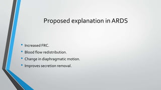
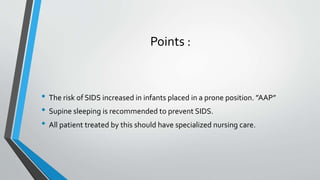
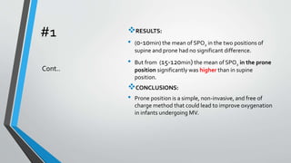
Prone Position in Neonate
Mohammad Abdulhameed Alobead
ID# 2130006217
Definitions
• Pre-term ?
Infant born before 37 weeks of gestational age.
• Supine position ?
Lying on the back, face or front upward.
• Prone position ?
Lying on the stomach, face downward.
• SIDS?
Sudden Infant Death Syndrome.
Proposed explanation in ARDS
• Increased FRC.
• Blood flow redistribution.
• Change in diaphragmatic motion.
• Improves secretion removal.
Points :
• The risk of SIDS increased in infants placed in a prone position. ”AAP”
• Supine sleeping is recommended to prevent SIDS.
• All patient treated by this should have specialized nursing care.
The effects of supine and prone
positions on oxygenation in
premature infants undergoing
mechanical ventilation
Iranian Journal Nursing Midwifery
Research. 2010
BACKGROUND:
• The use of high concentrations ofO2 in infants may
lead to chronic lung problems, using proper methods
of care in infants on MV is important in NICU.
Objectives:
• This study aimed to: investigate the effects of prone
and supine positions on oxygenation (SPO2) in
premature infants under mechanical ventilation and
comparing infants’ oxygenation in the two positions.
#1
#1 METHODS
• non randomized clinical trial study.
• On 32 preterm infants who are on MV.
• Inclusion criteria : (GA <37 weeks, age of less than 7
days, undergoing at least 4 hours of MV, using SIMV
mode).
• Firstly, they were placed in supine position for 120
minutes.
• Then in prone position for 120 minutes .
• SPO2 were monitored by pulse oximeter continuously
and was recorded every minute.
• Data analysis was done using Software SPSS.
Cont..
#1 RESULTS:
• (0-10min) the mean of SPO2 in the two positions of
supine and prone had no significant difference.
• But from (15-120min) the mean of SPO2 in the prone
position significantly was higher than in supine
position.
CONCLUSIONS:
• Prone position is a simple, non-invasive, and free of
charge method that could lead to improve oxygenation
in infants undergoing MV.
Cont..
#2 Background:
• Results of several studies suggest that prone position is
beneficial in improving the preterm infants’ cardio-
respiratory status.
• there is not any available clear study about the effect
of this position on cardio-respiratory rates of Nasal
Continuous PositiveAirway Pressure (NCPAP) treating
premature infants.
Objectives:
• This study aimed to comparing supine and prone
positions on cardio-respiratory rates of premature
infants with RDS who were treated using NCPAP.
Comparison the effect of Sleep
Positioning on Cardiorespiratory
Rate in NoninvasiveVentilated
Premature Infants.
Nurs Midwifery. 2013
#2 Methods:
• cross over study.
• on 44 premature 29-34 weeks gestation premature infants
who were receiving NCPAP in NICU.
• Infants were randomly assigned into two groups:
• 1st group was placed in prone at first and then in supine.
• 2nd group was first at supine and then prone.
• HR and RR were assessed 3 times in each position for 30
minutes.
• demographic data was analyzed using t test, Chi square
and Fisher exact test.
Cont…
#2 Results:
• There was a significant difference in HR and RR of premature
infants.
• HR and RR became lower at prone position than supine in
both groups.
Conclusion:
• So it can be concluded that prone position could decrease
infants HR and RR, but supine position might increase them.
• prone position in infants with respiratory complications
during receiving N-CPAP in NICU can be useful.
• Regarding prone position is a risk factor for SIDS, prone
position should be only used when the newborn is being
supervised carefully.
Cont…
#3
Objective:
• To evaluate the cardiovascular response to short-term
prone positioning in neonates.
Study design:
• 30 neonates (gestational age: 35±4 weeks;
postmenstrual age: 36±3 weeks) were enrolled.
• continuous monitoring of HR, stroke volume (SV) and
cardiac output (CO) by electrical velocimetry in
hemodynamically stable .
• Skin blood flow (SBF) assessed on the forehead or foot
using Laser Doppler technology. Systemic vascular
resistance (SVR) as mean blood pressure (BP)/CO.
• 10 min, supine, prone and back-to-supine position.
• Data were analyzed using repeated measures analysis of
variance.
Prone Positioning Decreases
Cardiac Output and Increases
SystemicVascular Resistance
in Neonates
Official Journal of the California
Perinatal Association,2015
#3 Results :
• HR did not change in response to positioning.
• in prone position, SV, CO and SBF decreased and SVR
index increased.
• Back-to-supine position, SV, CO, SBF and SVR index
returned to baseline.
Conclusion :
• Short-term prone positioning is associated with
decreased SV, CO and SBF and increased calculated
SVR index.
• systemic vascular tone increased with prone
positioning as evidenced by the increase in calculated
SVR index and a trend of an increase in BP
Cont…
#4 OBJECTIVE:
• To compare breathing patterns and lung function in
the supine, lateral, and prone positions in oxygen-
dependent preterm infants.
STUDY DESIGN:
• Respiratory function in preterm infants receiving
NCPAP for mild respiratory failure was evaluated by
respiratory inductive plethysmography.
• 19 Infants were randomized to supine, left lateral, and
prone positions for 3 hours.
• A nest provided a semiflexed posture for the infants
placed in the left lateral position.
• Vt, phase angle between abdominal and thoracic
movements, rib cage contribution toVt, and dynamic
elevation of end-expiratory lung volume were
measured.
Positioning effects on lung
function and breathing
pattern in premature
newborns.
J Pediatric. 2013
#4 RESULTS:
• Fraction of inspired O2 was similar in the 3 positions for 19
infants (mean gestational age, 27±2 weeks; mean birth
weight, 950±150 g; mean postnatal age, 17±5 days).
• Arterial O2 saturation andVt were higher in the left lateral
and prone positions than in the supine position.
• The phase angle between abdominal and thoracic
movements was lower and rib cage contribution toVt was
higher in the left lateral and prone positions than in the
supine position.
• Dynamic elevation of end-expiratory lung volume was
greater in the supine position than in the left lateral and
prone positions.
Cont…
#4
CONCLUSION:
• In oxygen-dependent preterm infants, both the left lateral
and prone positions improve lung function by optimizing
breathing strategy.
• In NICU , the left lateral position can be used as an
alternative to the prone position for mild respiratory failure.
Cont…
#5
Objective:
• To evaluate the influence of body position on the
displacement of nasal prongs in preterm infants.
Methods:
• prospective, randomized, crossover study.
• on 16 infants born at a mean GA of 29.7±2 weeks.
• on CPAP by nasal prongs.
• The main outcome was the number of times that the
nasal prongs were displaced following infant positioning
in the following body positions:
• prone, right lateral, left lateral, and supine, according to
a pre-established random order.
Influence of body position
on the displacement of nasal
prongs in preterm newborns
receiving continuous
positive airway pressure
Rev Paul Pediatr. 2015
#5
• cardiorespiratory variables (RR, HR and SPO2) were
evaluated for each body position.
• Data for each position were collected every 10 min, over
a period of 60 min.
• An occurrence was defined when the nasal prongs were
displaced from the nostrils after 3 min in the desired
position, requiring intervention of the examiner.
Cont…
#5
Cont…
Results:
• the occurrence of nasal prong displacement was only
observed in the prone position (9 infants - 56.2%) and in
the left lateral position (2 infants - 12.5%).
• The number of times that the prongs were displaced
was 11 in the prone position (7 within the first 10min).
• 2 in the left lateral position (1 within the first 10min).
• No clinically significant changes were observed in the
cardiorespiratory variables.
 Conclusions:
• Maintenance of the nasal prongs to
provide adequate noninvasive
respiratory support was harder in the
prone position.
References:
• American Academy of Pediatrics.
Link: http://pediatrics.aappublications.org/content/89/6/1120
• Egan’s Fundamentals Of Respiratory Care,10th Edition .
• 1st article :
http://www.ncbi.nlm.nih.gov/pmc/articles/PMC3203282/
• 2nd article :
http://www.ncbi.nlm.nih.gov/pmc/articles/PMC4228557/#A10318R4
• 3rd article :
http://www.medscape.com/viewarticle/846293
• 4th article :
http://www.ncbi.nlm.nih.gov/pubmed/23312684
• 5th article :
http://www.ncbi.nlm.nih.gov/pmc/articles/PMC4620954/
Evidences about Prone position in neonate

Evidences about Prone position in neonate

  • 1.
    Prone Position inNeonate Mohammad Abdulhameed Alobead ID# 2130006217
  • 2.
    Definitions • Pre-term ? Infantborn before 37 weeks of gestational age. • Supine position ? Lying on the back, face or front upward. • Prone position ? Lying on the stomach, face downward. • SIDS? Sudden Infant Death Syndrome.
  • 3.
    Proposed explanation inARDS • Increased FRC. • Blood flow redistribution. • Change in diaphragmatic motion. • Improves secretion removal.
  • 4.
    Points : • Therisk of SIDS increased in infants placed in a prone position. ”AAP” • Supine sleeping is recommended to prevent SIDS. • All patient treated by this should have specialized nursing care.
  • 5.
    The effects ofsupine and prone positions on oxygenation in premature infants undergoing mechanical ventilation Iranian Journal Nursing Midwifery Research. 2010 BACKGROUND: • The use of high concentrations ofO2 in infants may lead to chronic lung problems, using proper methods of care in infants on MV is important in NICU. Objectives: • This study aimed to: investigate the effects of prone and supine positions on oxygenation (SPO2) in premature infants under mechanical ventilation and comparing infants’ oxygenation in the two positions. #1
  • 6.
    #1 METHODS • nonrandomized clinical trial study. • On 32 preterm infants who are on MV. • Inclusion criteria : (GA <37 weeks, age of less than 7 days, undergoing at least 4 hours of MV, using SIMV mode). • Firstly, they were placed in supine position for 120 minutes. • Then in prone position for 120 minutes . • SPO2 were monitored by pulse oximeter continuously and was recorded every minute. • Data analysis was done using Software SPSS. Cont..
  • 7.
    #1 RESULTS: • (0-10min)the mean of SPO2 in the two positions of supine and prone had no significant difference. • But from (15-120min) the mean of SPO2 in the prone position significantly was higher than in supine position. CONCLUSIONS: • Prone position is a simple, non-invasive, and free of charge method that could lead to improve oxygenation in infants undergoing MV. Cont..
  • 8.
    #2 Background: • Resultsof several studies suggest that prone position is beneficial in improving the preterm infants’ cardio- respiratory status. • there is not any available clear study about the effect of this position on cardio-respiratory rates of Nasal Continuous PositiveAirway Pressure (NCPAP) treating premature infants. Objectives: • This study aimed to comparing supine and prone positions on cardio-respiratory rates of premature infants with RDS who were treated using NCPAP. Comparison the effect of Sleep Positioning on Cardiorespiratory Rate in NoninvasiveVentilated Premature Infants. Nurs Midwifery. 2013
  • 9.
    #2 Methods: • crossover study. • on 44 premature 29-34 weeks gestation premature infants who were receiving NCPAP in NICU. • Infants were randomly assigned into two groups: • 1st group was placed in prone at first and then in supine. • 2nd group was first at supine and then prone. • HR and RR were assessed 3 times in each position for 30 minutes. • demographic data was analyzed using t test, Chi square and Fisher exact test. Cont…
  • 10.
    #2 Results: • Therewas a significant difference in HR and RR of premature infants. • HR and RR became lower at prone position than supine in both groups. Conclusion: • So it can be concluded that prone position could decrease infants HR and RR, but supine position might increase them. • prone position in infants with respiratory complications during receiving N-CPAP in NICU can be useful. • Regarding prone position is a risk factor for SIDS, prone position should be only used when the newborn is being supervised carefully. Cont…
  • 11.
    #3 Objective: • To evaluatethe cardiovascular response to short-term prone positioning in neonates. Study design: • 30 neonates (gestational age: 35±4 weeks; postmenstrual age: 36±3 weeks) were enrolled. • continuous monitoring of HR, stroke volume (SV) and cardiac output (CO) by electrical velocimetry in hemodynamically stable . • Skin blood flow (SBF) assessed on the forehead or foot using Laser Doppler technology. Systemic vascular resistance (SVR) as mean blood pressure (BP)/CO. • 10 min, supine, prone and back-to-supine position. • Data were analyzed using repeated measures analysis of variance. Prone Positioning Decreases Cardiac Output and Increases SystemicVascular Resistance in Neonates Official Journal of the California Perinatal Association,2015
  • 12.
    #3 Results : •HR did not change in response to positioning. • in prone position, SV, CO and SBF decreased and SVR index increased. • Back-to-supine position, SV, CO, SBF and SVR index returned to baseline. Conclusion : • Short-term prone positioning is associated with decreased SV, CO and SBF and increased calculated SVR index. • systemic vascular tone increased with prone positioning as evidenced by the increase in calculated SVR index and a trend of an increase in BP Cont…
  • 13.
    #4 OBJECTIVE: • Tocompare breathing patterns and lung function in the supine, lateral, and prone positions in oxygen- dependent preterm infants. STUDY DESIGN: • Respiratory function in preterm infants receiving NCPAP for mild respiratory failure was evaluated by respiratory inductive plethysmography. • 19 Infants were randomized to supine, left lateral, and prone positions for 3 hours. • A nest provided a semiflexed posture for the infants placed in the left lateral position. • Vt, phase angle between abdominal and thoracic movements, rib cage contribution toVt, and dynamic elevation of end-expiratory lung volume were measured. Positioning effects on lung function and breathing pattern in premature newborns. J Pediatric. 2013
  • 14.
    #4 RESULTS: • Fractionof inspired O2 was similar in the 3 positions for 19 infants (mean gestational age, 27±2 weeks; mean birth weight, 950±150 g; mean postnatal age, 17±5 days). • Arterial O2 saturation andVt were higher in the left lateral and prone positions than in the supine position. • The phase angle between abdominal and thoracic movements was lower and rib cage contribution toVt was higher in the left lateral and prone positions than in the supine position. • Dynamic elevation of end-expiratory lung volume was greater in the supine position than in the left lateral and prone positions. Cont…
  • 15.
    #4 CONCLUSION: • In oxygen-dependentpreterm infants, both the left lateral and prone positions improve lung function by optimizing breathing strategy. • In NICU , the left lateral position can be used as an alternative to the prone position for mild respiratory failure. Cont…
  • 16.
    #5 Objective: • To evaluatethe influence of body position on the displacement of nasal prongs in preterm infants. Methods: • prospective, randomized, crossover study. • on 16 infants born at a mean GA of 29.7±2 weeks. • on CPAP by nasal prongs. • The main outcome was the number of times that the nasal prongs were displaced following infant positioning in the following body positions: • prone, right lateral, left lateral, and supine, according to a pre-established random order. Influence of body position on the displacement of nasal prongs in preterm newborns receiving continuous positive airway pressure Rev Paul Pediatr. 2015
  • 17.
    #5 • cardiorespiratory variables(RR, HR and SPO2) were evaluated for each body position. • Data for each position were collected every 10 min, over a period of 60 min. • An occurrence was defined when the nasal prongs were displaced from the nostrils after 3 min in the desired position, requiring intervention of the examiner. Cont…
  • 18.
    #5 Cont… Results: • the occurrenceof nasal prong displacement was only observed in the prone position (9 infants - 56.2%) and in the left lateral position (2 infants - 12.5%). • The number of times that the prongs were displaced was 11 in the prone position (7 within the first 10min). • 2 in the left lateral position (1 within the first 10min). • No clinically significant changes were observed in the cardiorespiratory variables.  Conclusions: • Maintenance of the nasal prongs to provide adequate noninvasive respiratory support was harder in the prone position.
  • 19.
    References: • American Academyof Pediatrics. Link: http://pediatrics.aappublications.org/content/89/6/1120 • Egan’s Fundamentals Of Respiratory Care,10th Edition . • 1st article : http://www.ncbi.nlm.nih.gov/pmc/articles/PMC3203282/ • 2nd article : http://www.ncbi.nlm.nih.gov/pmc/articles/PMC4228557/#A10318R4 • 3rd article : http://www.medscape.com/viewarticle/846293 • 4th article : http://www.ncbi.nlm.nih.gov/pubmed/23312684 • 5th article : http://www.ncbi.nlm.nih.gov/pmc/articles/PMC4620954/