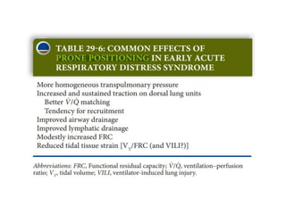
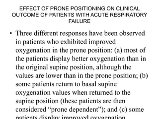
This document provides information on prone positioning for patients in the ICU and during occupational therapy. It discusses the physiologic effects of prone positioning on oxygenation, indications for prone positioning, types of proning procedures and techniques, assessing response, complications, and literature on the topic. Prone positioning can improve oxygenation for patients with conditions like ARDS by reducing lung compression and improving ventilation and perfusion. Proper patient positioning and monitoring during proning is important to prevent injuries and complications.


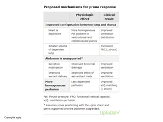
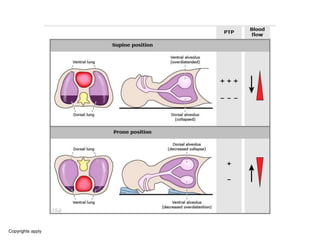
![Reduced lung compression — Lung compression by both the heart and the diaphragm
can be favorably affected by prone positioning.
• The effects of supine positioning – When an individual with ARDS is supine, the
heart compresses the medial posterior lung parenchyma and the diaphragm
compresses the posterior-caudal lung parenchyma.
• The latter is caused by the abdominal contents displacing the diaphragm cranially,
which can be exacerbated by a loss of diaphragmatic tone due to sedation and/or
paralysis or increased abdominal pressure.
• Compression by either the heart and/or the diaphragm may exaggerate dependent
lung collapse in the supine position, increasing hypoxemia (ie, worsening shunt)
and ventilator-associated lung injury [11].
• The effects of prone positioning – During prone ventilation, the heart becomes
dependent, lying on the sternum, potentially decreasing medial posterior lung
compression.
• In addition, the diaphragm is displaced caudally (especially in obese patients and
when the abdomen is left unsupported), decreasing compression of the posterior-
caudal lung parenchyma [12]. These effects improve ventilation and oxygenation
[13].](https://image.slidesharecdn.com/prone-220526052835-6ff4f64d/85/PRONE-pptx-12-320.jpg)