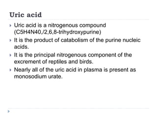
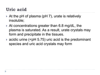
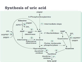
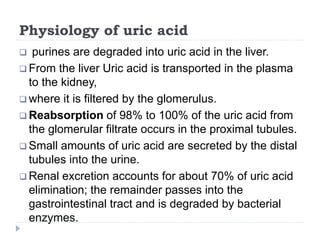
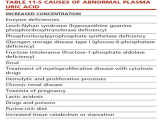
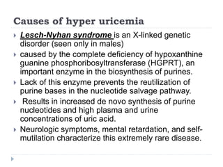
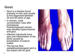
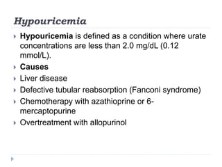
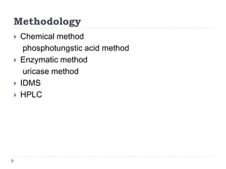
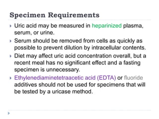
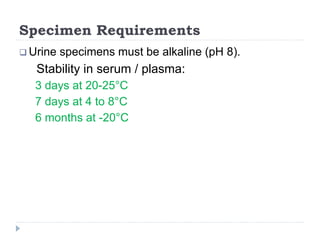
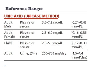
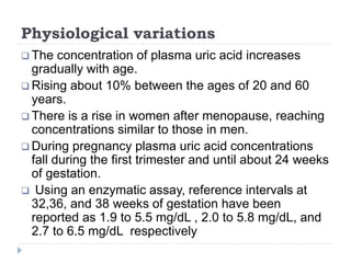
ā€«Ų§Ł„Ų±Ų­ŁŠŁ…ā€¬ ‫الرحمن‬ ‫هللا‬ ‫بسم‬
Hyperuricemia
Mahtab Ul Haq
MLT Final year
Objectives
 Uric acid
 Synthesis of uric acid
 Physiology of uric acid
 Hyper uricemia
 Causes of hyperuricemia
 hypouricemia
 Methodology
 Reference ranges
 Physiological variations
Uric acid
 Uric acid is a nitrogenous compound
(C5H4N40,/2,6,8-trihydroxypurine)
 It is the product of catabolism of the purine nucleic
acids.
 It is the principal nitrogenous component of the
excrement of reptiles and birds.
 Nearly all of the uric acid in plasma is present as
monosodium urate.
Uric acid
 At the pH of plasma (pH 7), urate is relatively
insoluble;
 At concentrations greater than 6.8 mg/dL, the
plasma is saturated. As a result, urate crystals may
form and precipitate in the tissues.
 acidic urine (<pH 5.75) uric acid is the predominant
species and uric acid crystals may form
Synthesis of uric acid
Physiology of uric acid
 purines are degraded into uric acid in the liver.
 From the liver Uric acid is transported in the plasma
to the kidney,
 where it is filtered by the glomerulus.
 Reabsorption of 98% to 100% of the uric acid from
the glomerular filtrate occurs in the proximal tubules.
 Small amounts of uric acid are secreted by the distal
tubules into the urine.
 Renal excretion accounts for about 70% of uric acid
elimination; the remainder passes into the
gastrointestinal tract and is degraded by bacterial
enzymes.
 The daily synthesis rate of uric acid is approximately
400 mg.
 Dietary sources contribute another 300mg.
 In men consuming a purine-free diet, the total body
pool of exchangeable urate is estimated at 1200 mg.
 In women it is estimated to be 600 mg.
 Patients with gouty arthritis and tissue deposition of
urate may have urate pools as large as 18,000 to
30,000 mg.
Hyper-uricemia
Hyperuricemia
 Hyperuricemia is most commonly defined as
plasma uric acid concentrations greater than 7.0
mg/dL (0.42 mmol/L) in men or greater than 6.0
mg/dL (0.36 mmol/L) in women.
Causes of hyper uricemia
 Lesch-Nyhan syndrome is an X-linked genetic
disorder (seen only in males)
 caused by the complete deficiency of hypoxanthine
guanine phosphoribosyltransferase (HGPRT), an
important enzyme in the biosynthesis of purines.
 Lack of this enzyme prevents the reutilization of
purine bases in the nucleotide salvage pathway.
 Results in increased de novo synthesis of purine
nucleotides and high plasma and urine
concentrations of uric acid.
 Neurologic symptoms, mental retardation, and self-
mutilation characterize this extremely rare disease.
Gout
 Gout is a disease found
primarily in men and usually
is first diagnosed between
30 and 50 years of age.
 In women, urate
concentration rises after
menopause.
Postmenopausal women
may develop hyperuricemia
and gout.
 Affected individuals have
pain and inflammation of the
joints caused by
precipitation of sodium
urates.
 The big toe (first
metatarsophalangeal) joint is
the classic site for gout.
Gout
 In 25% to 30% of these patients, hyperuricemia is
a result of overproduction of uric acid.
 Plasma uric acid concentration is usually greater
than 6.0 mg/dL.
 Kidney disease associated with hyperuricemia may
take one or more of several forms:
(1) gouty nephropathy with urate deposition in renal
parenchyma,
(2) acute intratubular deposition of urate crystals, and
(3) urate nephtolithiasis.
Causes of hyperuricemia
 Chronic renal disease causes increased uric acid
concentration because filtration and secretion are
impaired.
 toxemia of pregnancy (preeclampsia) probably
caused by uteroplacental tissue breakdown and
decreased kidney perfusion. Concentrations in
excess of 6.0 mg/dL (0.36 mmol/l) at 32 weeks
gestation have been noted to be associated with a
high perinatal mortality rate.'
 lactic acidosis, presumably as a result of
competition for binding sites in the renal tubules.
Hypouricemia
 Hypouricemia is defined as a condition where urate
concentrations are less than 2.0 mg/dL (0.12
mmol/L).
 Causes
 Liver disease
 Defective tubular reabsorption (Fanconi syndrome)
 Chemotherapy with azathioprine or 6-
mercaptopurine
 Overtreatment with allopurinol
Methodology
Methodology
 Chemical method
phosphotungstic acid method
 Enzymatic method
uricase method
 IDMS
 HPLC
Specimen Requirements
 Uric acid may be measured in heparinized plasma,
serum, or urine.
 Serum should be removed from cells as quickly as
possible to prevent dilution by intracellular contents.
 Diet may affect uric acid concentration overall, but a
recent meal has no significant effect and a fasting
specimen is unnecessary.
 Ethylenediaminetetraacetic acid (EDTA) or fluoride
additives should not be used for specimens that will
be tested by a uricase method.
Specimen Requirements
 Urine specimens must be alkaline (pH 8).
Stability in serum / plasma:
3 days at 20-25°C
7 days at 4 to 8°C
6 months at -20°C
Chemical Method
PHOSPHOTUNGSTIC ACID METHOD
Principle: Proteins in serum are precipitated with
tungstic acid. Uric acid in the supernatant reduces the
phosphotungstic acid into tungsten blue in an alkaline
medium of sodium bicarbonate.
The colour of the tungsten blue is proportional to the
uric acid concentration and is read at 660 nm.
Enzymatic Method
Interfering substances
 Proteins can cause high background absorbance,
reducing sensitivity.
 Hemoglobin and xanthine can cause negative
interference.
 Bilirubin and ascorbic acid,which destroy peroxide, if
present in sufficient quantity, can interfere.
 Commercial reagent preparations often include
potassium ferricyanide and ascorbate oxidase to
minimize these interferences.
Interfering substances
 Significant hemolysis may result in low values.
 Drugs such as salicylates and thiazides have been
shown to increase values for uric acid.
Reference Ranges
Physiological variations
 The concentration of plasma uric acid increases
gradually with age.
 Rising about 10% between the ages of 20 and 60
years.
 There is a rise in women after menopause, reaching
concentrations similar to those in men.
 During pregnancy plasma uric acid concentrations
fall during the first trimester and until about 24 weeks
of gestation.
 Using an enzymatic assay, reference intervals at
32,36, and 38 weeks of gestation have been
reported as 1.9 to 5.5 mg/dL , 2.0 to 5.8 mg/dL, and
2.7 to 6.5 mg/dL respectively
IDMS
 Detection of characteristic fragments following
ionization; quantification using isotopically labeled
compound
 Proposed reference method
HPLC
 HPLC methods using ion-exchange or reversed-
phase columns have been used to separate and
quantify uric acid.
 The column effluent is monitored at 293 nm to detect
the eluting uric acid.
 HPLC methods are specific and fast; mobile phases
are simple; and the retention time for uric acid is less
than 6 minutes.
Thank you

Hyperuricemia

  • 1.
  • 2.
  • 3.
    Objectives  Uric acid Synthesis of uric acid  Physiology of uric acid  Hyper uricemia  Causes of hyperuricemia  hypouricemia  Methodology  Reference ranges  Physiological variations
  • 4.
    Uric acid  Uricacid is a nitrogenous compound (C5H4N40,/2,6,8-trihydroxypurine)  It is the product of catabolism of the purine nucleic acids.  It is the principal nitrogenous component of the excrement of reptiles and birds.  Nearly all of the uric acid in plasma is present as monosodium urate.
  • 5.
    Uric acid  Atthe pH of plasma (pH 7), urate is relatively insoluble;  At concentrations greater than 6.8 mg/dL, the plasma is saturated. As a result, urate crystals may form and precipitate in the tissues.  acidic urine (<pH 5.75) uric acid is the predominant species and uric acid crystals may form
  • 6.
  • 7.
    Physiology of uricacid  purines are degraded into uric acid in the liver.  From the liver Uric acid is transported in the plasma to the kidney,  where it is filtered by the glomerulus.  Reabsorption of 98% to 100% of the uric acid from the glomerular filtrate occurs in the proximal tubules.  Small amounts of uric acid are secreted by the distal tubules into the urine.  Renal excretion accounts for about 70% of uric acid elimination; the remainder passes into the gastrointestinal tract and is degraded by bacterial enzymes.
  • 8.
     The dailysynthesis rate of uric acid is approximately 400 mg.  Dietary sources contribute another 300mg.  In men consuming a purine-free diet, the total body pool of exchangeable urate is estimated at 1200 mg.  In women it is estimated to be 600 mg.  Patients with gouty arthritis and tissue deposition of urate may have urate pools as large as 18,000 to 30,000 mg.
  • 9.
  • 10.
    Hyperuricemia  Hyperuricemia ismost commonly defined as plasma uric acid concentrations greater than 7.0 mg/dL (0.42 mmol/L) in men or greater than 6.0 mg/dL (0.36 mmol/L) in women.
  • 12.
    Causes of hyperuricemia  Lesch-Nyhan syndrome is an X-linked genetic disorder (seen only in males)  caused by the complete deficiency of hypoxanthine guanine phosphoribosyltransferase (HGPRT), an important enzyme in the biosynthesis of purines.  Lack of this enzyme prevents the reutilization of purine bases in the nucleotide salvage pathway.  Results in increased de novo synthesis of purine nucleotides and high plasma and urine concentrations of uric acid.  Neurologic symptoms, mental retardation, and self- mutilation characterize this extremely rare disease.
  • 13.
    Gout  Gout isa disease found primarily in men and usually is first diagnosed between 30 and 50 years of age.  In women, urate concentration rises after menopause. Postmenopausal women may develop hyperuricemia and gout.  Affected individuals have pain and inflammation of the joints caused by precipitation of sodium urates.  The big toe (first metatarsophalangeal) joint is the classic site for gout.
  • 14.
    Gout  In 25%to 30% of these patients, hyperuricemia is a result of overproduction of uric acid.  Plasma uric acid concentration is usually greater than 6.0 mg/dL.  Kidney disease associated with hyperuricemia may take one or more of several forms: (1) gouty nephropathy with urate deposition in renal parenchyma, (2) acute intratubular deposition of urate crystals, and (3) urate nephtolithiasis.
  • 15.
    Causes of hyperuricemia Chronic renal disease causes increased uric acid concentration because filtration and secretion are impaired.  toxemia of pregnancy (preeclampsia) probably caused by uteroplacental tissue breakdown and decreased kidney perfusion. Concentrations in excess of 6.0 mg/dL (0.36 mmol/l) at 32 weeks gestation have been noted to be associated with a high perinatal mortality rate.'  lactic acidosis, presumably as a result of competition for binding sites in the renal tubules.
  • 16.
    Hypouricemia  Hypouricemia isdefined as a condition where urate concentrations are less than 2.0 mg/dL (0.12 mmol/L).  Causes  Liver disease  Defective tubular reabsorption (Fanconi syndrome)  Chemotherapy with azathioprine or 6- mercaptopurine  Overtreatment with allopurinol
  • 17.
  • 18.
    Methodology  Chemical method phosphotungsticacid method  Enzymatic method uricase method  IDMS  HPLC
  • 19.
    Specimen Requirements  Uricacid may be measured in heparinized plasma, serum, or urine.  Serum should be removed from cells as quickly as possible to prevent dilution by intracellular contents.  Diet may affect uric acid concentration overall, but a recent meal has no significant effect and a fasting specimen is unnecessary.  Ethylenediaminetetraacetic acid (EDTA) or fluoride additives should not be used for specimens that will be tested by a uricase method.
  • 20.
    Specimen Requirements  Urinespecimens must be alkaline (pH 8). Stability in serum / plasma: 3 days at 20-25°C 7 days at 4 to 8°C 6 months at -20°C
  • 21.
    Chemical Method PHOSPHOTUNGSTIC ACIDMETHOD Principle: Proteins in serum are precipitated with tungstic acid. Uric acid in the supernatant reduces the phosphotungstic acid into tungsten blue in an alkaline medium of sodium bicarbonate. The colour of the tungsten blue is proportional to the uric acid concentration and is read at 660 nm.
  • 22.
  • 23.
    Interfering substances  Proteinscan cause high background absorbance, reducing sensitivity.  Hemoglobin and xanthine can cause negative interference.  Bilirubin and ascorbic acid,which destroy peroxide, if present in sufficient quantity, can interfere.  Commercial reagent preparations often include potassium ferricyanide and ascorbate oxidase to minimize these interferences.
  • 24.
    Interfering substances  Significanthemolysis may result in low values.  Drugs such as salicylates and thiazides have been shown to increase values for uric acid.
  • 25.
  • 26.
    Physiological variations  Theconcentration of plasma uric acid increases gradually with age.  Rising about 10% between the ages of 20 and 60 years.  There is a rise in women after menopause, reaching concentrations similar to those in men.  During pregnancy plasma uric acid concentrations fall during the first trimester and until about 24 weeks of gestation.  Using an enzymatic assay, reference intervals at 32,36, and 38 weeks of gestation have been reported as 1.9 to 5.5 mg/dL , 2.0 to 5.8 mg/dL, and 2.7 to 6.5 mg/dL respectively
  • 27.
    IDMS  Detection ofcharacteristic fragments following ionization; quantification using isotopically labeled compound  Proposed reference method
  • 28.
    HPLC  HPLC methodsusing ion-exchange or reversed- phase columns have been used to separate and quantify uric acid.  The column effluent is monitored at 293 nm to detect the eluting uric acid.  HPLC methods are specific and fast; mobile phases are simple; and the retention time for uric acid is less than 6 minutes.
  • 29.