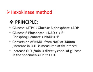
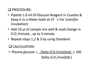
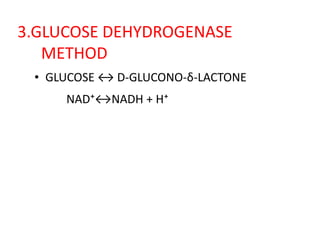
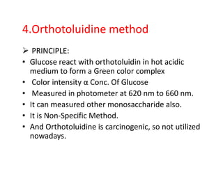
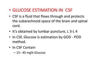
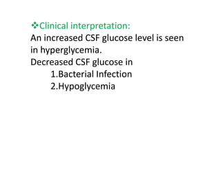
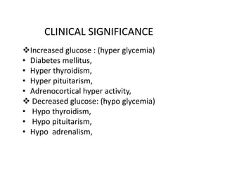
GLUCOSE ESTIMATION METHOD
INTRODUCTION:
• Glucose is a monosaccharide.
• It is central molecule in carbohydrate
metabolism.
• Stored as glycogen in liver and skeletal
muscle.
Entry of glucose into the cell
Two specific transport system are used :
1. Insulin –independent transport system:
• Carrier mediated uptake of glucose
• Not dependent on insulin.
• Present in hepatocytes, erythosytes & brain.
2. Insulin dependent transport system :
• Present in Skeletal muscle.
• For glucose estimation from any material,
blood is collect in fluoride containing vial.
• Fluoride inhibit glycolysis by inhibiting enolase
enzyme.
• In CSF, bacteria & other cells are also present
so analyzed immediately.
• For glucose estimation from urine, add 5ml
glacial acetic acid as preservative to inhibit
bacterial growth.
ENZYMETIC DETERMINATION
GOD POD METHOD:
PRINCIPLE:
Glucose + H₂O + O₂ GOD Gluconic acid + H₂O₂
4 Amino Phenazone + Phenol + H₂O₂ POD
Quinonimine – Pink colour compound
Intensity is determined at on 505 nm filter.
PROCEDURE
TEST STAN. BLANK
1)Glucose reagent (ml) 1.0 1.0 1.0
2)Serum(ml) 0.01 --- ----
3)Glucose standard(ml) --- 0.01 ----
4)Distilled water(ml) --- ---- 0.01
Mix & keep it for incubation at 37 ͦC for 15 min or at
room temperature for 30 min.
Measure the intensity of colour at 505 nm filter
(Green filter)
Calculation:
Concentration of Substance =
O.D. of Test-O.D. of Blank × Concentration of Std.
O.D. of Std.- O.D. of Blank
General Parameter:
General Parameter:
• Reaction type : End point
• Standard Conconcentration : 100 mg/dl
• Linearity is up to 500 mg/dl
• If sample value is 500mg/dl ,dilute the sample 1:2 with
distilled water & repeat assay
Hexokinase method
PRINCIPLE:
• Glucose +ATP↔Glucose 6 phosphate +ADP
• Glucose 6 Phosphate + NAD ↔ 6-
Phosphogluconate + NADH+H⁺
• Conversion of NADH from NAD at 340nm
,increase in O.D. is measured at fix interval
• Increase O.D. /min is directly conc. of glucose
in the specimen = Delta O.D.
PROCEDURE:
• Pipette 1.0 ml Of Glucose Reagent in Cuvette &
Keep It In a Water-bath at 37 ̊c For 1min(for
incubation)
• Add 10 μl of sample mix well & read change in
O.D /minute , up to 3 minute
• Repeat steps 1,2 & 3 by using Standard.
CALCULATION:
• Plasma glucose = Delta O.D./min(test) × 100
Delta O.D./min(Std.)
3.GLUCOSE DEHYDROGENASE
METHOD
• GLUCOSE ↔ D-GLUCONO-δ-LACTONE
NAD⁺↔NADH + H⁺
4.Orthotoluidine method
PRINCIPLE:
• Glucose react with orthotoluidin in hot acidic
medium to form a Green color complex
• Color intensity α Conc. Of Glucose
• Measured in photometer at 620 nm to 660 nm.
• It can measured other monosaccharide also.
• It is Non-Specific Method.
• And Orthotoluidine is carcinogenic, so not utilized
nowadays.
5. Folin Vui Method
• Time consuming method
• Non specific method , also measure fructose.
Blood is placed onto a test strip
& insert into the glucometer to
measure blood sugar levels
Glucometer
It is only type of dry chemistry
Advantage : Can do from capillary collection method. E.g. Heal Pick, Pinna Pick
Gives result with in second.
Disadvantage : Costly.
Slightly high result than actual.
MEASUREMENT OF GLUCOSE IN URINE
METHOD:
1.Qualitative
2.Quantitative
3.Semi- quantitative
1) QUALITATIVE METHOD:
• It is determination by Benedict test
2) QUANTITATIVE MATHOD:
• It Is Determination By Hexokinase & Glucose
Dehydrogenase
3)SEMI QUANTITATIVE MATHOD:
• It is determination by Glucose
Oxidase strip test
• E.g. Urine strip
Benedict's Test
This is a very simple and effective method of
the amount of glucose in the urine
Principle:
• Glucose(R-CHO)+ 2Cu⁺² +2H₂O→ Gluconic
acid(R-COOH) +Cu₂O +4H⁺
Procedure:
• 5 ml of Benedict's reagent + 8 to 10 drops
of urine Boiling the mixture & cool down it,
observe changes colour.
Result & Interpretation on Benedict Test
• Blue - sugar absent;
• Green - 0.5 gm% sugar = +1
• Yellow – 1.0 gm% sugar = +2
• Orange - 1.5 gm% sugar = +3
• Brick red – 2.0 % or more sugar = +4
Significant of Benedict Test
• If blood glucose level cross renal threshold,
than it excreted in urine. E.g. in diabetes
Mellitus
• If Renal threshold for glucose decrease in
renal failure, so in this case also benedict test
come positive.
• Each reducing substance gives positive test
• So Following substance can gives false positive
test E.g. Vitamin – C, B-Complex vitamin,
Salicylic acid
Glucose Oxidase Test
• Paper or plastic strips, called diastix .
• A color-chart is provided with the strips.
• Strip contain dye are O-tolidine,
tetramethylbenzidine , potassium iodide, 4-
amino phynazome,phenol.
• The dye changes colour on coming in contact
with the urine.
• After 30 to 60 seconds the colour of the strip
matched with the colours of the provided chart.
Oxidase Strip
• GLUCOSE ESTIMATION IN CSF
• CSF is a fluid that flows through and protects
the subarachnoid space of the brain and spinal
cord.
• It's obtained by lumbar puncture, L 3-L 4
• In CSF, Glucose is estimation by GOD - POD
method.
• In CSF Contain
– 15– 45 mg% Glucose
Clinical interpretation:
An increased CSF glucose level is seen
in hyperglycemia.
Decreased CSF glucose in
1.Bacterial Infection
2.Hypoglycemia
2.Hypoglycemia
CLINICAL SIGNIFICANCE
Increased glucose : (hyper glycemia)
• Diabetes mellitus,
• Hyper thyroidism,
• Hyper pituitarism,
• Adrenocortical hyper activity,
Decreased glucose: (hypo glycemia)
• Hypo thyroidism,
• Hypo pituitarism,
• Hypo adrenalism,
NORMAL RANGE
BLOOD :
• Random Blood Sugar : < 140 mg/dl
• Fasting Blood Sugar : 70 to 110 mg/dl
• Post Parendial Blood Sugar : <140 mg/dl
• CSF : 40 to 70 mg/dl (1/3 of plasma glucose)
• Urine : Absent
method_of_glucose_analysis_2018.pdf

method_of_glucose_analysis_2018.pdf

  • 1.
  • 2.
    INTRODUCTION: • Glucose isa monosaccharide. • It is central molecule in carbohydrate metabolism. • Stored as glycogen in liver and skeletal • Stored as glycogen in liver and skeletal muscle.
  • 3.
    Entry of glucoseinto the cell Two specific transport system are used : 1. Insulin –independent transport system: • Carrier mediated uptake of glucose • Not dependent on insulin. • Not dependent on insulin. • Present in hepatocytes, erythosytes & brain. 2. Insulin dependent transport system : • Present in Skeletal muscle.
  • 4.
    • For glucoseestimation from any material, blood is collect in fluoride containing vial. • Fluoride inhibit glycolysis by inhibiting enolase enzyme. • In CSF, bacteria & other cells are also present • In CSF, bacteria & other cells are also present so analyzed immediately. • For glucose estimation from urine, add 5ml glacial acetic acid as preservative to inhibit bacterial growth.
  • 5.
    ENZYMETIC DETERMINATION GOD PODMETHOD: PRINCIPLE: Glucose + H₂O + O₂ GOD Gluconic acid + H₂O₂ 4 Amino Phenazone + Phenol + H₂O₂ POD 4 Amino Phenazone + Phenol + H₂O₂ POD Quinonimine – Pink colour compound Intensity is determined at on 505 nm filter.
  • 6.
    PROCEDURE TEST STAN. BLANK 1)Glucosereagent (ml) 1.0 1.0 1.0 2)Serum(ml) 0.01 --- ---- 3)Glucose standard(ml) --- 0.01 ---- 4)Distilled water(ml) --- ---- 0.01 Mix & keep it for incubation at 37 ͦC for 15 min or at room temperature for 30 min. Measure the intensity of colour at 505 nm filter (Green filter)
  • 8.
    Calculation: Concentration of Substance= O.D. of Test-O.D. of Blank × Concentration of Std. O.D. of Std.- O.D. of Blank General Parameter: General Parameter: • Reaction type : End point • Standard Conconcentration : 100 mg/dl • Linearity is up to 500 mg/dl • If sample value is 500mg/dl ,dilute the sample 1:2 with distilled water & repeat assay
  • 9.
    Hexokinase method PRINCIPLE: • Glucose+ATP↔Glucose 6 phosphate +ADP • Glucose 6 Phosphate + NAD ↔ 6- Phosphogluconate + NADH+H⁺ Phosphogluconate + NADH+H⁺ • Conversion of NADH from NAD at 340nm ,increase in O.D. is measured at fix interval • Increase O.D. /min is directly conc. of glucose in the specimen = Delta O.D.
  • 10.
    PROCEDURE: • Pipette 1.0ml Of Glucose Reagent in Cuvette & Keep It In a Water-bath at 37 ̊c For 1min(for incubation) • Add 10 μl of sample mix well & read change in O.D /minute , up to 3 minute O.D /minute , up to 3 minute • Repeat steps 1,2 & 3 by using Standard. CALCULATION: • Plasma glucose = Delta O.D./min(test) × 100 Delta O.D./min(Std.)
  • 11.
    3.GLUCOSE DEHYDROGENASE METHOD • GLUCOSE↔ D-GLUCONO-δ-LACTONE NAD⁺↔NADH + H⁺
  • 12.
    4.Orthotoluidine method PRINCIPLE: • Glucosereact with orthotoluidin in hot acidic medium to form a Green color complex • Color intensity α Conc. Of Glucose • Color intensity α Conc. Of Glucose • Measured in photometer at 620 nm to 660 nm. • It can measured other monosaccharide also. • It is Non-Specific Method. • And Orthotoluidine is carcinogenic, so not utilized nowadays.
  • 13.
    5. Folin VuiMethod • Time consuming method • Non specific method , also measure fructose.
  • 14.
    Blood is placedonto a test strip & insert into the glucometer to measure blood sugar levels Glucometer It is only type of dry chemistry Advantage : Can do from capillary collection method. E.g. Heal Pick, Pinna Pick Gives result with in second. Disadvantage : Costly. Slightly high result than actual.
  • 15.
    MEASUREMENT OF GLUCOSEIN URINE METHOD: 1.Qualitative 2.Quantitative 3.Semi- quantitative 3.Semi- quantitative 1) QUALITATIVE METHOD: • It is determination by Benedict test
  • 16.
    2) QUANTITATIVE MATHOD: •It Is Determination By Hexokinase & Glucose Dehydrogenase 3)SEMI QUANTITATIVE MATHOD: • It is determination by Glucose • It is determination by Glucose Oxidase strip test • E.g. Urine strip
  • 17.
    Benedict's Test This isa very simple and effective method of the amount of glucose in the urine Principle: • Glucose(R-CHO)+ 2Cu⁺² +2H₂O→ Gluconic acid(R-COOH) +Cu₂O +4H⁺ acid(R-COOH) +Cu₂O +4H⁺ Procedure: • 5 ml of Benedict's reagent + 8 to 10 drops of urine Boiling the mixture & cool down it, observe changes colour.
  • 18.
    Result & Interpretationon Benedict Test • Blue - sugar absent; • Green - 0.5 gm% sugar = +1 • Yellow – 1.0 gm% sugar = +2 • Orange - 1.5 gm% sugar = +3 • Orange - 1.5 gm% sugar = +3 • Brick red – 2.0 % or more sugar = +4
  • 20.
    Significant of BenedictTest • If blood glucose level cross renal threshold, than it excreted in urine. E.g. in diabetes Mellitus • If Renal threshold for glucose decrease in renal failure, so in this case also benedict test renal failure, so in this case also benedict test come positive. • Each reducing substance gives positive test • So Following substance can gives false positive test E.g. Vitamin – C, B-Complex vitamin, Salicylic acid
  • 21.
    Glucose Oxidase Test •Paper or plastic strips, called diastix . • A color-chart is provided with the strips. • Strip contain dye are O-tolidine, tetramethylbenzidine , potassium iodide, 4- amino phynazome,phenol. amino phynazome,phenol. • The dye changes colour on coming in contact with the urine. • After 30 to 60 seconds the colour of the strip matched with the colours of the provided chart.
  • 22.
  • 23.
    • GLUCOSE ESTIMATIONIN CSF • CSF is a fluid that flows through and protects the subarachnoid space of the brain and spinal cord. • It's obtained by lumbar puncture, L 3-L 4 • In CSF, Glucose is estimation by GOD - POD • In CSF, Glucose is estimation by GOD - POD method. • In CSF Contain – 15– 45 mg% Glucose
  • 24.
    Clinical interpretation: An increasedCSF glucose level is seen in hyperglycemia. Decreased CSF glucose in 1.Bacterial Infection 2.Hypoglycemia 2.Hypoglycemia
  • 25.
    CLINICAL SIGNIFICANCE Increased glucose: (hyper glycemia) • Diabetes mellitus, • Hyper thyroidism, • Hyper pituitarism, • Adrenocortical hyper activity, • Adrenocortical hyper activity, Decreased glucose: (hypo glycemia) • Hypo thyroidism, • Hypo pituitarism, • Hypo adrenalism,
  • 26.
    NORMAL RANGE BLOOD : •Random Blood Sugar : < 140 mg/dl • Fasting Blood Sugar : 70 to 110 mg/dl • Post Parendial Blood Sugar : <140 mg/dl • Post Parendial Blood Sugar : <140 mg/dl • CSF : 40 to 70 mg/dl (1/3 of plasma glucose) • Urine : Absent