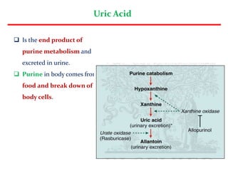
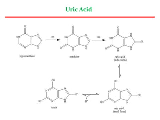
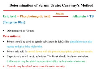
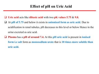
Uric Acid
Uric Acid
 Is the end product of
purine metabolism and
excreted in urine.
 Purine in body comes from
food and break down of
body cells.
Uric Acid
Determination of Serum Urate: Caraway’s Method
Uric Acid + Phosphotungstic Acid Allantoin + TB
(Tungsten Blue)
Alkaline
 OD measured at 700 nm.
Precautions:
 Serum should be used as certain substances in RBCs like glutathione can also
reduce and give false high color.
 Serum uric acid is carried down with the protein precipitate giving low results.
 Inspect and discard turbid solutions. The blank should be almost colorless.
Lithium salt may be added to prevent turbidity in final colored solution.
 Cyanide may be added to increase the color intensity.
Enzymatic Method: High Specificity
Uric Acid + 2H2O + O2 Allantoin + H2O2 + CO2
2H2O2 + 4-AAP +TBHB Quinoneimine + H2O
4-AAP: 4-Aminoantipyrine
TBHB: 2,4,6-Tribromo-3-hydroxy benzoic acid
OD at 500-540 nm.
Uricase
Peroxidase
The urate ion has characteristic absorption peak in the UV range (at
293 nm). The reaction products allantoin and H2O2 have little
absorption at this wavelength so that a derease in absorbance after
uricase activity is a specific measure of urate concentration.
Normal Serum Value
 3.5-7.2 mg/dl (Males);
 2.5-6.2 mg/dl (Females)
 Elevated level of uric acid in blood is one of the markers
of kidney dysfunction.
 High blood uric acid: HYPERURICEMIA
 Low blood uric acid: HYPOURICEMIA
Uric Acid: Serum Values
Renal Handling of Uric Acid
 Glomerular filtration of virtually all the uric acid in capillary
plasma.
 Reabsorption in PCT of about 98 % of the filtered Uric Acid.
 Subsequent secretion of nearly 50 % reabsorbed uric acid in
proximal PCT.
 Further reabsorption of 40 % in DCT.
 The net excretion of uric acid is 8 to 12 % of the amount
filtered. The urate pool in the body is about 1.2 g in men & 600
mg in females.
Effect of pH on Uric Acid
 Uric acid acts like dibasic acid with two pK values 5.75 & 9.8.
 At pH of 5.75 and below it exists in unionized form as uric acid. Due to
acidification in renal tubules, pH decreases to this level or below Hence in the
urine excreted as uric acid.
 Plasma has a pH of around 7.4. At this pH uric acid is present in ionised
form i.e salt form as monosodium urate that is 10 times more soluble than
uric acid.
Hyperuricemia
 Renal disorder leading to decreased excretion in urine e.g. renal failure
(90%).
 Increased production of uric acid (10%)
 Primary Gout: Defect in Uric Acid Metabolism
 HGPRTase deficiency
 PRPP Synthetase Overactivity
 Glucoe-6-Phosphatase Deficiency
 Adenosine deaminase Deficiency
 Secondary Gout: Results from other diseases in which increased uric acid
production is a by product of the disease. However, the uric acid metabolism is as
such normal.
 Psoriasis
 Leukaemia, Polycythaemia, Pernicious anaemia, Chronic haemolytic anaemia,
 Metabolic disorders associated with overproduction of uric acid.
At physiological form, uric acid is found in more soluble form as
sodium urate.
 In severe hyperuricemia, crystal of sodium urate get deposited in
in the soft tissues, particularly in joints. Such deposits are
commonly known as tophi. This causes inflammation of joints
resulting in gouty arthritis.
 The prevalence of gout is about 3 per 1000 persons, mostly affecting
affecting males. Post menopausal women, however are as
susceptible as men for this disease.
 Historically, gout was found to be associated with high living, over
Gout
Gout: Pathophysiology
Gout
Gout
Uric Acid
Hypouricemia
 Serum Uric Acid < 2 mg/dl.
 Some Uricosuric drugs increase
excretion of urate by impairing the
tubular reabsorption such as
Salicylates in large doses, benemid,
benzofuran & sulphinpyrazone.
 An analogue (ALLOPURINOL) of hypoxanthine when given inhibits the enzyme
Xanthine Oxidase and thus reduces the amount of urate formed from Xanthine
and hypoxanthine.
 Liver diseases wherein synthesis of uric acid is altered like Cirrhosis & Wilson’s
Disease.
 Renal disease that decreases renal tubular Resorption like Fanconi’s Syndrome.
Urate In Urine
 Method similar to those for serum have been applied to urine.
 After 10 fold dilution, the urine can be analysed by either of the
methods used for serum urate estimation.
Precautions:
 Store the urine at 4ºC, adding the uricase-inhibitor azide.
 Mix the urine well and warm for a few minutes at 60ºC to
dissolve any urate in the deposit, before diluting & proceeding
with determination.

Uric Acid.pptx

  • 1.
  • 2.
    Uric Acid  Isthe end product of purine metabolism and excreted in urine.  Purine in body comes from food and break down of body cells.
  • 3.
  • 4.
    Determination of SerumUrate: Caraway’s Method Uric Acid + Phosphotungstic Acid Allantoin + TB (Tungsten Blue) Alkaline  OD measured at 700 nm. Precautions:  Serum should be used as certain substances in RBCs like glutathione can also reduce and give false high color.  Serum uric acid is carried down with the protein precipitate giving low results.  Inspect and discard turbid solutions. The blank should be almost colorless. Lithium salt may be added to prevent turbidity in final colored solution.  Cyanide may be added to increase the color intensity.
  • 5.
    Enzymatic Method: HighSpecificity Uric Acid + 2H2O + O2 Allantoin + H2O2 + CO2 2H2O2 + 4-AAP +TBHB Quinoneimine + H2O 4-AAP: 4-Aminoantipyrine TBHB: 2,4,6-Tribromo-3-hydroxy benzoic acid OD at 500-540 nm. Uricase Peroxidase The urate ion has characteristic absorption peak in the UV range (at 293 nm). The reaction products allantoin and H2O2 have little absorption at this wavelength so that a derease in absorbance after uricase activity is a specific measure of urate concentration.
  • 6.
    Normal Serum Value 3.5-7.2 mg/dl (Males);  2.5-6.2 mg/dl (Females)  Elevated level of uric acid in blood is one of the markers of kidney dysfunction.  High blood uric acid: HYPERURICEMIA  Low blood uric acid: HYPOURICEMIA Uric Acid: Serum Values
  • 7.
    Renal Handling ofUric Acid  Glomerular filtration of virtually all the uric acid in capillary plasma.  Reabsorption in PCT of about 98 % of the filtered Uric Acid.  Subsequent secretion of nearly 50 % reabsorbed uric acid in proximal PCT.  Further reabsorption of 40 % in DCT.  The net excretion of uric acid is 8 to 12 % of the amount filtered. The urate pool in the body is about 1.2 g in men & 600 mg in females.
  • 8.
    Effect of pHon Uric Acid  Uric acid acts like dibasic acid with two pK values 5.75 & 9.8.  At pH of 5.75 and below it exists in unionized form as uric acid. Due to acidification in renal tubules, pH decreases to this level or below Hence in the urine excreted as uric acid.  Plasma has a pH of around 7.4. At this pH uric acid is present in ionised form i.e salt form as monosodium urate that is 10 times more soluble than uric acid.
  • 9.
    Hyperuricemia  Renal disorderleading to decreased excretion in urine e.g. renal failure (90%).  Increased production of uric acid (10%)  Primary Gout: Defect in Uric Acid Metabolism  HGPRTase deficiency  PRPP Synthetase Overactivity  Glucoe-6-Phosphatase Deficiency  Adenosine deaminase Deficiency  Secondary Gout: Results from other diseases in which increased uric acid production is a by product of the disease. However, the uric acid metabolism is as such normal.  Psoriasis  Leukaemia, Polycythaemia, Pernicious anaemia, Chronic haemolytic anaemia,
  • 11.
     Metabolic disordersassociated with overproduction of uric acid. At physiological form, uric acid is found in more soluble form as sodium urate.  In severe hyperuricemia, crystal of sodium urate get deposited in in the soft tissues, particularly in joints. Such deposits are commonly known as tophi. This causes inflammation of joints resulting in gouty arthritis.  The prevalence of gout is about 3 per 1000 persons, mostly affecting affecting males. Post menopausal women, however are as susceptible as men for this disease.  Historically, gout was found to be associated with high living, over Gout
  • 12.
  • 13.
  • 14.
  • 15.
  • 16.
    Hypouricemia  Serum UricAcid < 2 mg/dl.  Some Uricosuric drugs increase excretion of urate by impairing the tubular reabsorption such as Salicylates in large doses, benemid, benzofuran & sulphinpyrazone.  An analogue (ALLOPURINOL) of hypoxanthine when given inhibits the enzyme Xanthine Oxidase and thus reduces the amount of urate formed from Xanthine and hypoxanthine.  Liver diseases wherein synthesis of uric acid is altered like Cirrhosis & Wilson’s Disease.  Renal disease that decreases renal tubular Resorption like Fanconi’s Syndrome.
  • 17.
    Urate In Urine Method similar to those for serum have been applied to urine.  After 10 fold dilution, the urine can be analysed by either of the methods used for serum urate estimation. Precautions:  Store the urine at 4ºC, adding the uricase-inhibitor azide.  Mix the urine well and warm for a few minutes at 60ºC to dissolve any urate in the deposit, before diluting & proceeding with determination.