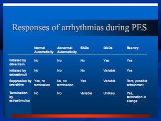
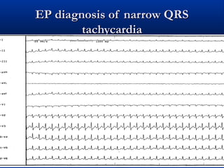
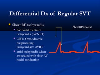
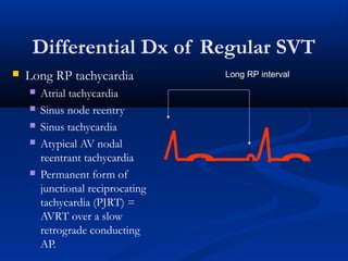
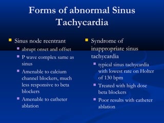
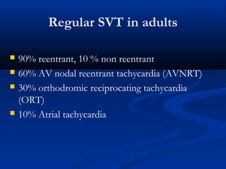
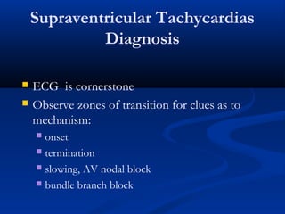
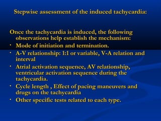
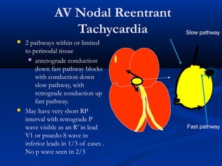
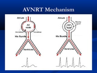
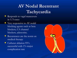
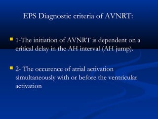
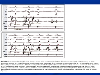
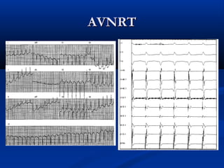
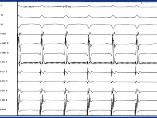
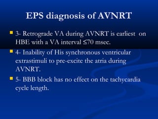
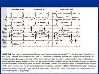
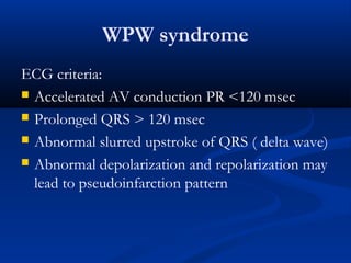
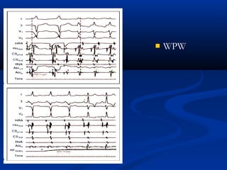
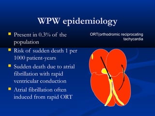
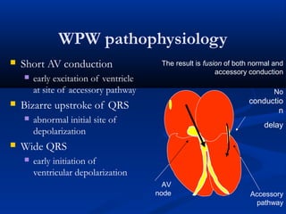
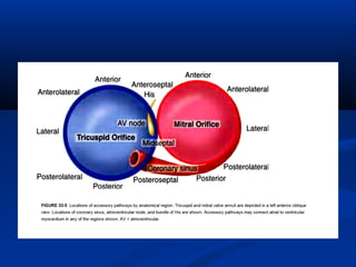
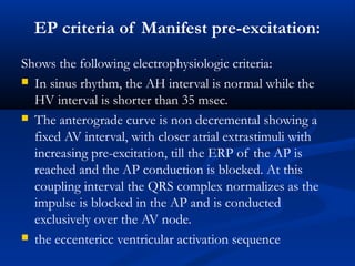
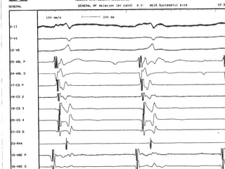
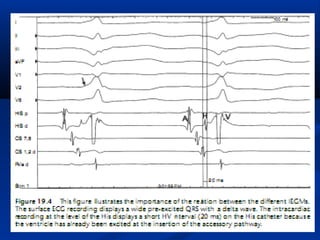
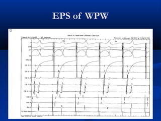
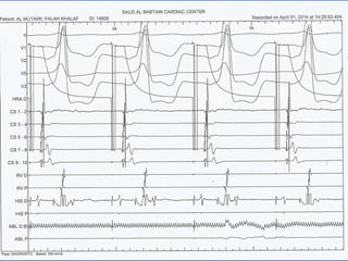
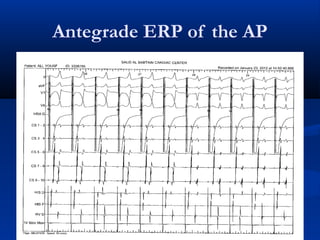
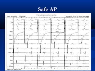
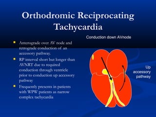
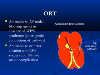
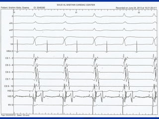
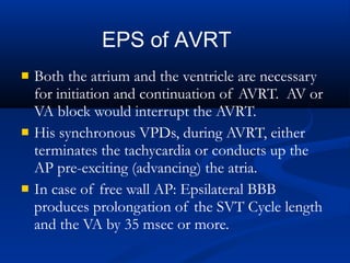
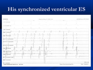
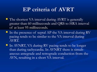
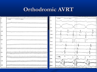
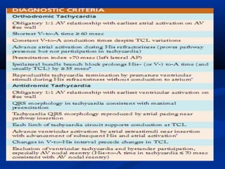
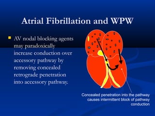
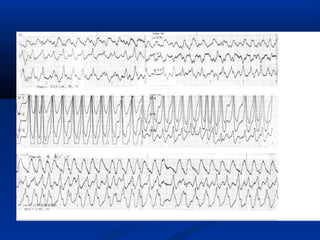
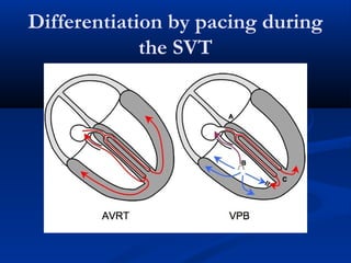
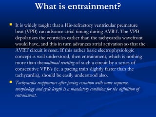
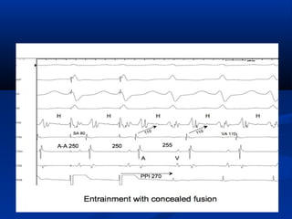
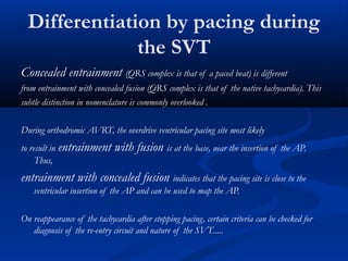
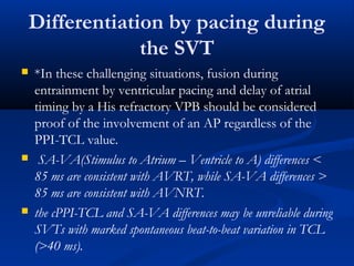
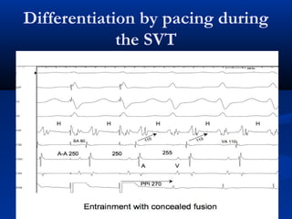
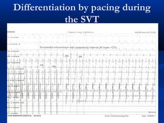
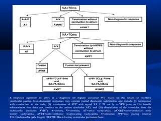
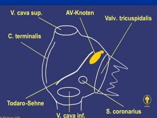
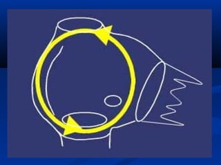
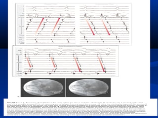
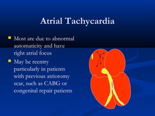
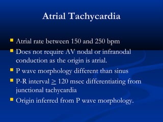
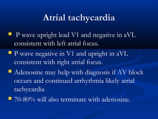
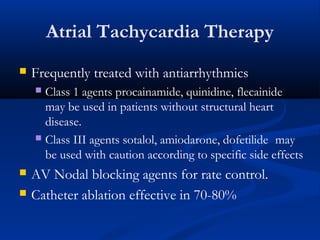
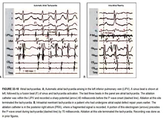
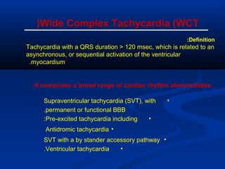
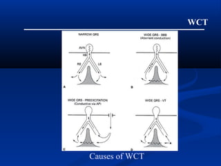
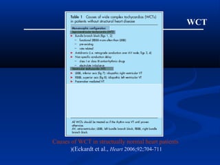
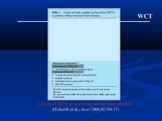
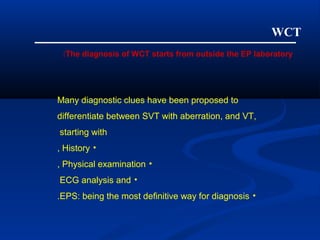
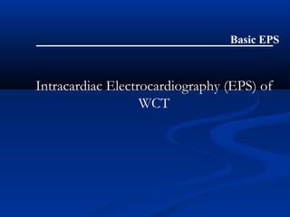
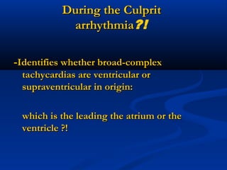
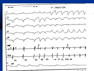
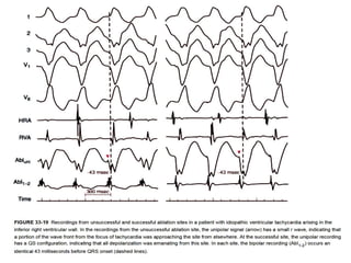
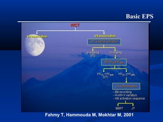
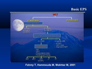
Basics of Electrophysiologic
Study, (part4)
Dr. Salah Atta, MD
Cosultant Electrophysiologist, SBCC
Professor of Cardiology
Assiut University, Egypt
Role of EPS in the Diagnosis and
Management of Arrhythmias
Mechanisms of Arrhythmia
 Abnormal automaticity
 automatic impulse generation from unusual site
or overtakes sinus node
 Triggered activity
 secondary depolarization during or after
repolarization
 Dig toxicity, Torsades de Pointes
 Reentry
 90 % of arrhythmias
Reentry
 Most common
mechanism
 Requires two separate
paths of conduction
 Requires an area of
slow conduction
 Requires unidirectional
block
EP diagnosis of narrow QRSEP diagnosis of narrow QRS
tachycardiatachycardia
Differential Dx of Regular SVT
 Short RP tachycardia
 AV nodal reentrant
tachycardia (AVNRT)
 ORT( Orthodromic
reciprocating
tachycardia)= AVRT
 atrial tachycardia when
associated with slow AV
nodal conduction
Short RP interval
Differential Dx of Regular SVT
 Long RP tachycardia
 Atrial tachycardia
 Sinus node reentry
 Sinus tachycardia
 Atypical AV nodal
reentrant tachycardia
 Permanent form of
junctional reciprocating
tachycardia (PJRT) =
AVRT over a slow
retrograde conducting
AP.
Long RP interval
Forms of abnormal Sinus
Tachycardia
 Sinus node reentrant
 abrupt onset and offset
 P wave complex same as
sinus
 Amenable to calcium
channel blockers, much
less responsive to beta
blockers
 Amenable to catheter
ablation
 Syndrome of
inappropriate sinus
tachycardia
 typical sinus tachycardia
with lowest rate on Holter
of 130 bpm
 Treated with high dose
beta blockers
 Poor results with catheter
ablation
Regular SVT in adults
 90% reentrant, 10 % non reentrant
 60% AV nodal reentrant tachycardia (AVNRT)
 30% orthodromic reciprocating tachycardia
(ORT)
 10% Atrial tachycardia
Supraventricular Tachycardias
Diagnosis
 ECG is cornerstone
 Observe zones of transition for clues as to
mechanism:
 onset
 termination
 slowing, AV nodal block
 bundle branch block
Stepwise assessment of the induced tachycardia:Stepwise assessment of the induced tachycardia:
Once the tachycardia is induced, the followingOnce the tachycardia is induced, the following
observations help establish the mechanism:observations help establish the mechanism:
• Mode of initiation and termination.Mode of initiation and termination.
• A-V relationship: 1:1 or variable, V-A relation andA-V relationship: 1:1 or variable, V-A relation and
intervalinterval
• Atrial activation sequence, AV relationship,Atrial activation sequence, AV relationship,
ventricular activation sequence during theventricular activation sequence during the
tachycardia.tachycardia.
• Cycle length , Effect of pacing maneuvers andCycle length , Effect of pacing maneuvers and
drugs on the tachycardiadrugs on the tachycardia
• Other specific tests related to each type.Other specific tests related to each type.
AV Nodal Reentrant
Tachycardia
 2 pathways within or limited
to perinodal tissue
 anterograde conduction
down fast pathway blocks
with conduction down
slow pathway, with
retrograde conduction up
fast pathway.
 May have very short RP
interval with retrograde P
wave visible as an R’ in lead
V1 or psuedo-S wave in
inferior leads in 1/3 of cases .
No p wave seen in 2/3
Slow pathway
Fast pathway
AVNRT Mechanism
AV Nodal Reentrant
Tachycardia
 Responds to vagal maneuvers
in 1/3 cases
 Very responsive to AV nodal
blocking agents such as beta
blockers, CA channel
blockers, adenosine.
 Recurrences are the norm on
medical therapy
 Catheter ablation 95%
successful with 1% major
complication rate
EPS Diagnostic criteria of AVNRT:
 1-The initiation of AVNRT is dependent on a
critical delay in the AH interval (AH jump).
 2- The occurence of atrial activation
simultaneously with or before the ventricular
activation
AVNRTAVNRT
EPS diagnosis of AVNRT
 3- Retrograde VA during AVNRT is earliest on
HBE with a VA interval ≤70 msec.
 4- Inability of His synchronous ventricular
extrastimuli to pre-excite the atria during
AVNRT.
 5- BBB block has no effect on the tachycardia
cycle length.
WPW syndrome
ECG criteria:
 Accelerated AV conduction PR <120 msec
 Prolonged QRS > 120 msec
 Abnormal slurred upstroke of QRS ( delta wave)
 Abnormal depolarization and repolarization may
lead to pseudoinfarction pattern
 WPW
WPW epidemiology
 Present in 0.3% of the
population
 Risk of sudden death 1 per
1000 patient-years
 Sudden death due to atrial
fibrillation with rapid
ventricular conduction
 Atrial fibrillation often
induced from rapid ORT
ORT(orthodromic reciprocating
tachycardia
WPW pathophysiology
 Short AV conduction
 early excitation of ventricle
at site of accessory pathway
 Bizarre upstroke of QRS
 abnormal initial site of
depolarization
 Wide QRS
 early initiation of
ventricular depolarization
The result is fusion of both normal and
accessory conduction
No
conductio
n
delay
AV
node Accessory
pathway
EP criteria of Manifest pre-excitation:
Shows the following electrophysiologic criteria:
 In sinus rhythm, the AH interval is normal while the
HV interval is shorter than 35 msec.
 The anterograde curve is non decremental showing a
fixed AV interval, with closer atrial extrastimuli with
increasing pre-excitation, till the ERP of the AP is
reached and the AP conduction is blocked. At this
coupling interval the QRS complex normalizes as the
impulse is blocked in the AP and is conducted
exclusively over the AV node.
 the eccentericc ventricular activation sequence
EPS of WPW
Antegrade ERP of the AP
Safe AP
Concealed pre-excitation
(unidirectional retrograde
conduction over the AP):
 Has the same electrophysiologic properties of
manifest APs in the retrograde direction.
Anterogradely, conduction is decremental as the
impulses proceed over the AV node.
Orthodromic Reciprocating
Tachycardia
 Anterograde over AV node and
retrograde conduction of an
accessory pathway.
 RP interval short but longer than
AVNRT due to required
conduction through ventricle
prior to conduction up accessory
pathway
 Frequently presents in patients
with WPW patients as narrow
complex tachycardia
Up
accessory
pathway
Conduction down AVnode
ORT
 Amenable to AV nodal
blocking agents in
absence of WPW
syndrome (anterograde
conduction of pathway)
 Amenable to catheter
ablation with 95%
success and 1% rate
major complication
Conduction down AVnode
Up
accessory
pathway
 AVRT is induced and terminated by VPD, APDs or
pacing.
 The retrograde atrial activation sequence during AVRT
is eccentric and fixed independent of the SVT cycle
length.
 During ventricular pacing with extrastimulation, the
retrograde curve is also non decremental. The
retrograde atrial activation sequence simulates that
during orthodromic AVRT.
 ParaHisian pacing: constant V-A interval.
EPS of AVRT
 Both the atrium and the ventricle are necessary
for initiation and continuation of AVRT. AV or
VA block would interrupt the AVRT.
 His synchronous VPDs, during AVRT, either
terminates the tachycardia or conducts up the
AP pre-exciting (advancing) the atria.
 In case of free wall AP: Epsilateral BBB
produces prolongation of the SVT Cycle length
and the VA by 35 msec or more.
EPS of AVRT
His synchronized ventricular ES
EP criteria of AVRT
 The shortest VA interval during AVRT is generally
greater than 60 milliseconds and QRS to HRA interval
of at least 95 milliseconds.
 In the presence of septal AP the VA interval during RV
pacing tends to be similar to the VA interval during
AVRT.
 In AVNRT, VA during RV pacing tends to be longer
than during tachycardia. In AVNRT there is simult-
aneous antegrade and retrograde conduction from the
AVN, resulting in a short VA interval.
Orthodromic AVRTOrthodromic AVRT
Atrial Fibrillation and WPW
 AV nodal blocking agents
may paradoxically
increase conduction over
accessory pathway by
removing concealed
retrograde penetration
into accessory pathway.
Concealed penetration into the pathway
causes intermittent block of pathway
conduction
Management of Atrial
Fibrillation with WPW
 Avoid AV nodal blockers
 IV procainamide to slow accessory pathway
conduction
 Amiodarone if decreased LVEF
 DC cardioversion if symptomatic with
hypotension
Management of Patients with
WPW
 All patients with symptomatic AF & WPW
should be evaluated with EPS
 Accessory pathways capable of conducting faster
than 240 BPM should be ablated (ERP<250ms)
 Patients with inducible arrhythmias involving
pathway should be ablated
 WPW patients in high risk professions should be
evaluated.
Differentiation by pacing during
the SVT
What is entrainment?
 It is widely taught that a His-refractory ventricular premature
beat (VPB) can advance atrial timing during AVRT. The VPB
depolarizes the ventricles earlier than the tachycardia wavefront
would have, and this in turn advances atrial activation so that the
AVRT circuit is reset. If this rather basic electrophysiologic
concept is well understood, then entrainment, which is nothing
more than thecontinual resetting of such a circuit by a series of
consecutive VPB's (ie. a pacing train slightly faster than the
tachycardia), should be easily understood also.
 Tachycardia reappearnce after pacing cessation with same sequence,
morphology and cycle length is a mandatory condition for the definition of
entrainment.
Concealed entrainment (QRS complex is that of a paced beat) is different
from entrainment with concealed fusion (QRS complex is that of the native tachycardia). This
subtle distinction in nomenclature is commonly overlooked .
During orthodromic AVRT, the overdrive ventricular pacing site most likely
to result in entrainment with fusion is at the base, near the insertion of the AP.
Thus,
entrainment with concealed fusion indicates that the pacing site is close to the
ventricular insertion of the AP and can be used to map the AP.
On reappearance of the tachycardia after stopping pacing, certain criteria can be checked for
diagnosis of the re-entry circuit and nature of the SVT.....
Differentiation by pacing during
the SVT
A cPPI-TCL (post pacing interval – tachycardia cycle length) >
110 ms is consistent with AVNRT,
while a cPPI-TCL < 110 ms is consistent with AVRT employing
a non-left sided AP.
A cPPI-TCL > 110 ms can occur with AVRT employing a
left sided AP simply because the RVA pacing site is far
from such a circuit. During a long RP interval SVT, a
cPPI-TCL > 110 ms should also prompt conside-ration
of orthodromic AVRT employing a slowly conducting
AP with decremental conduction properties.*
Differentiation by pacing during
the SVT
 *In these challenging situations, fusion during
entrainment by ventricular pacing and delay of atrial
timing by a His refractory VPB should be considered
proof of the involvement of an AP regardless of the
PPI-TCL value.
 SA-VA(Stimulus to Atrium – Ventricle to A) differences <
85 ms are consistent with AVRT, while SA-VA differences >
85 ms are consistent with AVNRT.
 the cPPI-TCL and SA-VA differences may be unreliable during
SVTs with marked spontaneous beat-to-beat variation in TCL
(>40 ms).
Differentiation by pacing during
the SVT
Differentiation by pacing during
the SVT
Differentiation by pacing during
the SVT
A proposed algorithm to arrive at a diagnosis for regular sustained SVT based on the results of overdrive
ventricular pacing. Non-diagnostic responses may contain partial diagnostic information and include (i) termination
with conduction to the atria, (ii) termination of SVT with septal VA ≥ 70 ms by a VPB prior to His bundle
refractoriness that does not conduct to the atrium (excludes AT), and (iii) dissociation of the ventricles from the
tachycardia (excludes AVRT). A=atrium; V=ventricle; AT=atrial tachycardia; AVNRT=atrioventricular node
reentry tachycardia; AVRT=atrioventricular reciprocating tachycardia; S=stimulus; PPI=post pacing interval;
TCL=tachycardia cycle length; HRVPB=His refractory ventricular premature beat.
Atrial Flutter
An atrial flutter (Macrore-entry in the right
atrium with atrial rate: 300/min) with two
to one conduction to the ventricles with
LBBB aberration was evident.
Atrial Tachycardia
 Most are due to abnormal
automaticity and have
right atrial focus
 May be reentry
particularly in patients
with previous atriotomy
scar, such as CABG or
congenital repair patients
Atrial Tachycardia
 Atrial rate between 150 and 250 bpm
 Does not require AV nodal or infranodal
conduction as the origin is atrial.
 P wave morphology different than sinus
 P-R interval > 120 msec differentiating from
junctional tachycardia
 Origin inferred from P wave morphology.
Atrial tachycardia
 P wave upright lead V1 and negative in aVL
consistent with left atrial focus.
 P wave negative in V1 and upright in aVL
consistent with right atrial focus.
 Adenosine may help with diagnosis if AV block
occurs and continued arrhythmia likely atrial
tachycardia
 70-80% will also terminate with adenosine.
Atrial Tachycardia Therapy
 Frequently treated with antiarrhythmics
 Class 1 agents procainamide, quinidine, flecainide
may be used in patients without structural heart
disease.
 Class III agents sotalol, amiodarone, dofetilide may
be used with caution according to specific side effects
 AV Nodal blocking agents for rate control.
 Catheter ablation effective in 70-80%
EP Diagnosis of
Wide-Complex Tachycardia
(WCT)
Basic EPS
Definition:
Tachycardia with a QRS duration > 120 msec, which is related to an
asynchronous, or sequential activation of the ventricular
myocardium.
Wide Complex Tachycardia (WCT(
It comprises a broad range of cardiac rhythm abnormalities:
•Supraventricular tachycardia (SVT), with
permanent or functional BBB.
•Pre-excited tachycardia including:
•Antidromic tachycardia
•SVT with a by stander accessory pathway
•Ventricular tachycardia.
WCT
Causes of WCT
WCT
Causes of WCT in structually normal heart patients
(Eckardt et al., Heart 2006;92;704-711(
WCT
Causes of WCT in structually normal heart patients
(Eckardt et al., Heart 2006;92;704-711(
WCT
Many diagnostic clues have been proposed to
differentiate between SVT with aberration, and VT,
starting with
•History,
•Physical examination,
•ECG analysis and
•EPS: being the most definitive way for diagnosis.
The diagnosis of WCT starts from outside the EP laboratory:
Intracardiac Electrocardiography (EPS) of
WCT
Basic EPS
--Identifies whether broad-complexIdentifies whether broad-complex
tachycardias are ventricular ortachycardias are ventricular or
supraventricular in origin:supraventricular in origin:
which is the leading the atrium or thewhich is the leading the atrium or the
ventricle ?!ventricle ?!
During the CulpritDuring the Culprit
arrhythmiaarrhythmia?!
Basic EPS
Fahmy T, Hammouda M, Mokhtar M, 2001
Basic EPS
Fahmy T, Hammouda M, Mokhtar M, 2001
Thank you

ELectrophysiology basics part4

  • 1.
    Basics of Electrophysiologic Study,(part4) Dr. Salah Atta, MD Cosultant Electrophysiologist, SBCC Professor of Cardiology Assiut University, Egypt
  • 2.
    Role of EPSin the Diagnosis and Management of Arrhythmias
  • 3.
    Mechanisms of Arrhythmia Abnormal automaticity  automatic impulse generation from unusual site or overtakes sinus node  Triggered activity  secondary depolarization during or after repolarization  Dig toxicity, Torsades de Pointes  Reentry  90 % of arrhythmias
  • 4.
    Reentry  Most common mechanism Requires two separate paths of conduction  Requires an area of slow conduction  Requires unidirectional block
  • 6.
    EP diagnosis ofnarrow QRSEP diagnosis of narrow QRS tachycardiatachycardia
  • 7.
    Differential Dx ofRegular SVT  Short RP tachycardia  AV nodal reentrant tachycardia (AVNRT)  ORT( Orthodromic reciprocating tachycardia)= AVRT  atrial tachycardia when associated with slow AV nodal conduction Short RP interval
  • 8.
    Differential Dx ofRegular SVT  Long RP tachycardia  Atrial tachycardia  Sinus node reentry  Sinus tachycardia  Atypical AV nodal reentrant tachycardia  Permanent form of junctional reciprocating tachycardia (PJRT) = AVRT over a slow retrograde conducting AP. Long RP interval
  • 9.
    Forms of abnormalSinus Tachycardia  Sinus node reentrant  abrupt onset and offset  P wave complex same as sinus  Amenable to calcium channel blockers, much less responsive to beta blockers  Amenable to catheter ablation  Syndrome of inappropriate sinus tachycardia  typical sinus tachycardia with lowest rate on Holter of 130 bpm  Treated with high dose beta blockers  Poor results with catheter ablation
  • 10.
    Regular SVT inadults  90% reentrant, 10 % non reentrant  60% AV nodal reentrant tachycardia (AVNRT)  30% orthodromic reciprocating tachycardia (ORT)  10% Atrial tachycardia
  • 11.
    Supraventricular Tachycardias Diagnosis  ECGis cornerstone  Observe zones of transition for clues as to mechanism:  onset  termination  slowing, AV nodal block  bundle branch block
  • 12.
    Stepwise assessment ofthe induced tachycardia:Stepwise assessment of the induced tachycardia: Once the tachycardia is induced, the followingOnce the tachycardia is induced, the following observations help establish the mechanism:observations help establish the mechanism: • Mode of initiation and termination.Mode of initiation and termination. • A-V relationship: 1:1 or variable, V-A relation andA-V relationship: 1:1 or variable, V-A relation and intervalinterval • Atrial activation sequence, AV relationship,Atrial activation sequence, AV relationship, ventricular activation sequence during theventricular activation sequence during the tachycardia.tachycardia. • Cycle length , Effect of pacing maneuvers andCycle length , Effect of pacing maneuvers and drugs on the tachycardiadrugs on the tachycardia • Other specific tests related to each type.Other specific tests related to each type.
  • 13.
    AV Nodal Reentrant Tachycardia 2 pathways within or limited to perinodal tissue  anterograde conduction down fast pathway blocks with conduction down slow pathway, with retrograde conduction up fast pathway.  May have very short RP interval with retrograde P wave visible as an R’ in lead V1 or psuedo-S wave in inferior leads in 1/3 of cases . No p wave seen in 2/3 Slow pathway Fast pathway
  • 14.
  • 15.
    AV Nodal Reentrant Tachycardia Responds to vagal maneuvers in 1/3 cases  Very responsive to AV nodal blocking agents such as beta blockers, CA channel blockers, adenosine.  Recurrences are the norm on medical therapy  Catheter ablation 95% successful with 1% major complication rate
  • 16.
    EPS Diagnostic criteriaof AVNRT:  1-The initiation of AVNRT is dependent on a critical delay in the AH interval (AH jump).  2- The occurence of atrial activation simultaneously with or before the ventricular activation
  • 18.
  • 20.
    EPS diagnosis ofAVNRT  3- Retrograde VA during AVNRT is earliest on HBE with a VA interval ≤70 msec.  4- Inability of His synchronous ventricular extrastimuli to pre-excite the atria during AVNRT.  5- BBB block has no effect on the tachycardia cycle length.
  • 22.
    WPW syndrome ECG criteria: Accelerated AV conduction PR <120 msec  Prolonged QRS > 120 msec  Abnormal slurred upstroke of QRS ( delta wave)  Abnormal depolarization and repolarization may lead to pseudoinfarction pattern
  • 23.
  • 24.
    WPW epidemiology  Presentin 0.3% of the population  Risk of sudden death 1 per 1000 patient-years  Sudden death due to atrial fibrillation with rapid ventricular conduction  Atrial fibrillation often induced from rapid ORT ORT(orthodromic reciprocating tachycardia
  • 25.
    WPW pathophysiology  ShortAV conduction  early excitation of ventricle at site of accessory pathway  Bizarre upstroke of QRS  abnormal initial site of depolarization  Wide QRS  early initiation of ventricular depolarization The result is fusion of both normal and accessory conduction No conductio n delay AV node Accessory pathway
  • 27.
    EP criteria ofManifest pre-excitation: Shows the following electrophysiologic criteria:  In sinus rhythm, the AH interval is normal while the HV interval is shorter than 35 msec.  The anterograde curve is non decremental showing a fixed AV interval, with closer atrial extrastimuli with increasing pre-excitation, till the ERP of the AP is reached and the AP conduction is blocked. At this coupling interval the QRS complex normalizes as the impulse is blocked in the AP and is conducted exclusively over the AV node.  the eccentericc ventricular activation sequence
  • 30.
  • 32.
  • 33.
  • 34.
    Concealed pre-excitation (unidirectional retrograde conductionover the AP):  Has the same electrophysiologic properties of manifest APs in the retrograde direction. Anterogradely, conduction is decremental as the impulses proceed over the AV node.
  • 35.
    Orthodromic Reciprocating Tachycardia  Anterogradeover AV node and retrograde conduction of an accessory pathway.  RP interval short but longer than AVNRT due to required conduction through ventricle prior to conduction up accessory pathway  Frequently presents in patients with WPW patients as narrow complex tachycardia Up accessory pathway Conduction down AVnode
  • 36.
    ORT  Amenable toAV nodal blocking agents in absence of WPW syndrome (anterograde conduction of pathway)  Amenable to catheter ablation with 95% success and 1% rate major complication Conduction down AVnode Up accessory pathway
  • 37.
     AVRT isinduced and terminated by VPD, APDs or pacing.  The retrograde atrial activation sequence during AVRT is eccentric and fixed independent of the SVT cycle length.  During ventricular pacing with extrastimulation, the retrograde curve is also non decremental. The retrograde atrial activation sequence simulates that during orthodromic AVRT.  ParaHisian pacing: constant V-A interval. EPS of AVRT
  • 39.
     Both theatrium and the ventricle are necessary for initiation and continuation of AVRT. AV or VA block would interrupt the AVRT.  His synchronous VPDs, during AVRT, either terminates the tachycardia or conducts up the AP pre-exciting (advancing) the atria.  In case of free wall AP: Epsilateral BBB produces prolongation of the SVT Cycle length and the VA by 35 msec or more. EPS of AVRT
  • 40.
  • 41.
    EP criteria ofAVRT  The shortest VA interval during AVRT is generally greater than 60 milliseconds and QRS to HRA interval of at least 95 milliseconds.  In the presence of septal AP the VA interval during RV pacing tends to be similar to the VA interval during AVRT.  In AVNRT, VA during RV pacing tends to be longer than during tachycardia. In AVNRT there is simult- aneous antegrade and retrograde conduction from the AVN, resulting in a short VA interval.
  • 42.
  • 44.
    Atrial Fibrillation andWPW  AV nodal blocking agents may paradoxically increase conduction over accessory pathway by removing concealed retrograde penetration into accessory pathway. Concealed penetration into the pathway causes intermittent block of pathway conduction
  • 46.
    Management of Atrial Fibrillationwith WPW  Avoid AV nodal blockers  IV procainamide to slow accessory pathway conduction  Amiodarone if decreased LVEF  DC cardioversion if symptomatic with hypotension
  • 47.
    Management of Patientswith WPW  All patients with symptomatic AF & WPW should be evaluated with EPS  Accessory pathways capable of conducting faster than 240 BPM should be ablated (ERP<250ms)  Patients with inducible arrhythmias involving pathway should be ablated  WPW patients in high risk professions should be evaluated.
  • 48.
  • 49.
    What is entrainment? It is widely taught that a His-refractory ventricular premature beat (VPB) can advance atrial timing during AVRT. The VPB depolarizes the ventricles earlier than the tachycardia wavefront would have, and this in turn advances atrial activation so that the AVRT circuit is reset. If this rather basic electrophysiologic concept is well understood, then entrainment, which is nothing more than thecontinual resetting of such a circuit by a series of consecutive VPB's (ie. a pacing train slightly faster than the tachycardia), should be easily understood also.  Tachycardia reappearnce after pacing cessation with same sequence, morphology and cycle length is a mandatory condition for the definition of entrainment.
  • 51.
    Concealed entrainment (QRScomplex is that of a paced beat) is different from entrainment with concealed fusion (QRS complex is that of the native tachycardia). This subtle distinction in nomenclature is commonly overlooked . During orthodromic AVRT, the overdrive ventricular pacing site most likely to result in entrainment with fusion is at the base, near the insertion of the AP. Thus, entrainment with concealed fusion indicates that the pacing site is close to the ventricular insertion of the AP and can be used to map the AP. On reappearance of the tachycardia after stopping pacing, certain criteria can be checked for diagnosis of the re-entry circuit and nature of the SVT..... Differentiation by pacing during the SVT
  • 52.
    A cPPI-TCL (postpacing interval – tachycardia cycle length) > 110 ms is consistent with AVNRT, while a cPPI-TCL < 110 ms is consistent with AVRT employing a non-left sided AP. A cPPI-TCL > 110 ms can occur with AVRT employing a left sided AP simply because the RVA pacing site is far from such a circuit. During a long RP interval SVT, a cPPI-TCL > 110 ms should also prompt conside-ration of orthodromic AVRT employing a slowly conducting AP with decremental conduction properties.* Differentiation by pacing during the SVT
  • 53.
     *In thesechallenging situations, fusion during entrainment by ventricular pacing and delay of atrial timing by a His refractory VPB should be considered proof of the involvement of an AP regardless of the PPI-TCL value.  SA-VA(Stimulus to Atrium – Ventricle to A) differences < 85 ms are consistent with AVRT, while SA-VA differences > 85 ms are consistent with AVNRT.  the cPPI-TCL and SA-VA differences may be unreliable during SVTs with marked spontaneous beat-to-beat variation in TCL (>40 ms). Differentiation by pacing during the SVT
  • 54.
  • 55.
  • 56.
    A proposed algorithmto arrive at a diagnosis for regular sustained SVT based on the results of overdrive ventricular pacing. Non-diagnostic responses may contain partial diagnostic information and include (i) termination with conduction to the atria, (ii) termination of SVT with septal VA ≥ 70 ms by a VPB prior to His bundle refractoriness that does not conduct to the atrium (excludes AT), and (iii) dissociation of the ventricles from the tachycardia (excludes AVRT). A=atrium; V=ventricle; AT=atrial tachycardia; AVNRT=atrioventricular node reentry tachycardia; AVRT=atrioventricular reciprocating tachycardia; S=stimulus; PPI=post pacing interval; TCL=tachycardia cycle length; HRVPB=His refractory ventricular premature beat.
  • 57.
    Atrial Flutter An atrialflutter (Macrore-entry in the right atrium with atrial rate: 300/min) with two to one conduction to the ventricles with LBBB aberration was evident.
  • 62.
    Atrial Tachycardia  Mostare due to abnormal automaticity and have right atrial focus  May be reentry particularly in patients with previous atriotomy scar, such as CABG or congenital repair patients
  • 63.
    Atrial Tachycardia  Atrialrate between 150 and 250 bpm  Does not require AV nodal or infranodal conduction as the origin is atrial.  P wave morphology different than sinus  P-R interval > 120 msec differentiating from junctional tachycardia  Origin inferred from P wave morphology.
  • 64.
    Atrial tachycardia  Pwave upright lead V1 and negative in aVL consistent with left atrial focus.  P wave negative in V1 and upright in aVL consistent with right atrial focus.  Adenosine may help with diagnosis if AV block occurs and continued arrhythmia likely atrial tachycardia  70-80% will also terminate with adenosine.
  • 65.
    Atrial Tachycardia Therapy Frequently treated with antiarrhythmics  Class 1 agents procainamide, quinidine, flecainide may be used in patients without structural heart disease.  Class III agents sotalol, amiodarone, dofetilide may be used with caution according to specific side effects  AV Nodal blocking agents for rate control.  Catheter ablation effective in 70-80%
  • 67.
    EP Diagnosis of Wide-ComplexTachycardia (WCT) Basic EPS
  • 68.
    Definition: Tachycardia with aQRS duration > 120 msec, which is related to an asynchronous, or sequential activation of the ventricular myocardium. Wide Complex Tachycardia (WCT( It comprises a broad range of cardiac rhythm abnormalities: •Supraventricular tachycardia (SVT), with permanent or functional BBB. •Pre-excited tachycardia including: •Antidromic tachycardia •SVT with a by stander accessory pathway •Ventricular tachycardia.
  • 69.
  • 70.
    WCT Causes of WCTin structually normal heart patients (Eckardt et al., Heart 2006;92;704-711(
  • 71.
    WCT Causes of WCTin structually normal heart patients (Eckardt et al., Heart 2006;92;704-711(
  • 72.
    WCT Many diagnostic clueshave been proposed to differentiate between SVT with aberration, and VT, starting with •History, •Physical examination, •ECG analysis and •EPS: being the most definitive way for diagnosis. The diagnosis of WCT starts from outside the EP laboratory:
  • 73.
  • 74.
    --Identifies whether broad-complexIdentifieswhether broad-complex tachycardias are ventricular ortachycardias are ventricular or supraventricular in origin:supraventricular in origin: which is the leading the atrium or thewhich is the leading the atrium or the ventricle ?!ventricle ?! During the CulpritDuring the Culprit arrhythmiaarrhythmia?!
  • 77.
    Basic EPS Fahmy T,Hammouda M, Mokhtar M, 2001
  • 78.
    Basic EPS Fahmy T,Hammouda M, Mokhtar M, 2001
  • 79.