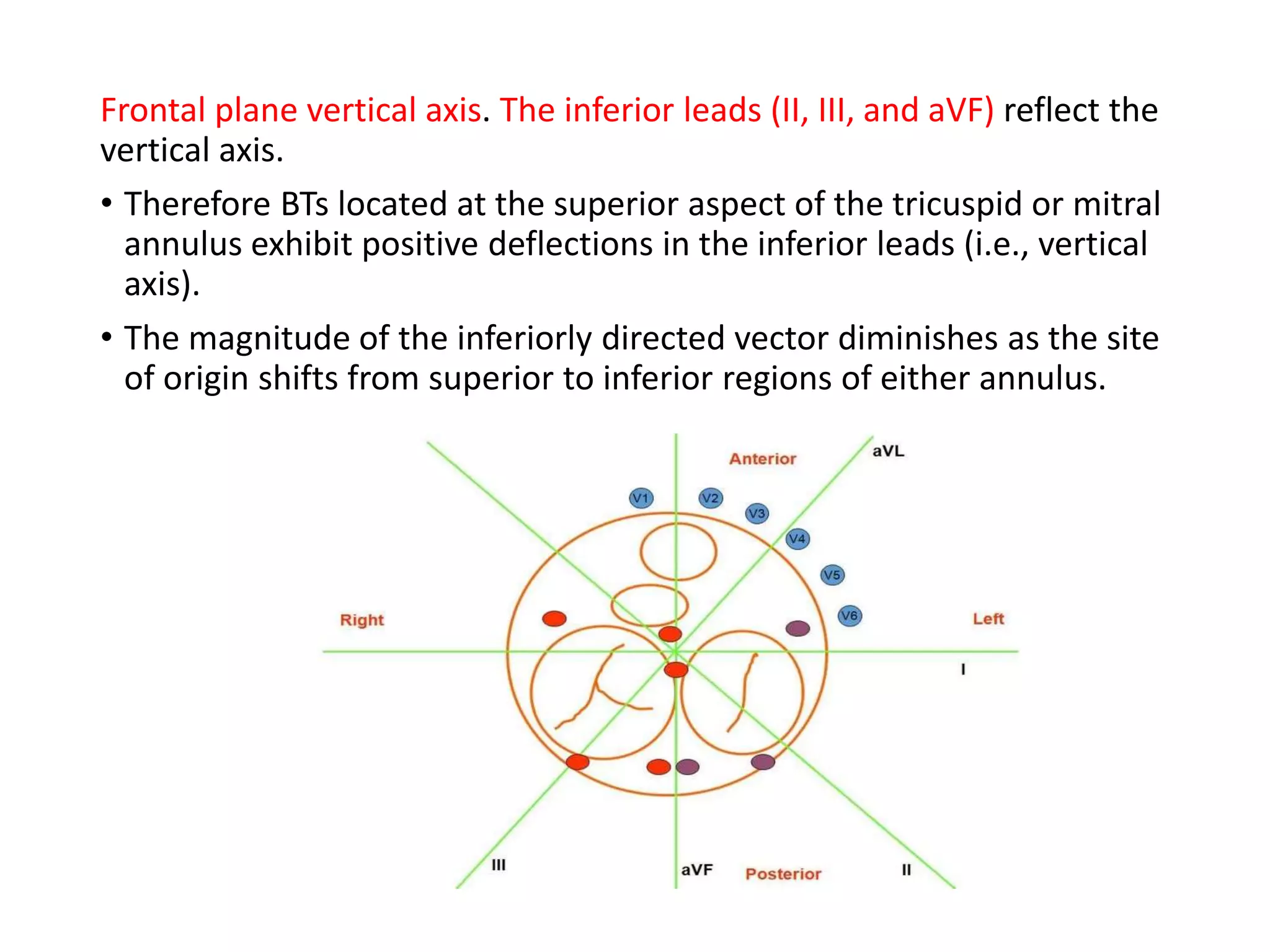
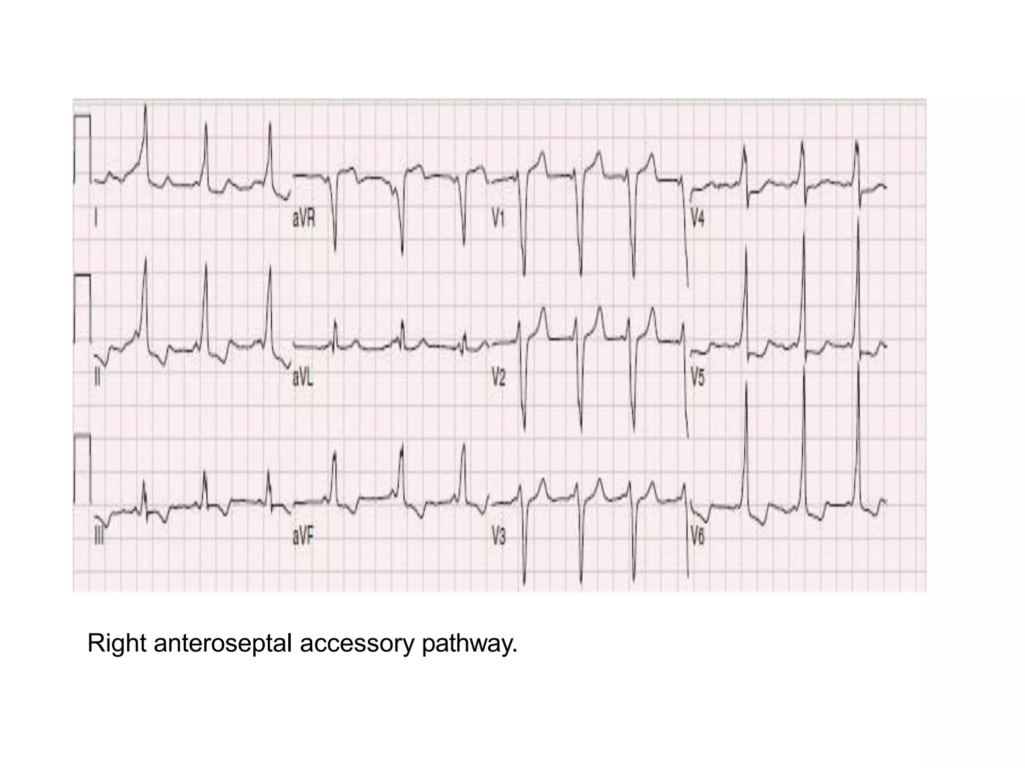
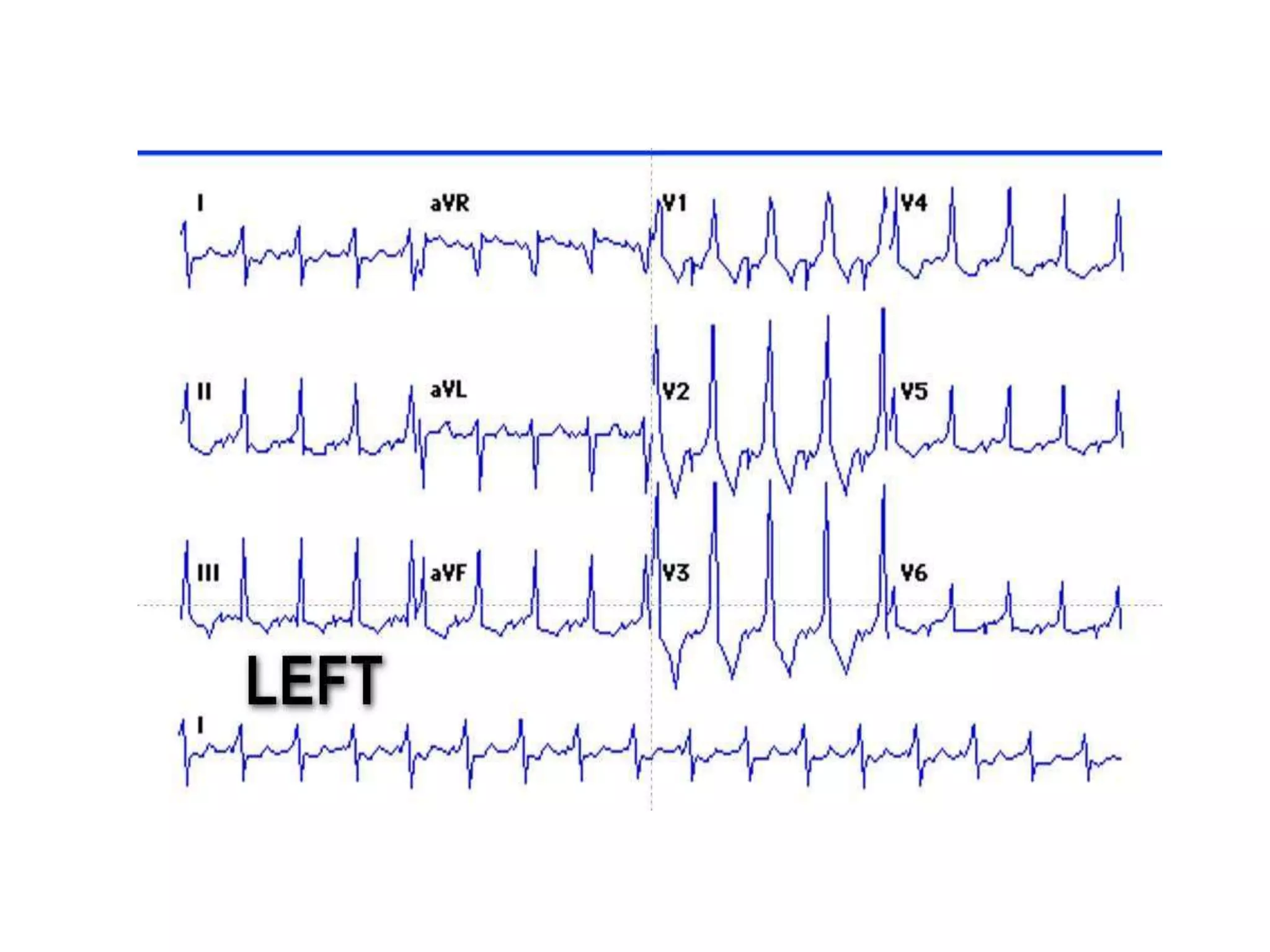
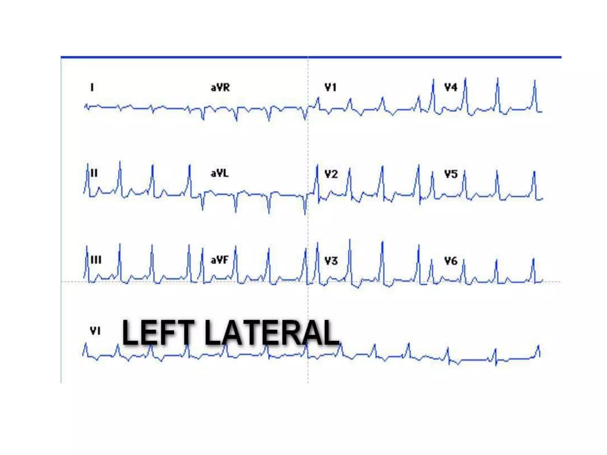
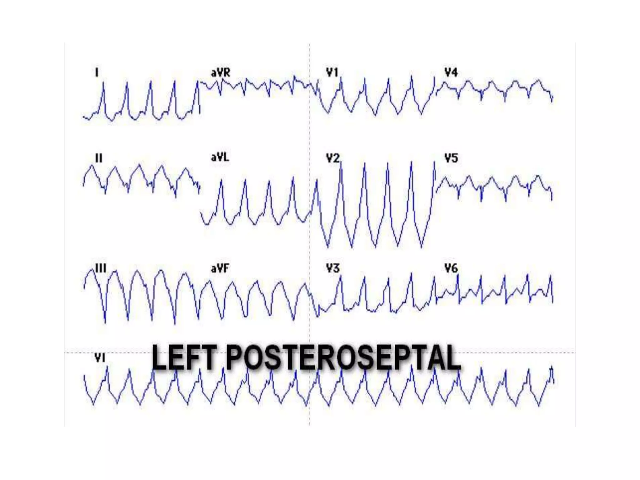
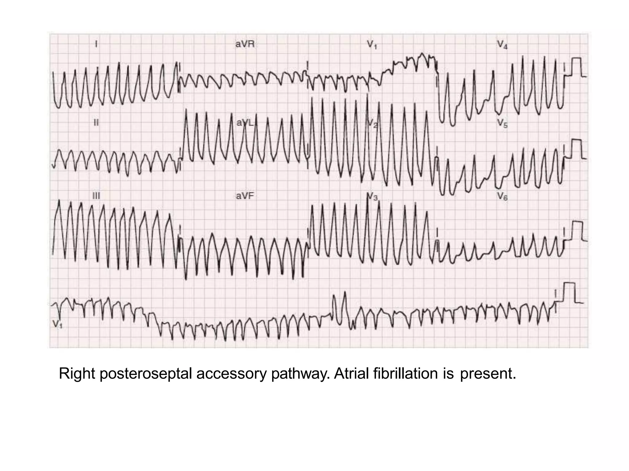
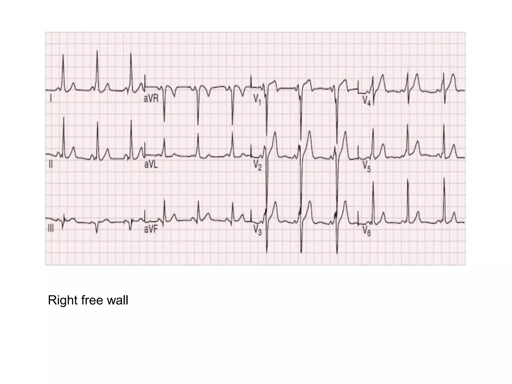
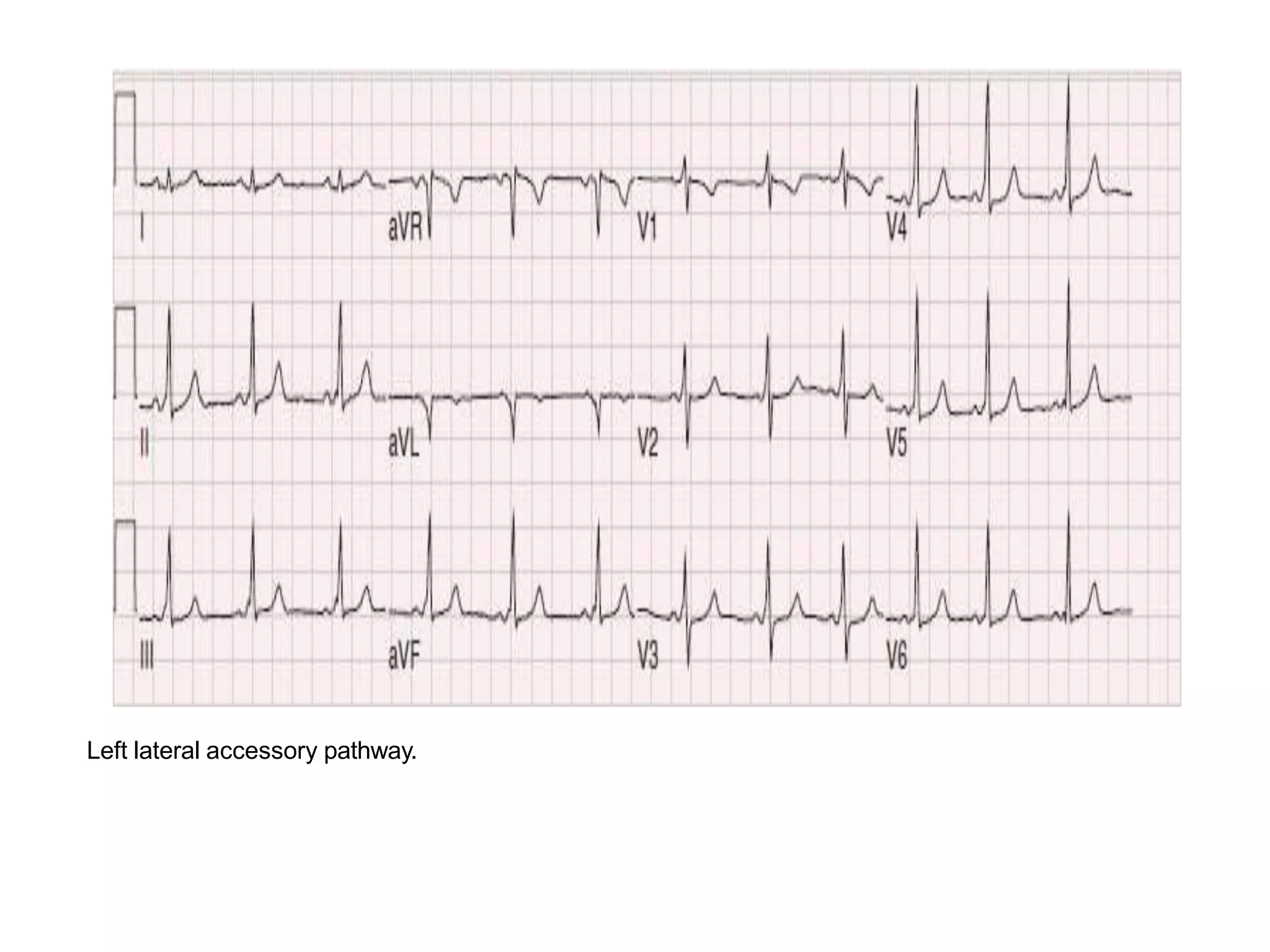
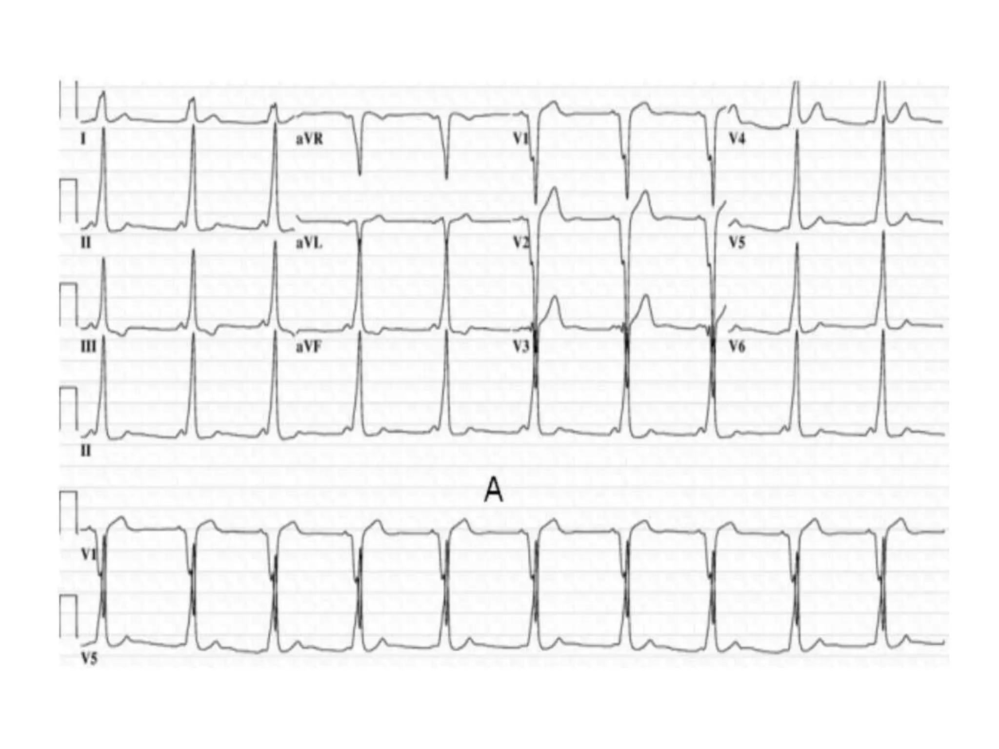
The document discusses localization of accessory pathways using electrocardiography. It describes:
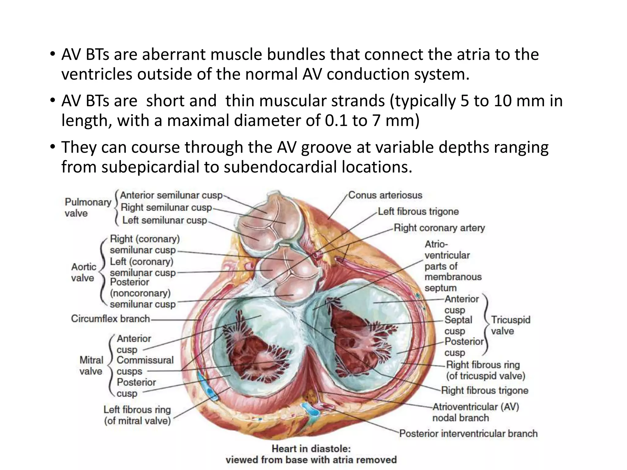
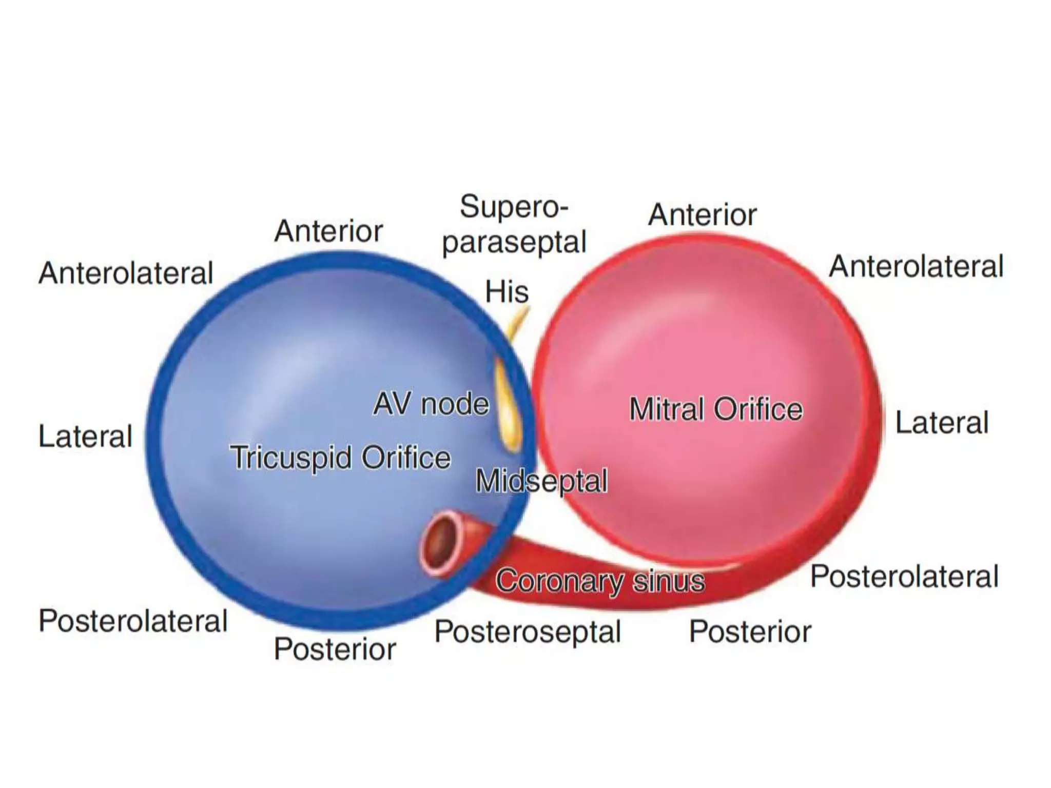
- Accessory pathways are aberrant muscle bundles connecting the atria and ventricles outside the normal conduction system.
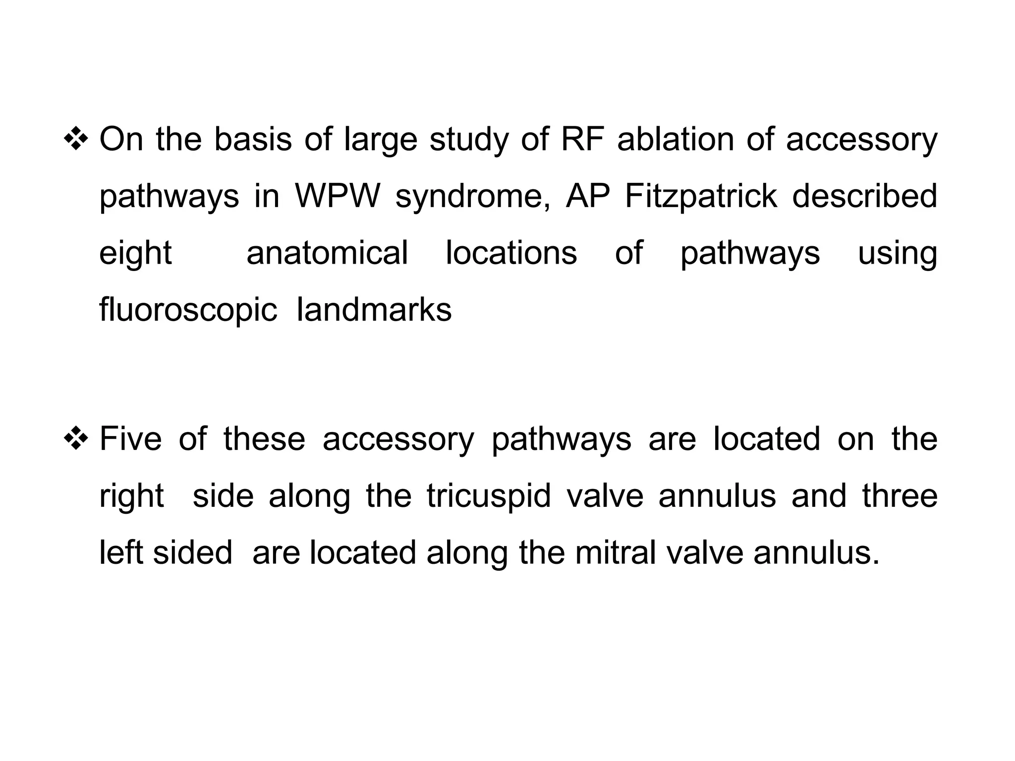
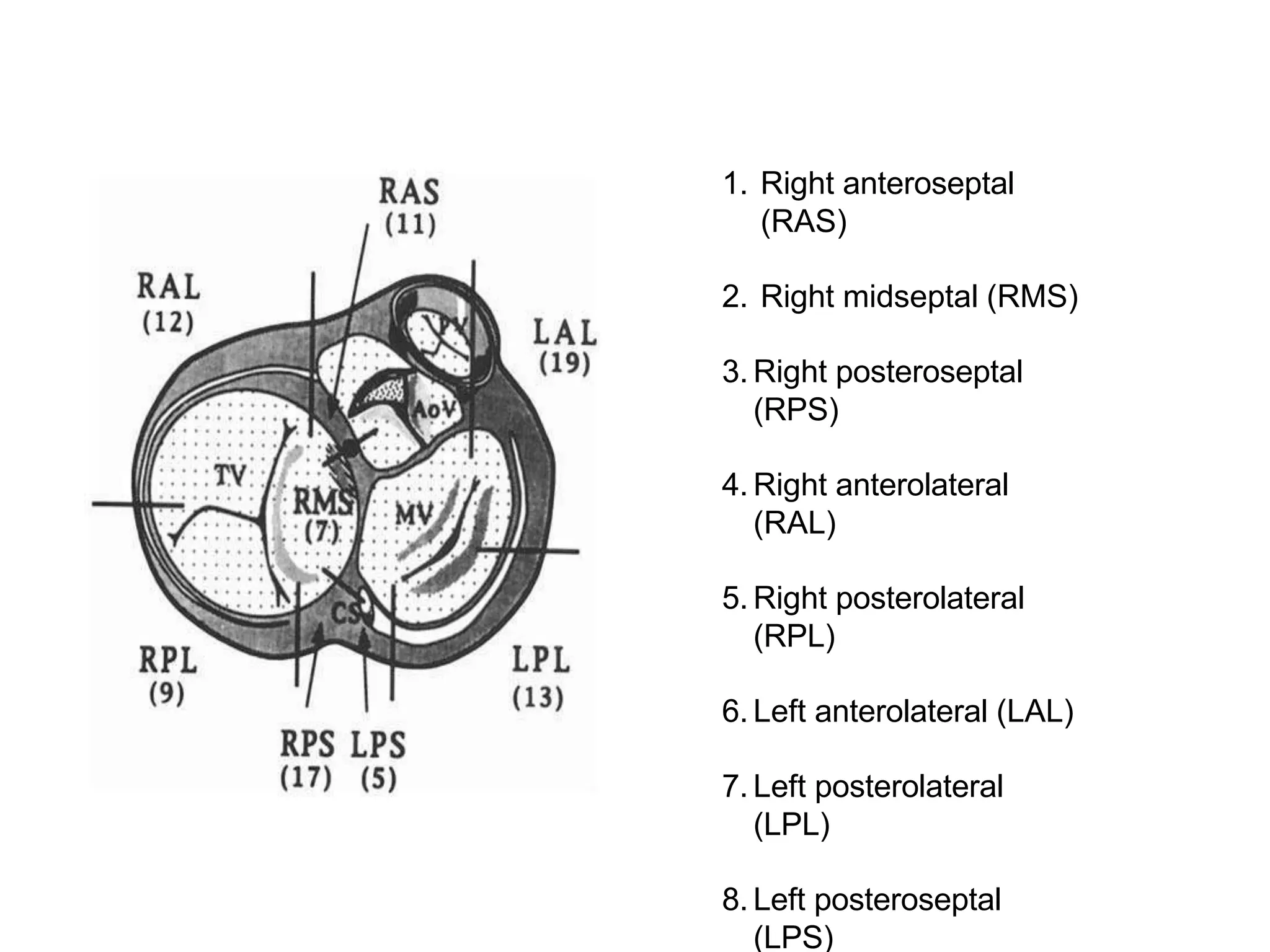
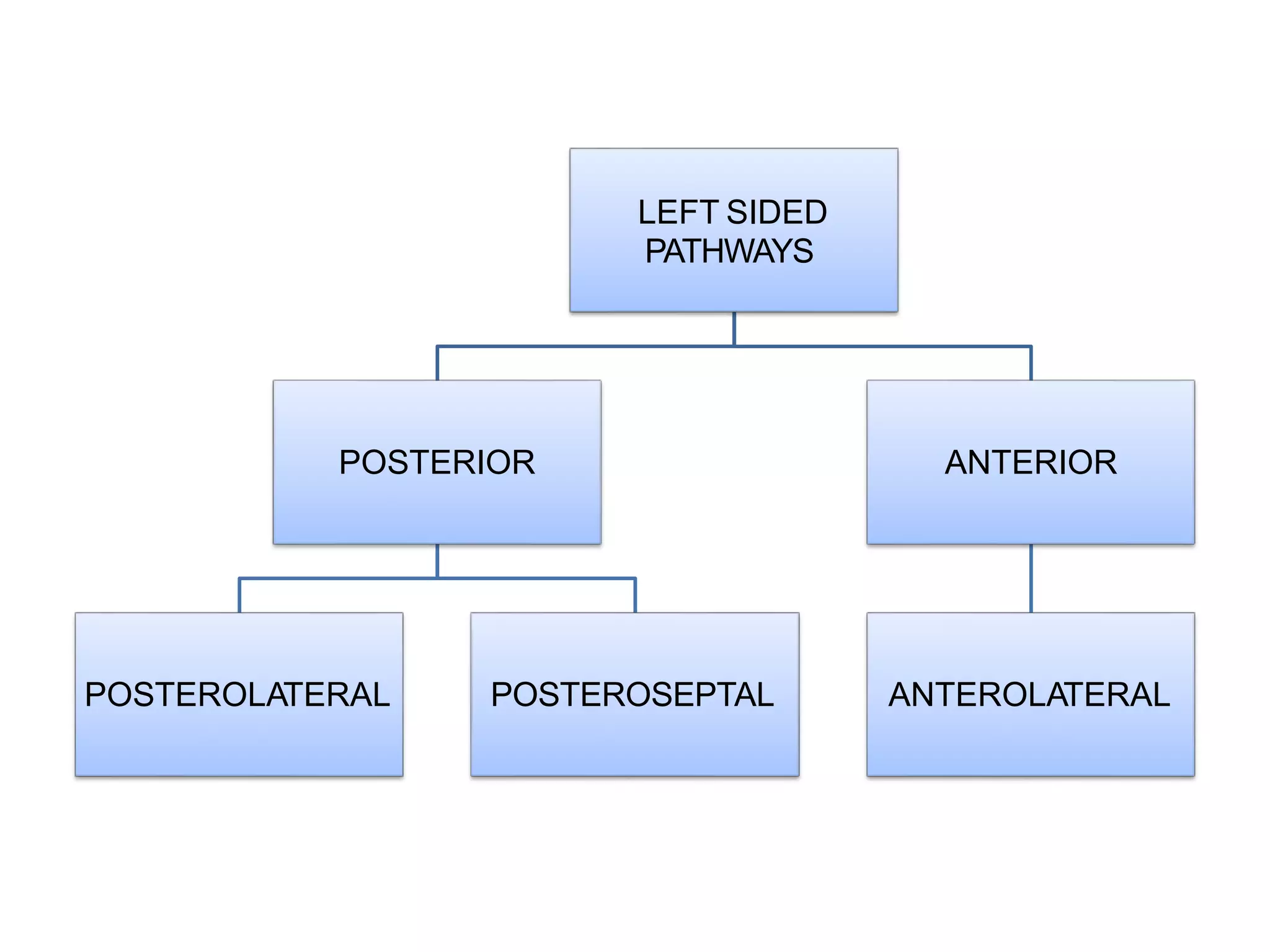
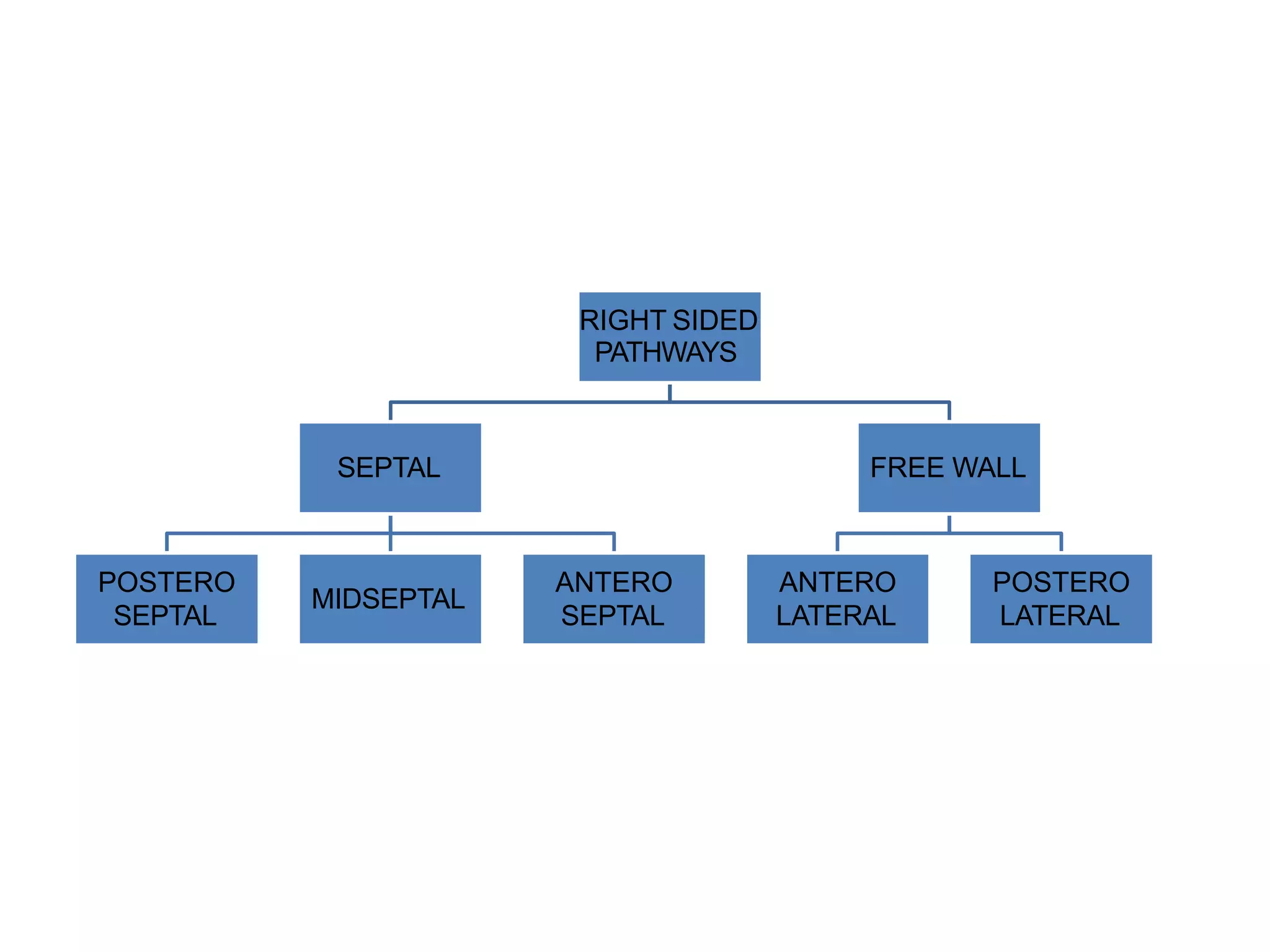
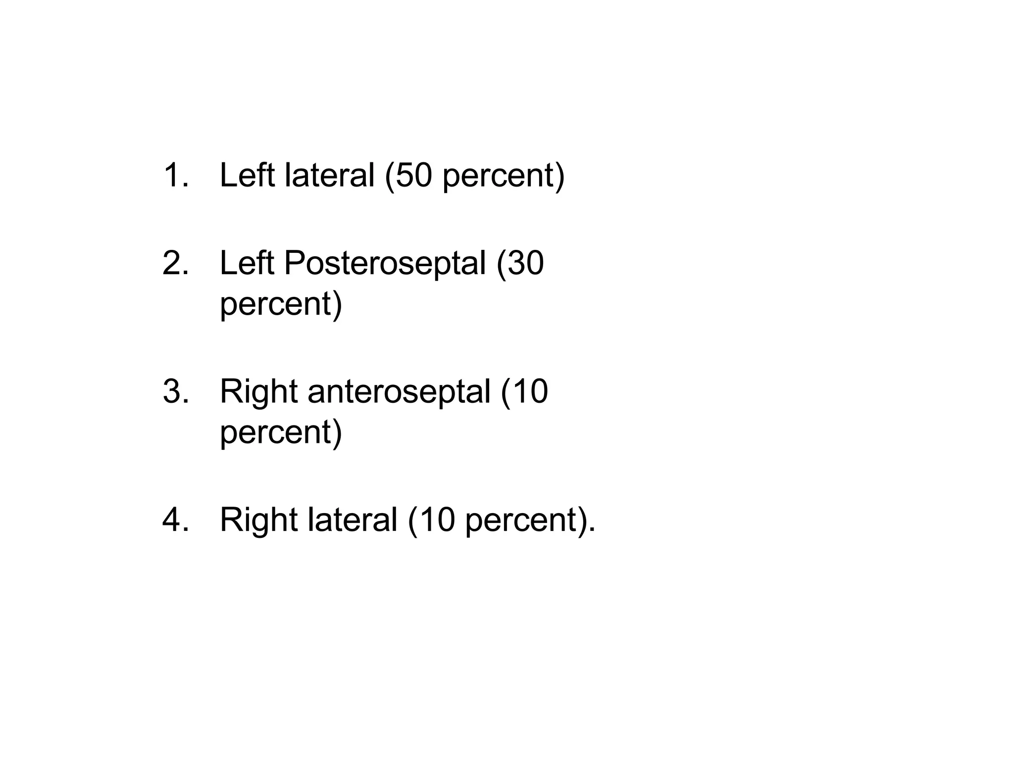
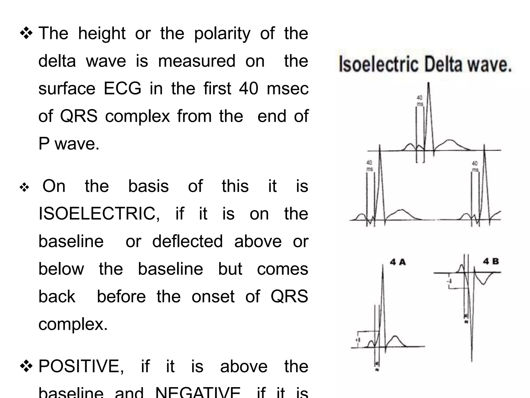
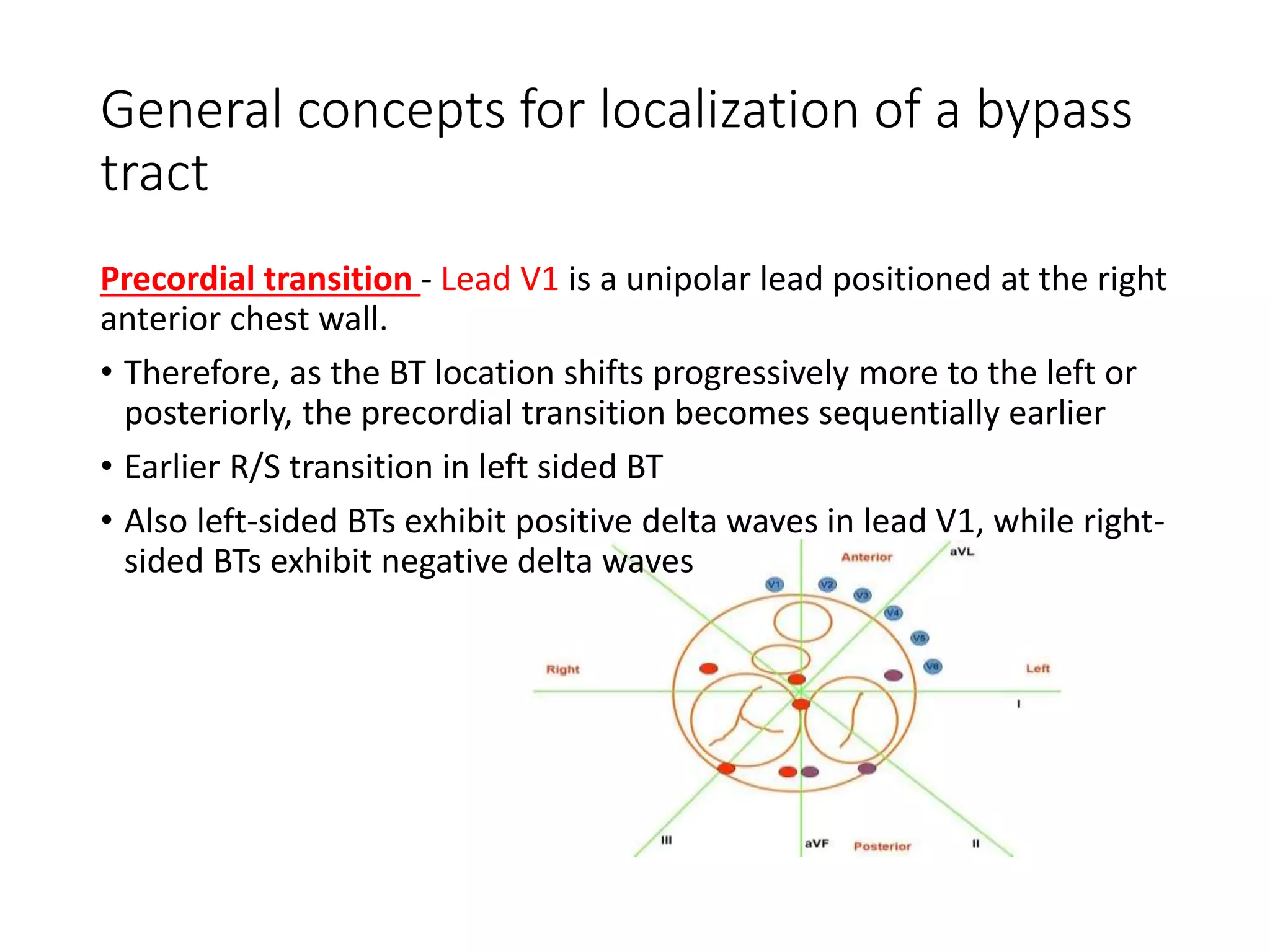
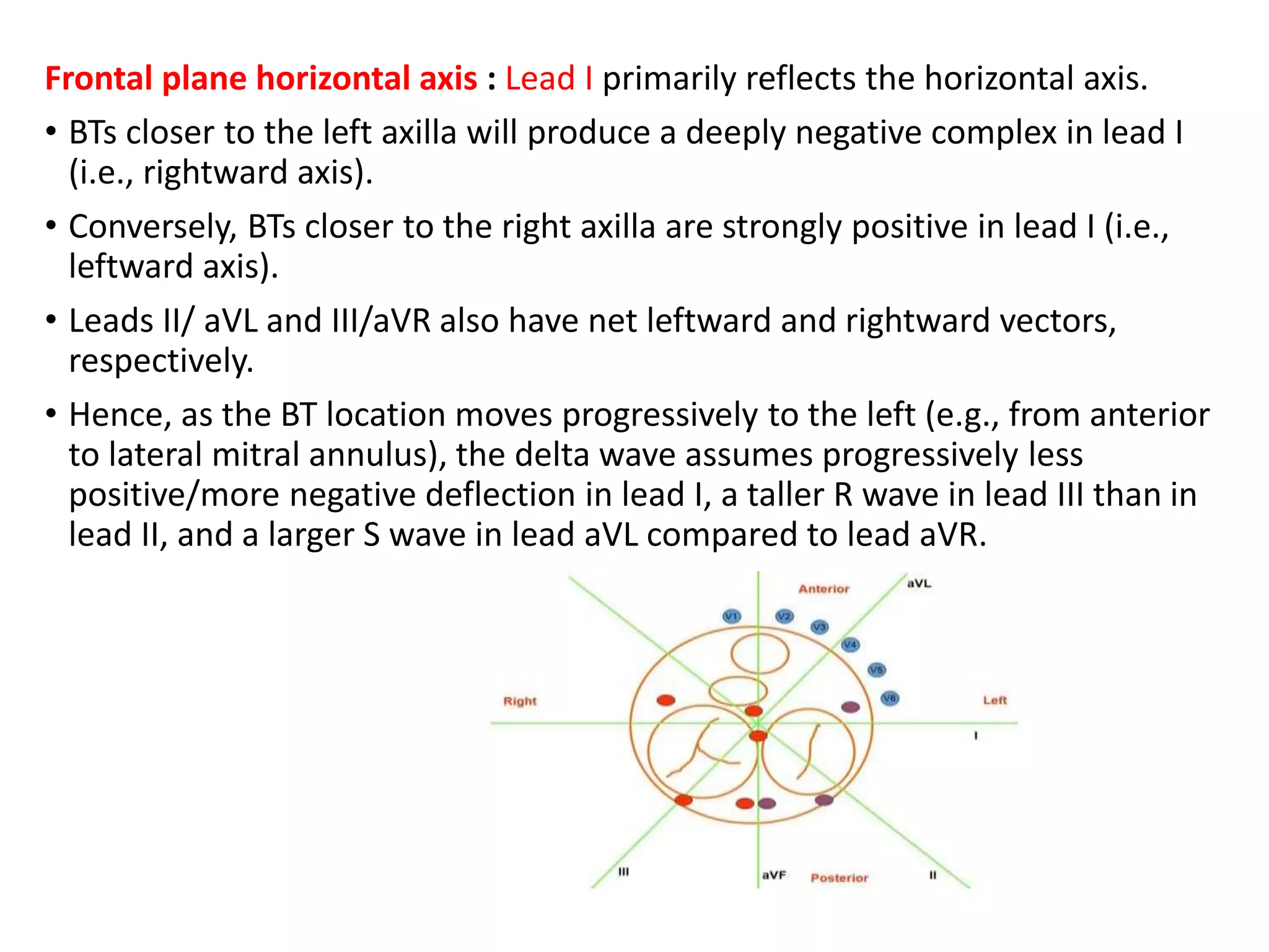
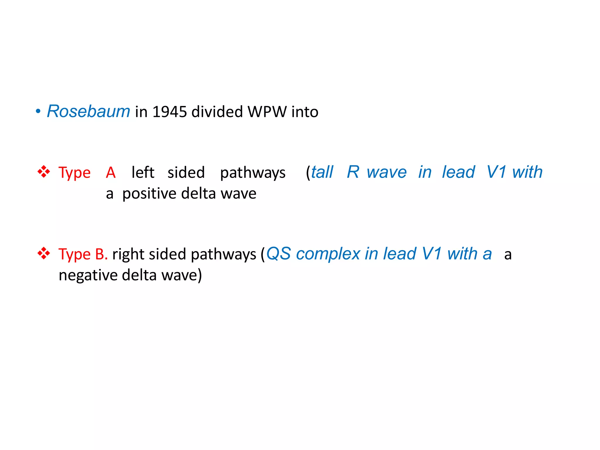
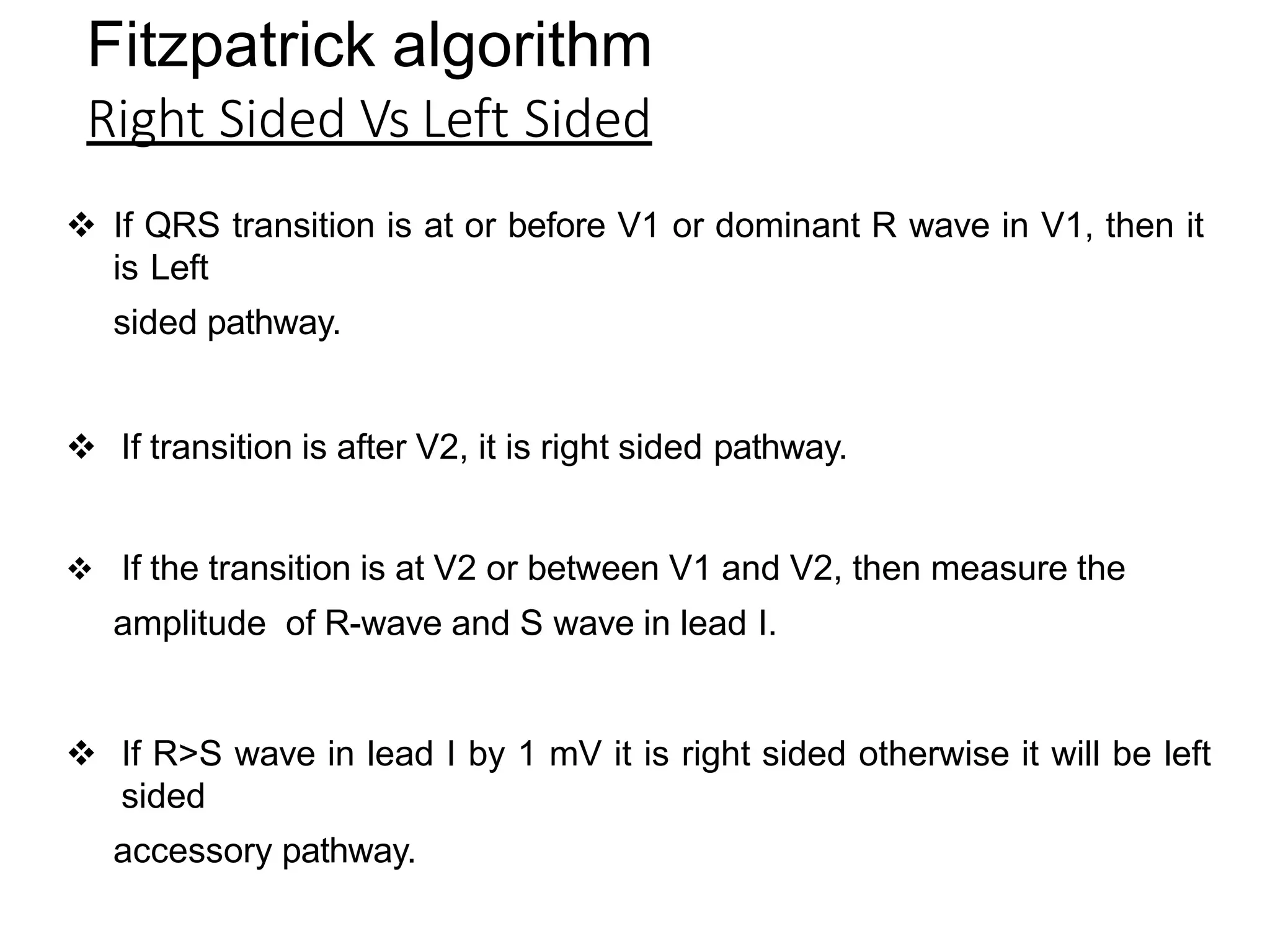
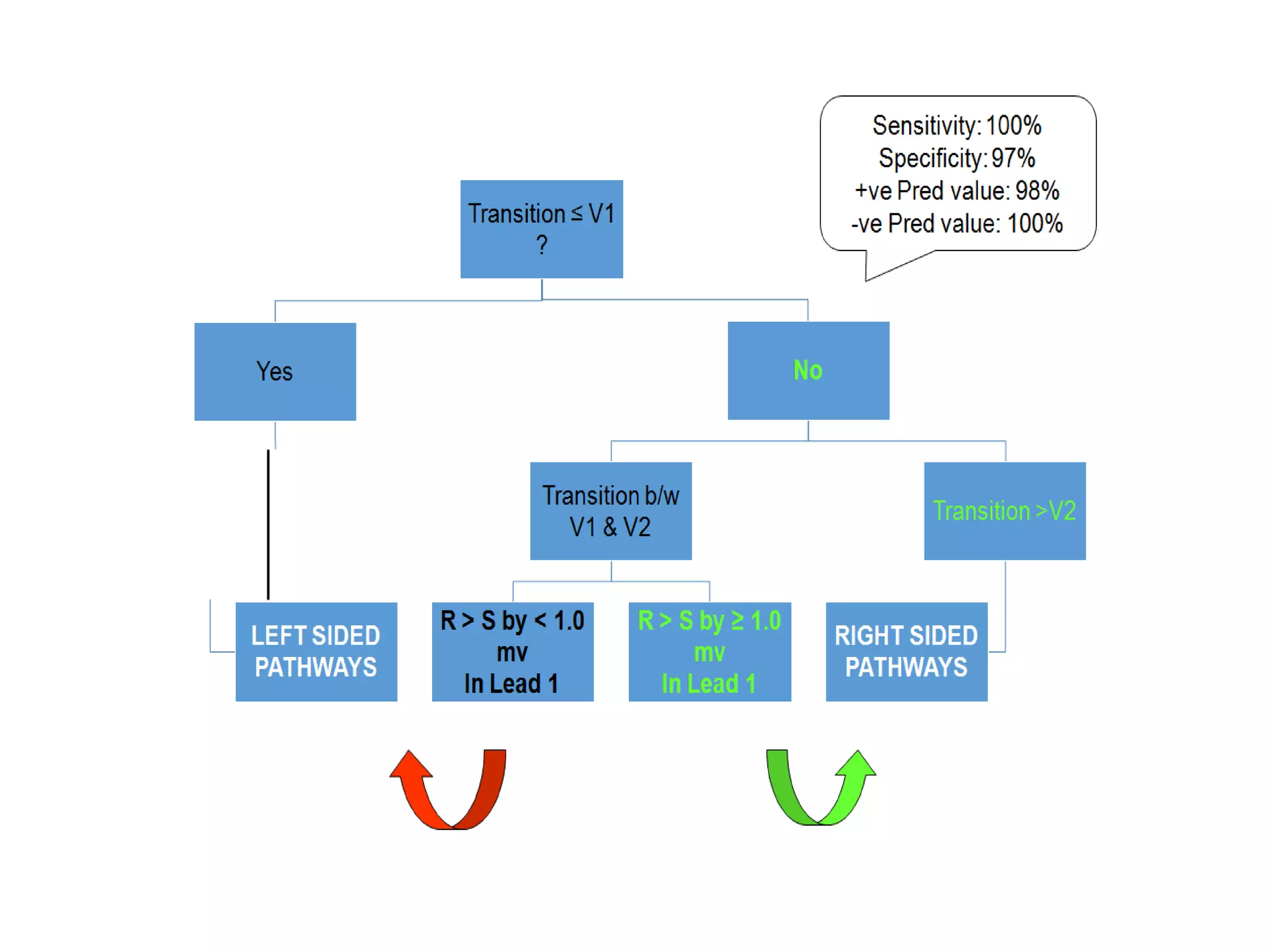
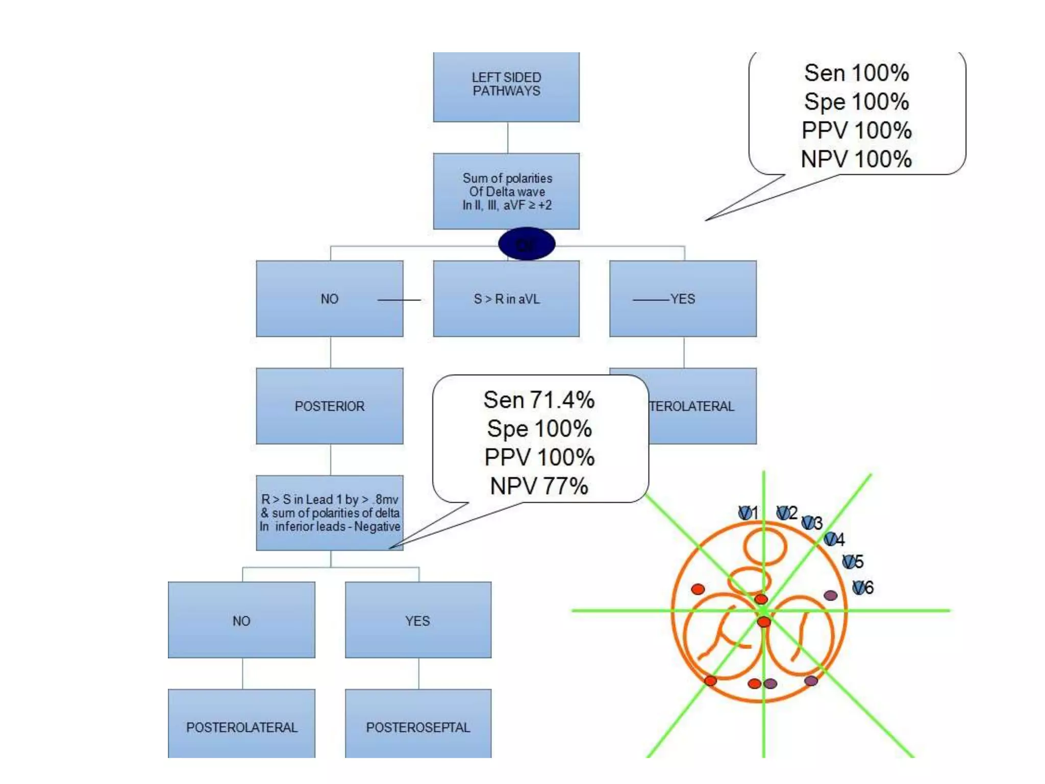
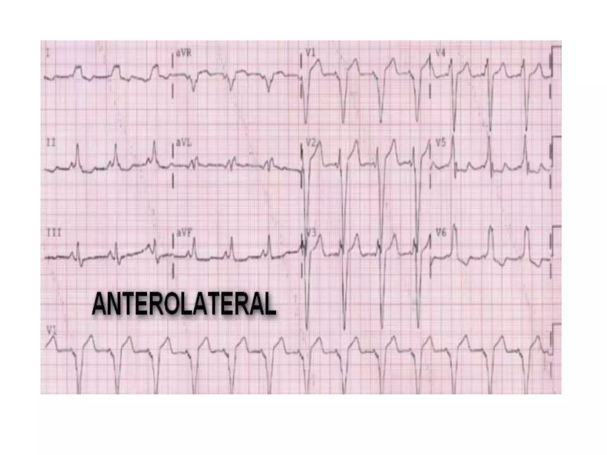
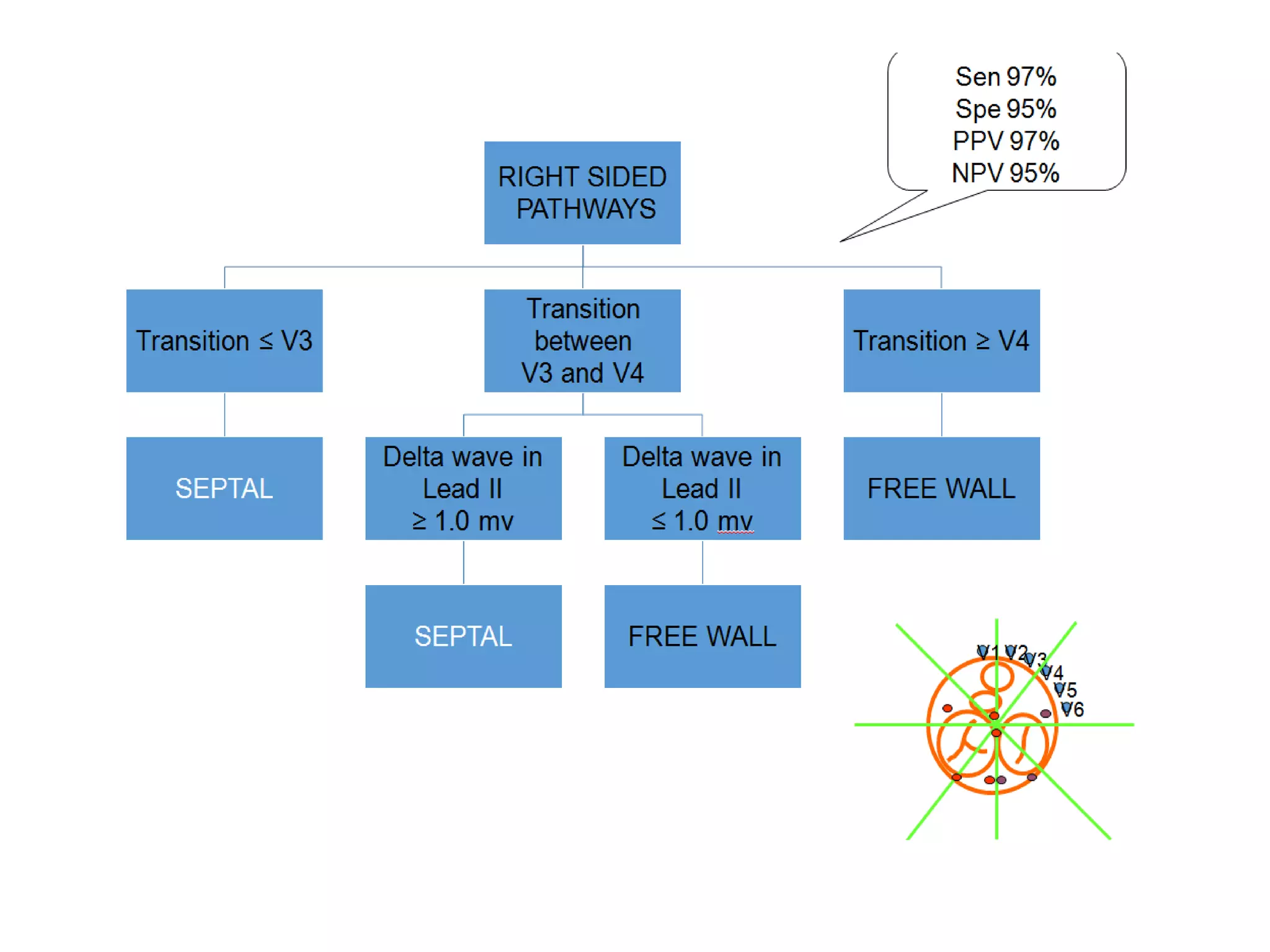
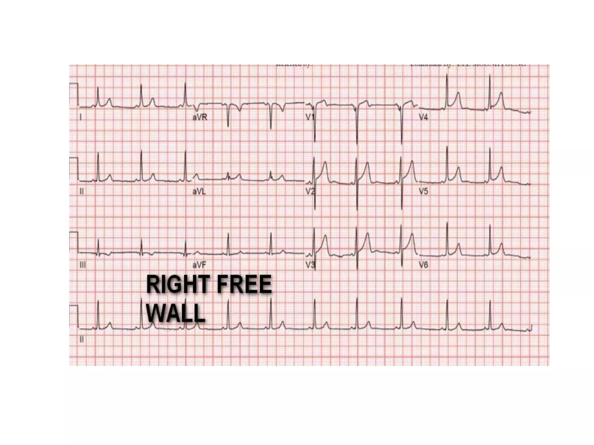
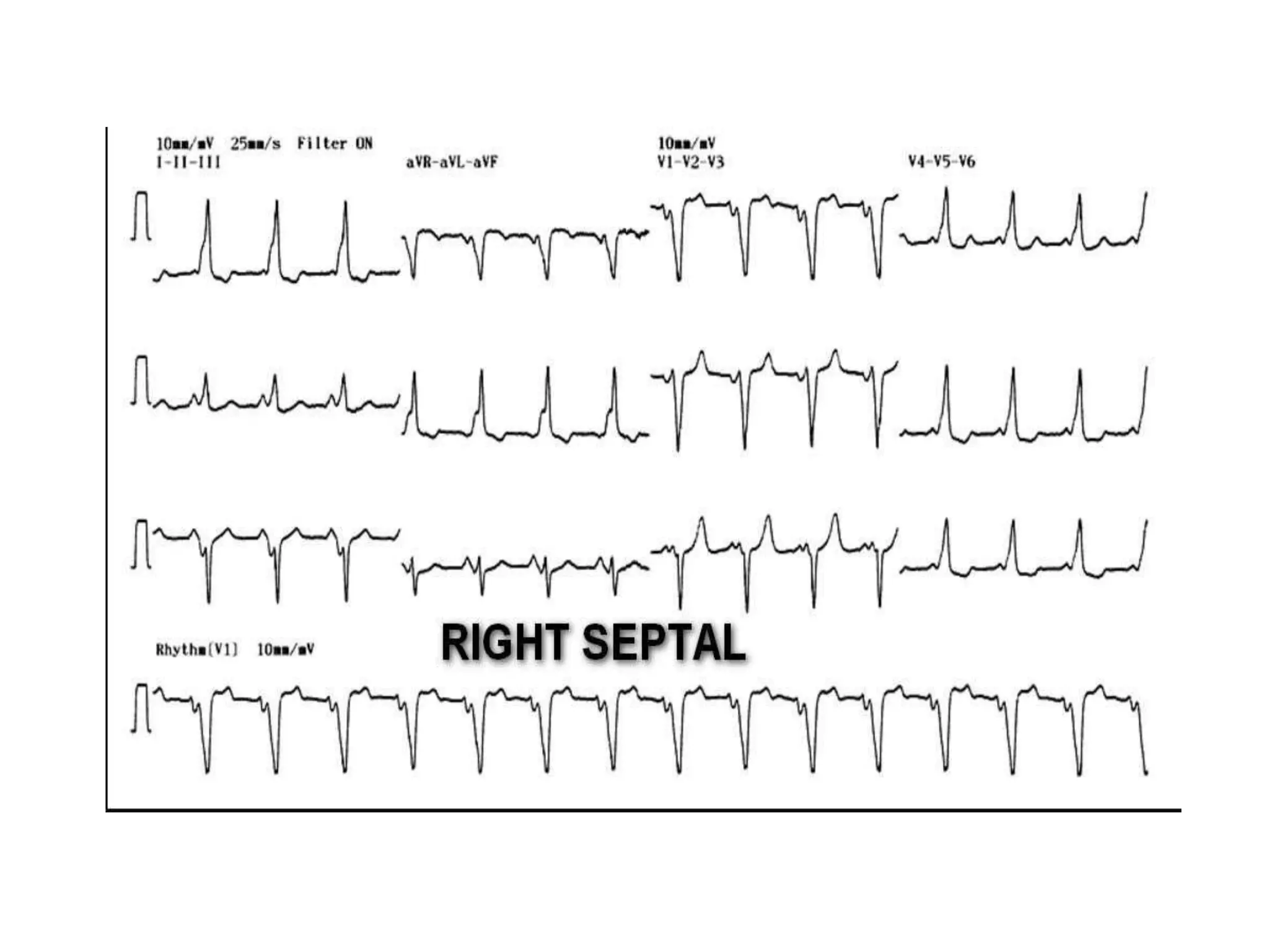
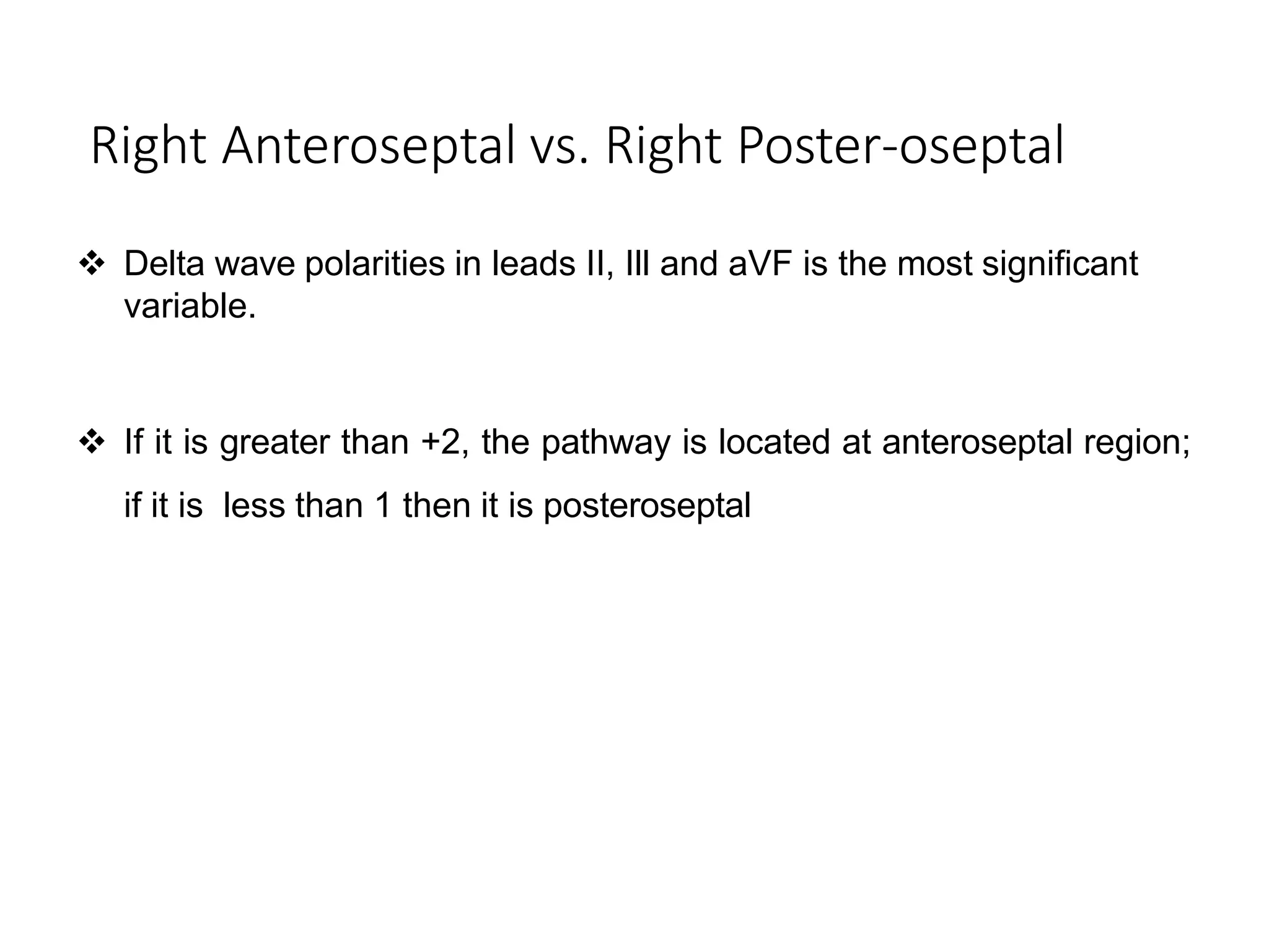
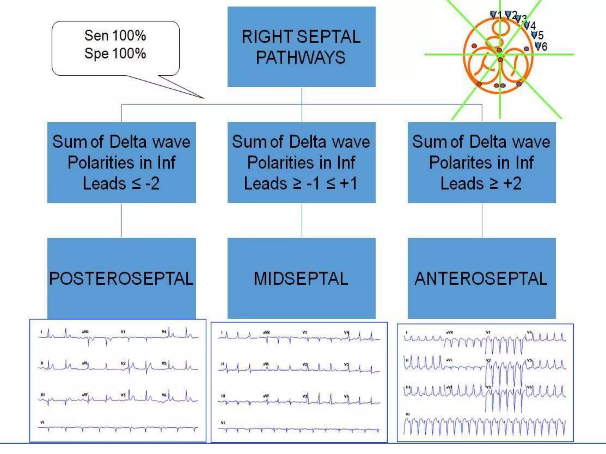
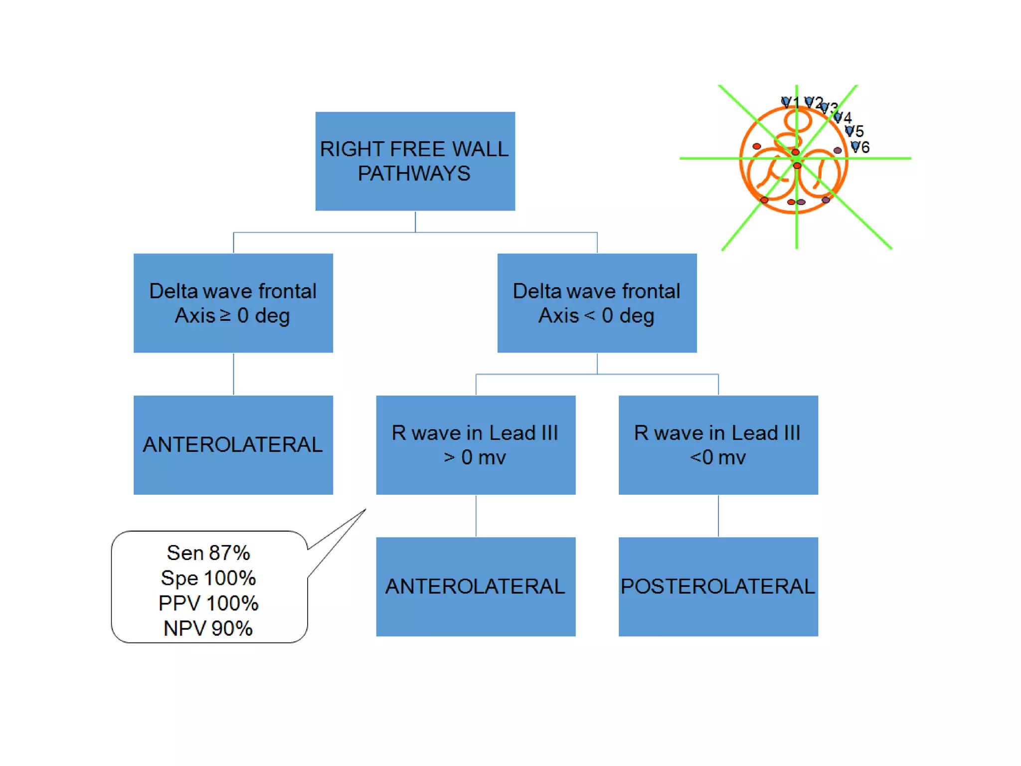
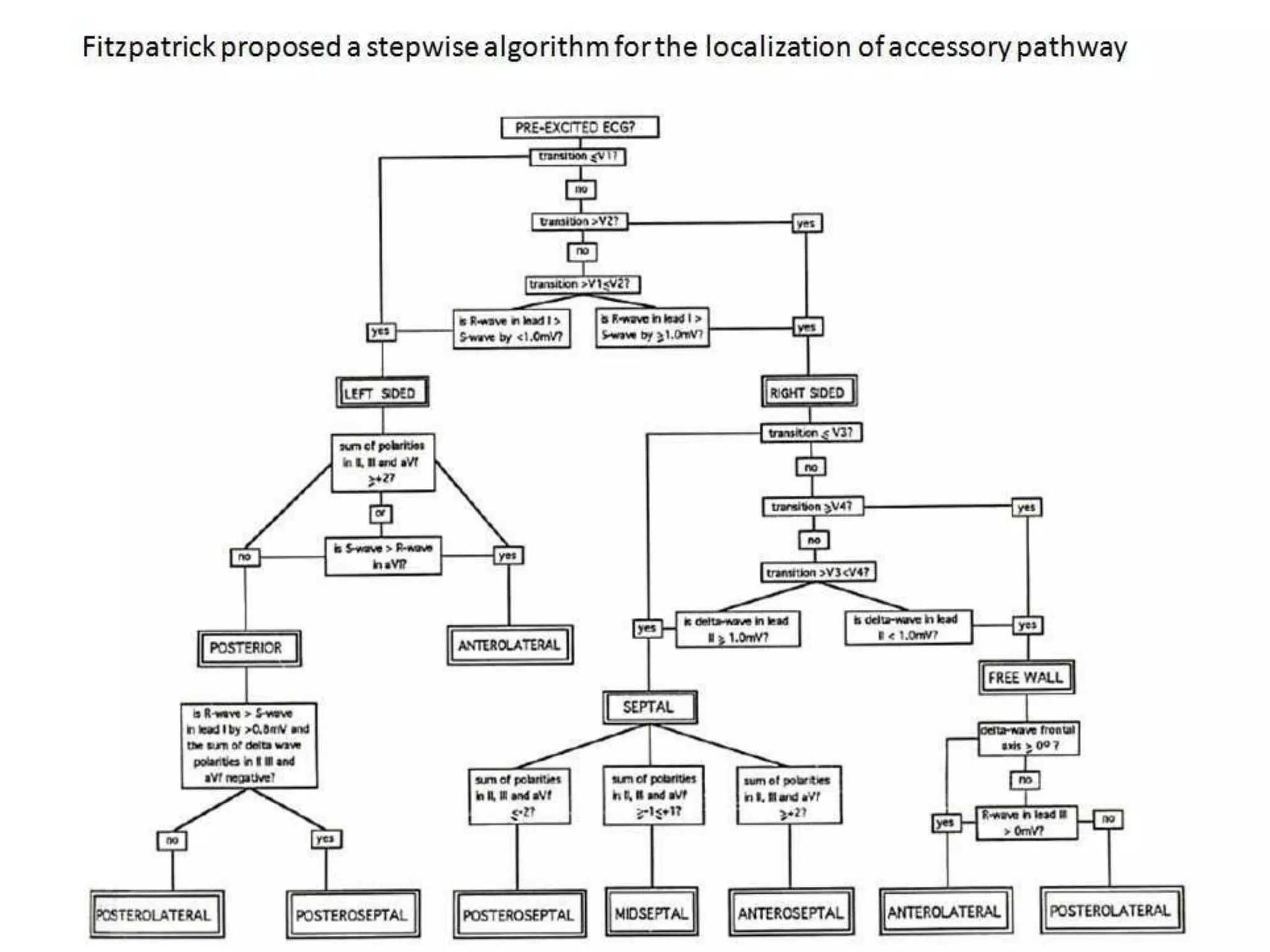
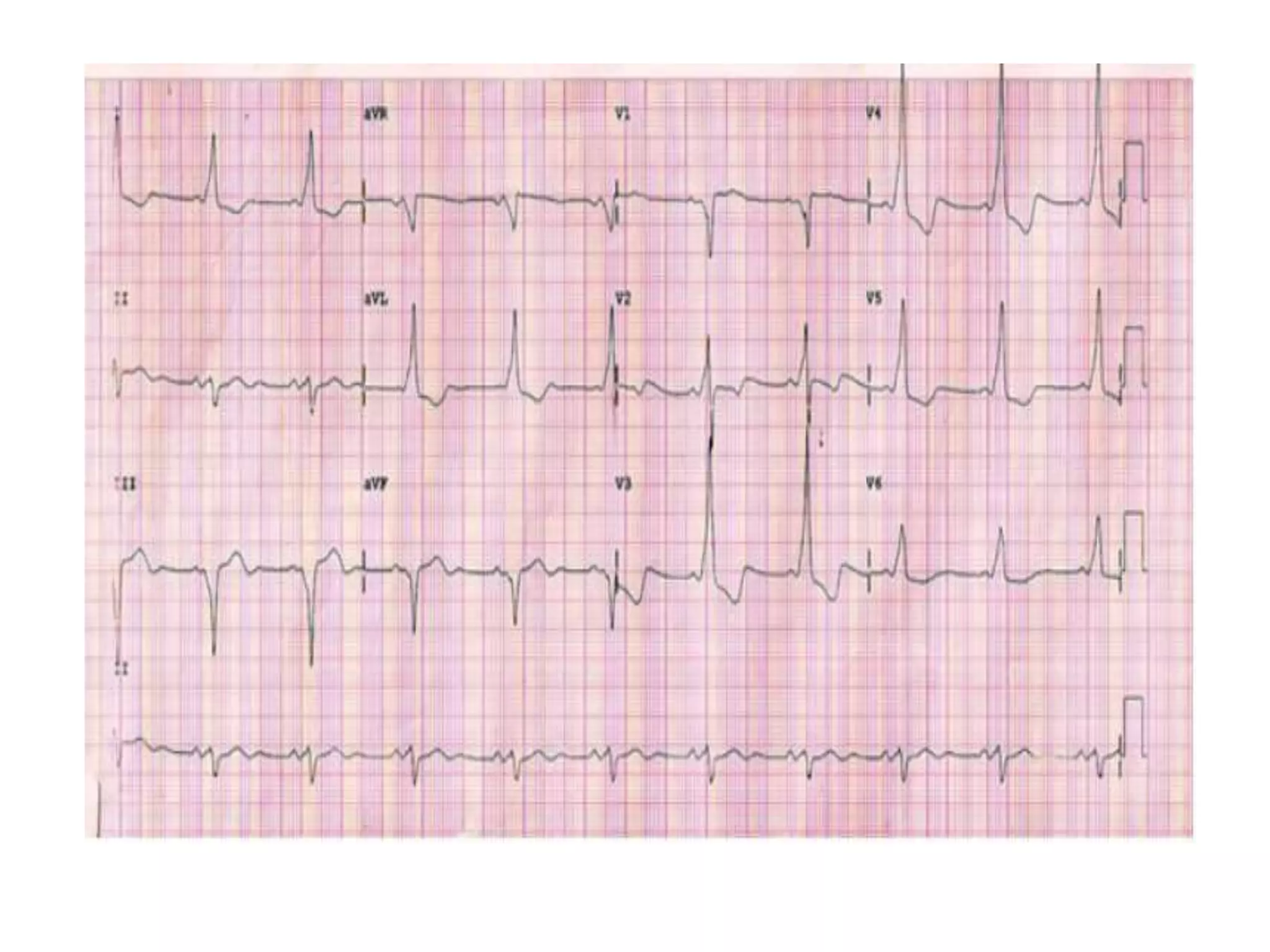
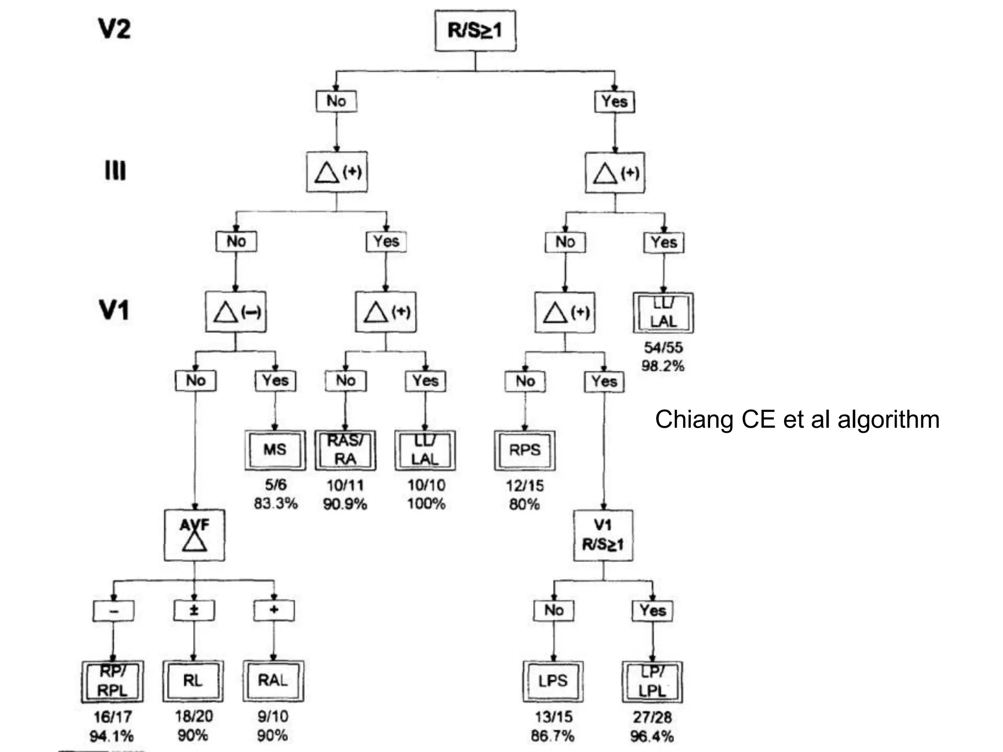
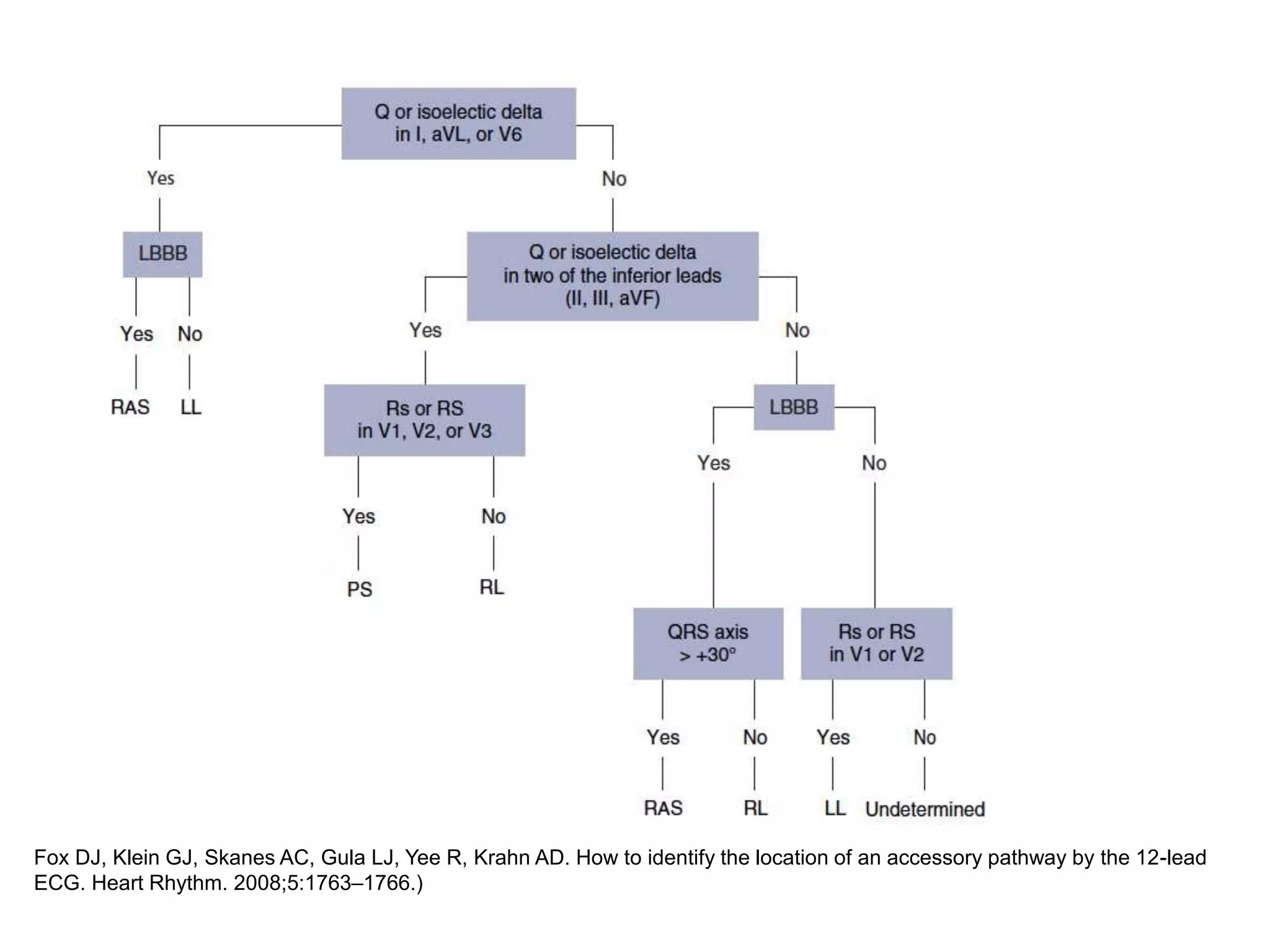
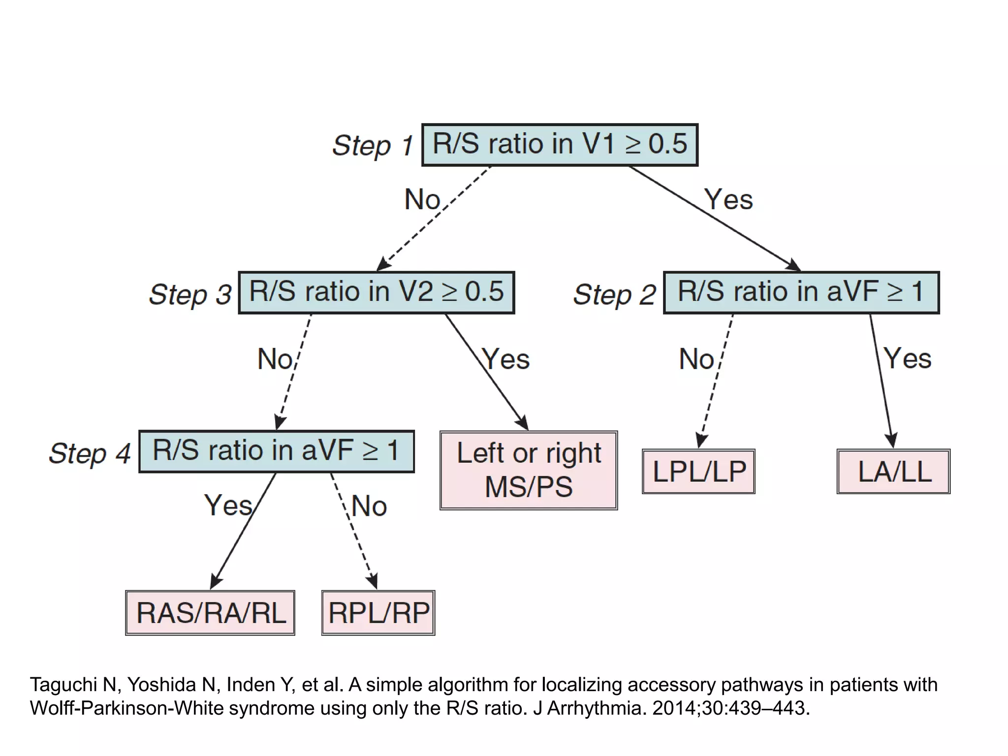
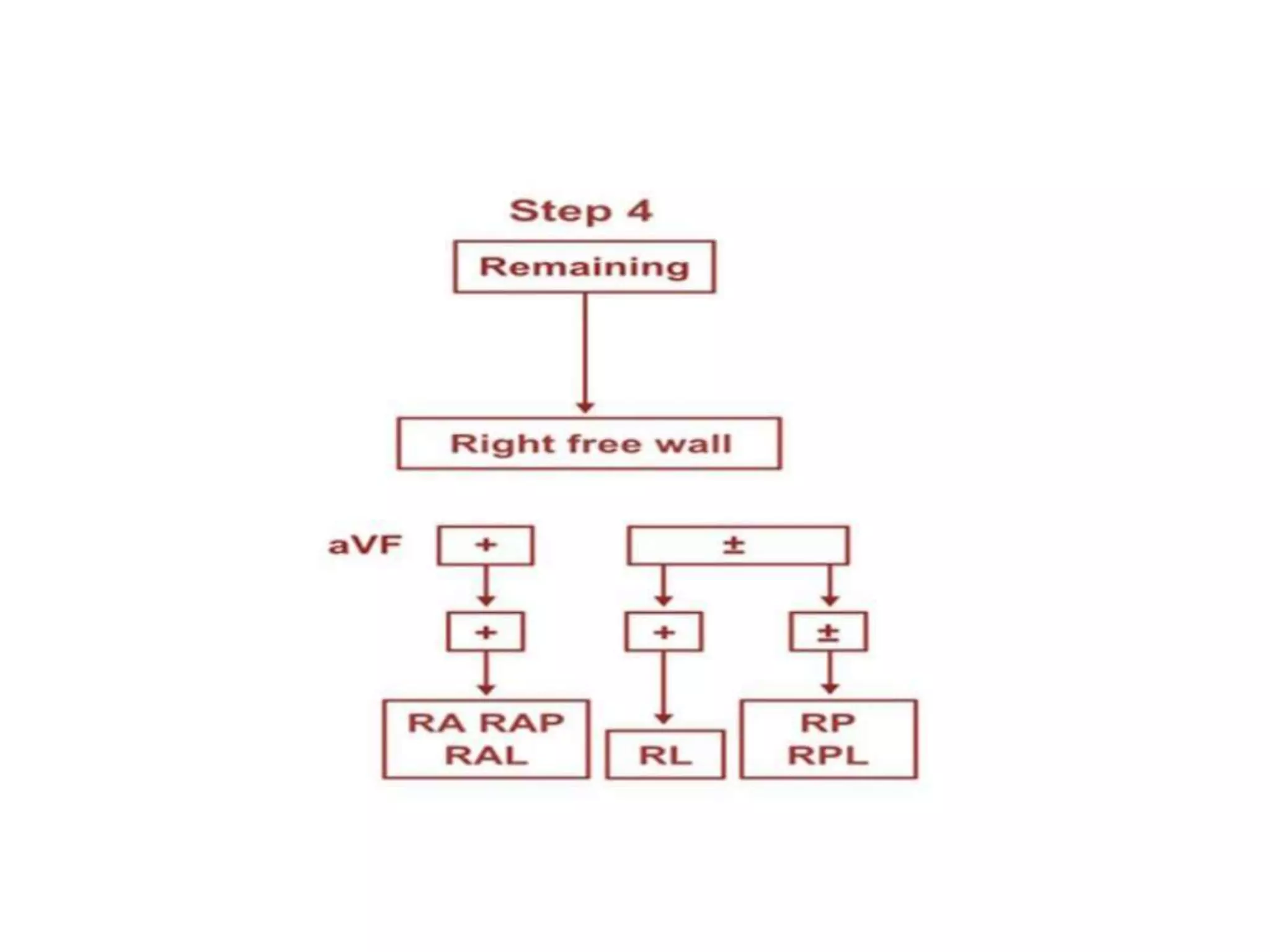
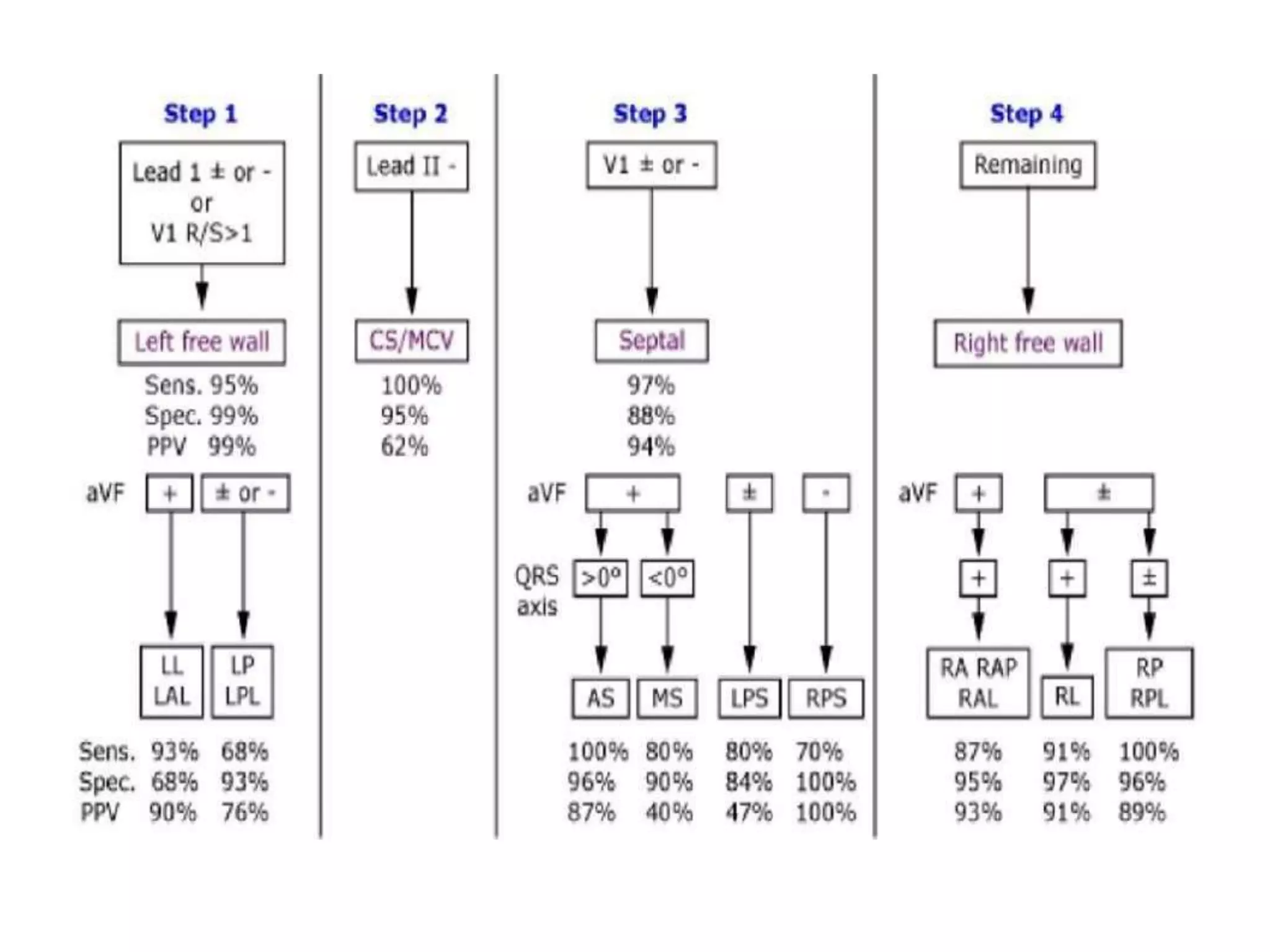
- Algorithms have been developed to predict accessory pathway location based on delta wave morphology and other ECG criteria. Fitzpatrick's algorithm divides pathways into 8 anatomical locations.
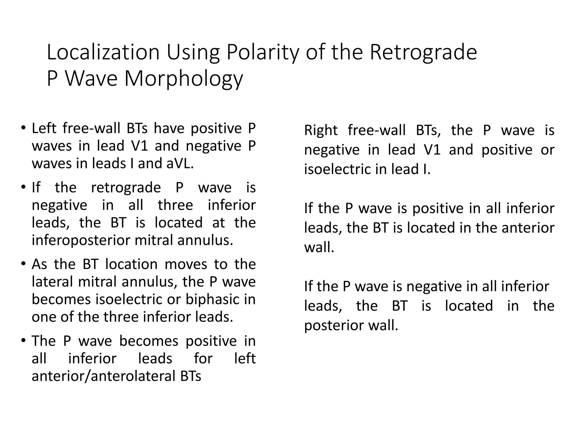
- Factors like precordial transition, delta wave polarity in various leads, and retrograde P wave morphology can provide clues about left vs. right, septal vs. free wall, and anteroseptal vs. posteroseptal localization.
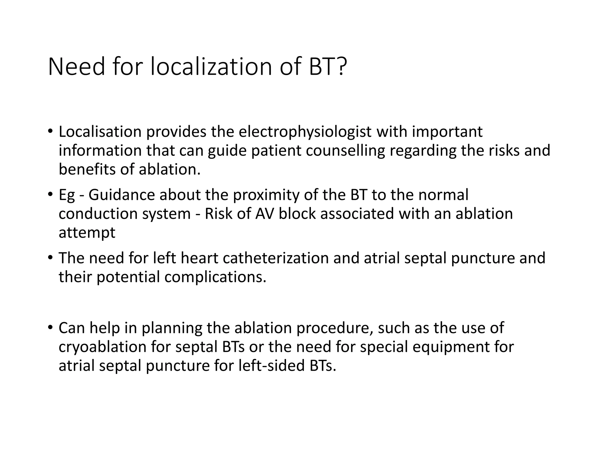
- Accurate localization guides patient counseling on ablation risks and assists planning the procedure
An electrocardiogram (ECG or EKG) records the electrical signal from your heart to check for different heart conditions. Electrodes are placed on your chest to record your heart's electrical signals, which cause your heart to beat. The signals are shown as waves on an attached computer monitor or printer
Some slides are taken from different textbooks of medicine like Davidson, Kumar and Clark and Oxford, and some from other presentations made by respected tutors. I'm barely responsible for compilation of various resources per my interest. These resources are free for use, and I do not claim any copyright. Hoping knowledge remains free for all, forever.
CRISPR-Cas9, a revolutionary gene-editing tool, holds immense potential to reshape medicine, agriculture, and our understanding of life. But like any powerful tool, it comes with ethical considerations.
Unveiling CRISPR: This naturally occurring bacterial defense system (crRNA & Cas9 protein) fights viruses. Scientists repurposed it for precise gene editing (correction, deletion, insertion) by targeting specific DNA sequences.
The Promise: CRISPR offers exciting possibilities:
Gene Therapy: Correcting genetic diseases like cystic fibrosis.
Agriculture: Engineering crops resistant to pests and harsh environments.
Research: Studying gene function to unlock new knowledge.
The Peril: Ethical concerns demand attention:
Off-target Effects: Unintended DNA edits can have unforeseen consequences.
Eugenics: Misusing CRISPR for designer babies raises social and ethical questions.
Equity: High costs could limit access to this potentially life-saving technology.
The Path Forward: Responsible development is crucial:
International Collaboration: Clear guidelines are needed for research and human trials.
Public Education: Open discussions ensure informed decisions about CRISPR.
Prioritize Safety and Ethics: Safety and ethical principles must be paramount.
CRISPR offers a powerful tool for a better future, but responsible development and addressing ethical concerns are essential. By prioritizing safety, fostering open dialogue, and ensuring equitable access, we can harness CRISPR's power for the benefit of all. (2998 characters)
Navigating Challenges: Mental Health, Legislation, and the Prison System in B...Guillermo Rivera
This conference will delve into the intricate intersections between mental health, legal frameworks, and the prison system in Bolivia. It aims to provide a comprehensive overview of the current challenges faced by mental health professionals working within the legislative and correctional landscapes. Topics of discussion will include the prevalence and impact of mental health issues among the incarcerated population, the effectiveness of existing mental health policies and legislation, and potential reforms to enhance the mental health support system within prisons.
One of the most developed cities of India, the city of Chennai is the capital of Tamilnadu and many people from different parts of India come here to earn their bread and butter. Being a metropolitan, the city is filled with towering building and beaches but the sad part as with almost every Indian city
Explore our infographic on 'Essential Metrics for Palliative Care Management' which highlights key performance indicators crucial for enhancing the quality and efficiency of palliative care services.
This visual guide breaks down important metrics across four categories: Patient-Centered Metrics, Care Efficiency Metrics, Quality of Life Metrics, and Staff Metrics. Each section is designed to help healthcare professionals monitor and improve care delivery for patients facing serious illnesses. Understand how to implement these metrics in your palliative care practices for better outcomes and higher satisfaction levels.
How many patients does case series should have In comparison to case reports.pdfpubrica101
Pubrica’s team of researchers and writers create scientific and medical research articles, which may be important resources for authors and practitioners. Pubrica medical writers assist you in creating and revising the introduction by alerting the reader to gaps in the chosen study subject. Our professionals understand the order in which the hypothesis topic is followed by the broad subject, the issue, and the backdrop.
https://pubrica.com/academy/case-study-or-series/how-many-patients-does-case-series-should-have-in-comparison-to-case-reports/
Antibiotic Stewardship by Anushri Srivastava.pptxAnushriSrivastav
Stewardship is the act of taking good care of something.
Antimicrobial stewardship is a coordinated program that promotes the appropriate use of antimicrobials (including antibiotics), improves patient outcomes, reduces microbial resistance, and decreases the spread of infections caused by multidrug-resistant organisms.
WHO launched the Global Antimicrobial Resistance and Use Surveillance System (GLASS) in 2015 to fill knowledge gaps and inform strategies at all levels.
ACCORDING TO apic.org,
Antimicrobial stewardship is a coordinated program that promotes the appropriate use of antimicrobials (including antibiotics), improves patient outcomes, reduces microbial resistance, and decreases the spread of infections caused by multidrug-resistant organisms.
ACCORDING TO pewtrusts.org,
Antibiotic stewardship refers to efforts in doctors’ offices, hospitals, long term care facilities, and other health care settings to ensure that antibiotics are used only when necessary and appropriate
According to WHO,
Antimicrobial stewardship is a systematic approach to educate and support health care professionals to follow evidence-based guidelines for prescribing and administering antimicrobials
In 1996, John McGowan and Dale Gerding first applied the term antimicrobial stewardship, where they suggested a causal association between antimicrobial agent use and resistance. They also focused on the urgency of large-scale controlled trials of antimicrobial-use regulation employing sophisticated epidemiologic methods, molecular typing, and precise resistance mechanism analysis.
Antimicrobial Stewardship(AMS) refers to the optimal selection, dosing, and duration of antimicrobial treatment resulting in the best clinical outcome with minimal side effects to the patients and minimal impact on subsequent resistance.
According to the 2019 report, in the US, more than 2.8 million antibiotic-resistant infections occur each year, and more than 35000 people die. In addition to this, it also mentioned that 223,900 cases of Clostridoides difficile occurred in 2017, of which 12800 people died. The report did not include viruses or parasites
VISION
Being proactive
Supporting optimal animal and human health
Exploring ways to reduce overall use of antimicrobials
Using the drugs that prevent and treat disease by killing microscopic organisms in a responsible way
GOAL
to prevent the generation and spread of antimicrobial resistance (AMR). Doing so will preserve the effectiveness of these drugs in animals and humans for years to come.
being to preserve human and animal health and the effectiveness of antimicrobial medications.
to implement a multidisciplinary approach in assembling a stewardship team to include an infectious disease physician, a clinical pharmacist with infectious diseases training, infection preventionist, and a close collaboration with the staff in the clinical microbiology laboratory
to prevent antimicrobial overuse, misuse and abuse.
to minimize the developme
The dimensions of healthcare quality refer to various attributes or aspects that define the standard of healthcare services. These dimensions are used to evaluate, measure, and improve the quality of care provided to patients. A comprehensive understanding of these dimensions ensures that healthcare systems can address various aspects of patient care effectively and holistically. Dimensions of Healthcare Quality and Performance of care include the following; Appropriateness, Availability, Competence, Continuity, Effectiveness, Efficiency, Efficacy, Prevention, Respect and Care, Safety as well as Timeliness.
CHAPTER 1 SEMESTER V PREVENTIVE-PEDIATRICS.pdfSachin Sharma
This content provides an overview of preventive pediatrics. It defines preventive pediatrics as preventing disease and promoting children's physical, mental, and social well-being to achieve positive health. It discusses antenatal, postnatal, and social preventive pediatrics. It also covers various child health programs like immunization, breastfeeding, ICDS, and the roles of organizations like WHO, UNICEF, and nurses in preventive pediatrics.
2. INTRODUCTION
First fully described by Wolf-Parkinson & White in 1930.
It is the commonest variety of Pre-excitation Syndrome
associated with an accessory AV connection, called Kent
Bundle or Paladino tracts.
The surface ECG is characterized by
1) Shortened PR interval for age (<120 milliseconds in
adults),
2)Prolonged QRS duration for age (>120 milliseconds),
with a slurred slow rising onset of the R wave upstroke
(Delta Wave).
3)Secondary ST and T wave changes which are directed
opposite to the major Delta wave and QRS vector.
3. • AV BTs are aberrant muscle bundles that connect the atria to the
ventricles outside of the normal AV conduction system.
• AV BTs are short and thin muscular strands (typically 5 to 10 mm in
length, with a maximal diameter of 0.1 to 7 mm)
• They can course through the AV groove at variable depths ranging
from subepicardial to subendocardial locations.
4. Direction of conduction
• 60% of AV BTs conduct both anterogradely and retrogradely
• AV BTs with only anterograde conduction are uncommon (less than 5%),
often cross the right AV groove, and frequently possess decremental
conduction properties.
• BTs that conduct only in the retrograde direction occur more frequently -
17% to 37%
• When the BT is capable of anterograde conduction, ventricular preexcitation
is usually evident during normal sinus rhythm (NSR), and the BT is referred to
as manifest.
• BTs capable of retrograde only conduction are referred to as concealed.
Orthodromic AVRT accounts for approximately 95% of AVRTs and 35% of all
paroxysmal SVTs, and 50% of the BTs that participate in orthodromic AVRT are
concealed
6. Need for localization of BT?
• Localisation provides the electrophysiologist with important
information that can guide patient counselling regarding the risks and
benefits of ablation.
• Eg - Guidance about the proximity of the BT to the normal
conduction system - Risk of AV block associated with an ablation
attempt
• The need for left heart catheterization and atrial septal puncture and
their potential complications.
• Can help in planning the ablation procedure, such as the use of
cryoablation for septal BTs or the need for special equipment for
atrial septal puncture for left-sided BTs.
7. On the basis of large study of RF ablation of accessory
pathways in WPW syndrome, AP Fitzpatrick described
eight anatomical locations of pathways using
fluoroscopic landmarks
Five of these accessory pathways are located on the
right side along the tricuspid valve annulus and three
left sided are located along the mitral valve annulus.
8. 1. Right anteroseptal
(RAS)
2. Right midseptal (RMS)
3. Right posteroseptal
(RPS)
4. Right anterolateral
(RAL)
5. Right posterolateral
(RPL)
6. Left anterolateral (LAL)
7. Left posterolateral
(LPL)
8. Left posteroseptal
(LPS)
12. 1. Left lateral (50 percent)
2. Left Posteroseptal (30
percent)
3. Right anteroseptal (10
percent)
4. Right lateral (10 percent).
13. Multiple bypass tracts
• Multiple AV BTs occur in 5% to 10% of patients.
• BTs are defined as multiple when they are separated by more than 1
to 3 cm at the AV junction.
• The most common combination of widely spaced multiple BTs is
posteroseptal and right free-wall BTs.
The incidence of multiple BTs is particularly high in patients with
• Antidromic AVRT (50% to 75%),
• Patients in whom AF resulted in VF
• Ebstein anomaly.
14. Localization of the accessory pathway
• Localization of the accessory
pathway
is
generally
of value
only
when considering catheter ablation.
• The pathway localization or the degree of preexcitation
otherwise does not predict the clinical course.
• Various algorithms (Chern – En Chiang’s, Ftizpatrick’s and
Xie’s algorithms) have been used for predicting accessory
pathway location using different electrographic criteria.
15. Localization Using the Delta Wave
Delta wave morphology reflects the ventricular insertion site of the BT
and, hence, is helpful in approximating the BT location
Retro grade p waves during orthodromic AVRT indicates the location of
atrial connection of the BT and can be used
16. The height or the polarity of the
delta wave is measured on the
surface ECG in the first 40 msec
of QRS complex from the end of
P wave.
On the basis of this it is
ISOELECTRIC, if it is on the
baseline or deflected above or
below the baseline but comes
back before the onset of QRS
complex.
POSITIVE, if it is above the
17. • QRS duration is significantly increased in right sided
than the left sided accessory pathways (145±17, range
100-180 msec; versus 131±15, range 110-164 msec).
18.
19. General concepts for localization of a bypass
tract
Precordial transition - Lead V1 is a unipolar lead positioned at the right
anterior chest wall.
• Therefore, as the BT location shifts progressively more to the left or
posteriorly, the precordial transition becomes sequentially earlier
• Earlier R/S transition in left sided BT
• Also left-sided BTs exhibit positive delta waves in lead V1, while right-
sided BTs exhibit negative delta waves
20. Frontal plane horizontal axis : Lead I primarily reflects the horizontal axis.
• BTs closer to the left axilla will produce a deeply negative complex in lead I
(i.e., rightward axis).
• Conversely, BTs closer to the right axilla are strongly positive in lead I (i.e.,
leftward axis).
• Leads II/ aVL and III/aVR also have net leftward and rightward vectors,
respectively.
• Hence, as the BT location moves progressively to the left (e.g., from anterior
to lateral mitral annulus), the delta wave assumes progressively less
positive/more negative deflection in lead I, a taller R wave in lead III than in
lead II, and a larger S wave in lead aVL compared to lead aVR.
21. Frontal plane vertical axis. The inferior leads (II, III, and aVF) reflect the
vertical axis.
• Therefore BTs located at the superior aspect of the tricuspid or mitral
annulus exhibit positive deflections in the inferior leads (i.e., vertical
axis).
• The magnitude of the inferiorly directed vector diminishes as the site
of origin shifts from superior to inferior regions of either annulus.
22. • Rosebaum in 1945 divided WPW into
Type A left sided pathways (tall R wave in lead V1 with
a positive delta wave
Type B. right sided pathways (QS complex in lead V1 with a a
negative delta wave)
23. Fitzpatrick algorithm
Right Sided Vs Left Sided
If QRS transition is at or before V1 or dominant R wave in V1, then it
is Left
sided pathway.
If transition is after V2, it is right sided pathway.
If the transition is at V2 or between V1 and V2, then measure the
amplitude of R-wave and S wave in lead I.
If R>S wave in lead I by 1 mV it is right sided otherwise it will be left
sided
accessory pathway.
24.
25. Left Anterolateral Vs Left Posterior
The most significant variable is delta wave polarities in the
inferior leads and the ratio of the R wave to the S wave in lead
avL.
Two or more than two positive delta wave in inferior leads or S
wave larger than R wave in aVL indicates anterolateral location
of the accessory pathway.
26. Left Posterolateral vs. Left Posteroseptal
The sum of the inferior delta wave polarities and the
amplitude of R wave to S wave in lead I are the best ECG
variables to differentiate the two sites.
If the R wave is greater than S wave in lead 1 by 0.8 mV
and delta waves are negative in inferior leads, the
pathway is located at left posteroseptal site otherwise it
will be left posterolateral location of the accessory
pathway.
27.
28. Right Septal vs. Right Free Wall
QRS transition is the most significant variable and delta wave
amplitude in lead II can assist to discriminate where the first variable
is equivocal.
QRS transition at or before V3 indicates a septal location, whereas
transition at or after V4 indicates free wall location (Anterolateral or
poserolateral) of the accessory pathways.
If the transition is between V3 & V4 then look for the amplitude of
delta wave in lead II; if it is equal or more than 1 mV then septal
location otherwise lateral location (97% sensitivity & 95%
specificity).
29.
30.
31.
32.
33. Right Anteroseptal vs. Right Poster-oseptal
Delta wave polarities in leads II, Ill and aVF is the most significant
variable.
If it is greater than +2, the pathway is located at anteroseptal region;
if it is less than 1 then it is posteroseptal
48. Fox DJ, Klein GJ, Skanes AC, Gula LJ, Yee R, Krahn AD. How to identify the location of an accessory pathway by the 12-lead
ECG. Heart Rhythm. 2008;5:1763–1766.)
49. Taguchi N, Yoshida N, Inden Y, et al. A simple algorithm for localizing accessory pathways in patients with
Wolff-Parkinson-White syndrome using only the R/S ratio. J Arrhythmia. 2014;30:439–443.
50. Arruda aproach
An algorithm developed by Arruda et.al utilizing the
surface
ECG has an overall sensitivity of 90% and specificity
of 99%.
51. Step 1: If the delta wave in lead 1 is negative or isoelectric or the R
wave is greater in amplitude than the S wave in V1 a left free wall
accessory pathway is present. If these criteria are fulfilled then lead
aVF is examined.
If the delta wave in lead aVF is positive, a left lateral,
anterolateral
accessory pathway is identified.
If the the delta wave in lead aVF is isoelectric or negative
then the
accessory pathway is located at the left posterior or posterolateral
region
52.
53. Step 2: lead II is examined.
• A negative delta wave in lead II identifies the
subepicardial coronary sinus or middle cardiac vein
accessory pathway.
• If the delta wave in lead II is isoelectric or positive,
proceed to step 3.
54.
55. Step 3:lead V1 is examined.
A negative or isoelectric delta wave in lead Vi identifies a septal
accessory pathway.
If these criteria are fulfilled, lead aVf is examined.
If the delta wave is negative, an accessory pathway is identified, which
is
located at the posteroseptal tricuspid annulus .
If the delta wave is isoelectric in lead aVF, the accessory pathway may
be located to either the posteroseptal tricuspid annulus or the
posteroseptal mitral annulus.
56. A positive delta wave in aVF identifies a pathway located within the
anteroseptal/right anterior paraseptal or midseptal tricuspid annulus
regions .
If the delta wave in V1 is positive after having excluded patients with
a left free-wall accessory pathway in Step 1, a right free wall
accessory AV pathway is identified.
Proceed to Step 4.
57.
58. Step 4: In patients with a right free –wall accessory pathways,
examine aVF.
A positive delta wave in aVF identifies a right anterior/
anterolateral
accessory pathway.
If the delta wave in aVF is isoelectric or negative, examine
lead II.
A positive delta wave in lead II identifies a right lateral
accessory pathway , and an isoelectric delta wave in lead II
identifies a right posterior/posterolateral pathway.
59.
60.
61. Localization Using Polarity of the Retrograde
P Wave Morphology
• Left free-wall BTs have positive P
waves in lead V1 and negative P
waves in leads I and aVL.
• If the retrograde P wave is
negative in all three inferior
leads, the BT is located at the
inferoposterior mitral annulus.
• As the BT location moves to the
lateral mitral annulus, the P wave
becomes isoelectric or biphasic in
one of the three inferior leads.
• The P wave becomes positive in
all inferior leads for left
anterior/anterolateral BTs
Right free-wall BTs, the P wave is
negative in lead V1 and positive or
isoelectric in lead I.
If the P wave is positive in all inferior
leads, the BT is located in the anterior
wall.
If the P wave is negative in all inferior
leads, the BT is located in the
posterior wall.
62. Limitations of algorithms
They are inherently limited by
1. Biological variability in anatomy - (e.g., Rotation of the heart within the
thorax),
2. Variable degree of preexcitation and QRS fusion
3. The presence of more than one manifest BT
4. Intrinsic ECG abnormalities (such as prior MI and ventricular hypertrophy)
5. Patient body habitus
6. Technical variability in ECG acquisition and electrode positioning