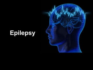
Epilepsy, active and impaired functions of the nervous system
•
2 likes•859 views

Active parts of the nervous system in epilepsy. Functions of the nervous system that are apparent and/or impaired in epilepsy patients.
Report
Share
Report
Share
Download to read offline

Recommended
Neurophysiology of epilepsy
The document discusses the basic neurophysiology of the brain related to epilepsy. It defines key terms like seizure and epileptogenesis. It describes the basic anatomy of the cortex including its layers. It explains concepts like synapses, action potentials, and the cellular mechanisms involved in seizure generation and propagation. These include factors that influence neuronal excitability both at the neuronal and network level. It also discusses the processes of focal seizure initiation, seizure propagation, and epileptogenesis.
Epileptogenesis - Mechanisms and Clinical Implications
Mechanisms of epileptogenesis, epileptogenicity, anatomical substrate, role of GABA, hippocampal circuitry, applications in clinical practice.
Issues in brainmapping...The secrets of conventional EEG
Issues in brainmapping...The secrets of conventional EEG
http://yassermetwally.com
http://yassermetwally.net
Epileptogenesis
Epileptogenesis is the process by which normal brain tissue is transformed into tissue capable of generating spontaneous recurrent seizures. It involves multiple mechanisms including genetic and acquired factors. The hippocampus is particularly susceptible to epileptogenesis due to its circuitry. Status epilepticus animal models are commonly used to study the process. Epileptogenesis occurs in acute, subacute, and chronic stages. Acute changes include increased expression of immediate early genes and post-translational modifications of proteins. Subacute changes involve neuronal death, alterations in neurotrophic factors and inflammation. Chronic changes include mossy fiber sprouting and neurotransmission alterations.
Recognition of abnormal EEG.
This lecture is all about the recognition of an abnormal EEG, its characteristics, its appearance and all about how to differentiate the abnormal activity with normal EEG background.
Epileptogenesis and drugs with mechaism
this includes all the pathe pathways(basic mechanism) of epileptogenesis,causes of drud induced epilepsy and applications
EEG & Evoked potentials
EEG measures the electrical activity of the brain through electrodes placed on the scalp. It can detect different wave patterns associated with different brain states. Evoked potentials involve stimulating a sensory pathway and measuring the electrical response along the pathway. This allows localization of lesions. Somatosensory evoked potentials involve stimulating a peripheral nerve like the median nerve and measuring the response along the pathway to detect spinal cord or brain injuries. Auditory evoked potentials involve measuring the brainstem response to a click stimulus to detect acoustic neuromas or other posterior fossa lesions. Both evoked potentials and EMG monitoring are used during surgery to detect injuries.
Physiopathology of epilepsy
Hyper-excitable neurons lead to excessive excitability in surrounding neurons, causing seizures (hyper-synchronization). This occurs due to an imbalance of excitatory vs inhibitory neurotransmitters - glutamate activation and lowered calcium channel thresholds increase neuronal excitation, while reduced GABA inhibition decreases the inhibitory surround. This disruption of the normal depolarization-afterhyperpolarization cycle in neurons results in a continuous firing state and seizure focus.
Periodic lateralized epileptiform discharges
Periodic Lateralized Epileptiform Discharges (PLEDs) are repeating waveforms seen on EEG that occur at regular intervals and are localized to one hemisphere. They are commonly seen after acute cortical injuries like stroke and infections. PLEDs are classified based on their pattern and presence of additional rhythmic discharges. They indicate unstable brain physiology resulting from seizures, injury or metabolic disturbances. While not strictly ictal, PLEDs are associated with increased risk of clinical seizures. Prognosis depends on the underlying cause, with acute severe strokes having the worst outcomes. Treatment involves antiepileptic drugs mainly if clinical seizures are present.
Abnormal EEG patterns
1. The document defines abnormal EEG patterns (AEPs) and describes two main types: non-epileptiform and epileptiform.
2. Non-epileptiform patterns include slow waves, which can be focal or diffuse, and amplitude/frequency asymmetry. Epileptiform patterns consist of spike and sharp waves that can be focal or generalized.
3. Specific AEPs are described such as benign rolandic epilepsy, 3/sec spike-wave, periodic lateralized epileptiform discharges, and others associated with various neurological conditions.
Epilepogenesis
Epileptogenesis is the process by which the brain becomes epileptic. It occurs in three phases - an initial injury, a latent period of neuronal changes, and chronic epilepsy. During the latent period, various molecular pathways are dysregulated, including mTOR and REST, and neuronal circuits like the dentate gate and temporoammonic pathway are altered. These changes involve loss of inhibitory interneurons and abnormal sprouting, leading to recurrent seizures. Understanding epileptogenesis may help develop new treatments targeting the latent period to prevent epilepsy.
Encephalopathy with EEG based Grading
This document discusses encephalopathy and summarizes key points about its causes, features on EEG, and types. Encephalopathy is defined as altered brain function resulting in impaired consciousness. It can be caused by metabolic, toxic, infectious, hepatic or other issues. On EEG, encephalopathy typically shows generalized slowing and reduced reactivity. Specific patterns like triphasic waves indicate metabolic encephalopathy. The document outlines different types of encephalopathy and their associated EEG findings to help evaluate severity and guide treatment.
Periodic Lateralizing Epileptiform Discharges
Periodic Lateralized Epileptiform Discharges (PLEDs) are a pattern seen on EEG consisting of unilateral focal spikes that occur periodically every 1-2 seconds. PLEDs indicate acute or subacute focal pathology in the brain and are associated with an 80% risk of seizures. Common causes include strokes, tumors, infections like herpes simplex encephalitis. PLEDs usually last from days to weeks and resolve once the underlying condition is treated.
EEG & Epilepsy syndromes report [Autosaved]
This document provides information about electroencephalography (EEG) and how it is used to diagnose and classify epilepsy. It discusses how EEG detects abnormal brain wave patterns associated with seizures. Specific EEG patterns can help distinguish between different types of partial and generalized seizures. Partial seizures are localized to one area of the brain, while generalized seizures involve both hemispheres. Common seizure types discussed include simple and complex partial, absence, myoclonic, clonic, tonic, and tonic-clonic seizures. The EEG patterns that correspond to each seizure type are described to aid in diagnosis and classification of epilepsy.
Epilepsy and Seizures, e-Medicine Article
This document provides an overview of epilepsy and seizures, including:
1) Epilepsy is defined as a brain disorder characterized by recurrent, unprovoked seizures. Seizures are caused by abnormal neuronal activity in the brain.
2) There are many potential causes of seizures, including genetic factors, head trauma, tumors, drug or alcohol withdrawal, and other medical conditions.
3) Epilepsy affects people of all ages, with the lifetime risk of having a seizure being around 9% and the lifetime risk of an epilepsy diagnosis being around 3%.
Anti epileptogenesis
Epileptogenesis is the process by which a brain network that was previously normal is functionally altered toward increased seizure susceptibility, thus having an enhanced probability to generate spontaneous recurrent seizures (SRSs). The process of epileptogenesis occurs in 3 phases: the occurrence of a precipitating injury; a 'latent' period of epileptogenesis and chronic, established epilepsy. Structural and molecular changes associated with epileptogenesis include selective neuronal loss,axonal and dendritic reorganisation, neurogenesis, altered expression of neurotransmitters, and changes at glial architecture. Antiepileptogenesis can be complete or partial. Complete prevention aborts the development of epilepsy while partial prevention can delay the development of epilepsy or reduce its severity. Targeting signaling pathways that alter the expression of genes involved in epileptogenesis may provide novel therapeutic approaches for preventing epileptogenesis. The mTOR and REST pathways are exciting new potential targets for intervention in the epileptogenic process.
Neuromonitoring in anesthesia
Neuromonitoring techniques can monitor the brain's function, cerebral blood flow and intracranial pressure, and brain oxygenation and metabolism. Electroencephalography (EEG) measures electrical brain activity and is useful for detecting ischemia. Evoked potentials like somatosensory evoked potentials (SSEPs) monitor sensory pathways from stimulus to cortex. Jugular venous oximetry and near infrared spectroscopy (NIRS) provide noninvasive monitoring of cerebral oxygenation. These techniques guide anesthesia management and detect intraoperative brain injury.
PLEDS
1. PLEDs (Periodic Lateralized Epileptiform Discharges) are a pattern seen on EEG characterized by periodic discharges that are lateralized to one hemisphere.
2. They are commonly seen in conditions involving acute brain injury or inflammation such as stroke, encephalitis, tumors, or hypoxic ischemic encephalopathy.
3. PLEDs are associated with a risk of seizures but generally indicate an unstable brain state that will improve over time as the underlying condition resolves. Prognosis depends on the specific cause.
Nervous System(CNS)
The document discusses the nervous system. It describes the nervous system as having two main parts: the central nervous system (CNS) and the peripheral nervous system. The CNS is made up of the brain and spinal cord, while the peripheral nervous system consists of nerves that connect the CNS to other parts of the body and organs. The document also discusses the different types of nervous tissue, specific parts of the brain and spinal cord, and various nervous system disorders.
Generalised periodic epileptiform discharges
This presentation looks at generalised periodic epileptiform discharges and the various disorders like Creutzfeldt Jacob disease (CJD), SSPE and metabolic encephalopathies in which it is seen. SIRPID is also discussed. Triphasic waves are described. Radermacker complexes in SSPE are described.
Abnormal focal eeg patterns
This presentation looks at abnormal EEG patterns with examples for each. Benign variants, artifacts and focal ictal patterns are not part of this presentation.
neonatal cerebral function monitoring
The document discusses the cerebral function monitor (CFM), also known as amplitude-integrated EEG. The CFM is a device that measures brain activity through a single lead placed on the head. It was initially developed in the 1960s for adults but was later introduced for neonates in the 1980s. The CFM can help detect seizures, monitor the effects of drugs/therapy, and aid in predicting outcomes for conditions like hypoxic-ischemic encephalopathy. It provides a simplified view of brain activity through amplitude and variability measurements. General patterns seen include normal/abnormal voltage levels and the presence/absence of sleep-wake cycling.
EEG in neurology and psychiatry
This presentation gives basic account of characteristic EEG patterns seen in common psychiatric and neurological conditions.
Healthy and unhealthy nmj
The neuromuscular junction is a chemical synapse between motor neurons and muscle fibers. Alterations in structure and function of the NMJ can cause disorders. Myasthenia gravis is an autoimmune disorder where antibodies attack acetylcholine receptors, damaging the postsynaptic membrane. Symptoms include fatigable weakness. Treatment involves cholinesterase inhibitors, immunosuppressants, IVIG, plasmapheresis, and sometimes thymectomy. Lambert-Eaton myasthenic syndrome is caused by antibodies against voltage-gated calcium channels, reducing acetylcholine release. It presents with proximal weakness and reduced reflexes.
normal eeg
This document provides an overview of normal EEG patterns in adults. It begins with a brief history of EEG and then describes the basic electrical activity generated by the brain and how EEG recordings work. It outlines the normal frequency bands seen in EEG - delta, theta, alpha, beta and gamma. Specific normal EEG patterns like the alpha rhythm, vertex waves, sleep spindles and K-complexes are described. It also discusses benign variants and activation procedures. In summary, the document serves as a reference for the typical EEG patterns seen in healthy, awake and sleeping adults.
Spinalshock 151204153237-lva1-app6891
Spinal shock is a temporary loss of motor and sensory function below the level of spinal cord injury. It results from the loss of descending pathways from the brain to the spinal cord. Spinal shock occurs in four phases as reflexes are initially lost and then return over weeks. During the first phase, all reflexes are absent as the spinal cord below the injury becomes unresponsive. In the second phase lasting 1-3 days, cutaneous reflexes begin to recover due to denervation supersensitivity. The third phase from 1-4 weeks sees early hyperreflexia as new synapses form. Finally from 1-12 months, spasticity develops as long axon synapses mature. The duration and severity of spinal
Abnormal EEG patterns
This document discusses various abnormal EEG patterns, including both epileptic and non-epileptic abnormalities. It provides details on specific epileptic patterns such as benign rolandic epilepsy, 3Hz spike and wave patterns indicative of absence seizures, periodic lateralized epileptiform discharges, and other conditions including Lennox Gastaut syndrome and Creutzfeldt-Jakob disease. It describes the characteristic EEG findings, clinical symptoms, age of onset and other factors for each of these abnormal patterns.
Evoked potentials, clinical importance & physiological basis of consciousness...
This document summarizes key aspects of electroencephalography (EEG), including its uses and physiological basis. EEG is used to diagnose epilepsy and study sleep disorders. It records electrical activity in the brain through scalp electrodes. Specific EEG rhythms like alpha, beta, theta, and delta waves are associated with different brain states. Evoked potentials are EEG responses to sensory stimuli and are clinically used to assess hearing, vision, and somatosensory pathways. Epilepsy is classified as focal or generalized seizures. EEG patterns in epilepsy include sharp waves in focal seizures and high-voltage discharges in generalized seizures. Non-rapid eye movement (REM) and REM sleep have distinct EEG signatures and functions in brain and body restoration.
Seizure and nursing care.
Seizures are episodes of abnormal brain activity resulting from excessive neuronal discharge. Epilepsy is characterized by recurrent seizures and is caused by various factors like brain tumors, genetic predisposition, trauma and infections. Seizures can be classified as partial or generalized based on the area of brain involved. Diagnostic tests include EEG, MRI and blood tests. Treatment involves medications, surgery, vagus nerve stimulation or lifestyle modifications. Nursing care focuses on safety during seizures and education about managing the condition.
Epilepsy and its treatment.pptx
This document provides information on epilepsy, including its definition, types of seizures, causes, diagnosis, treatment, and mechanisms of action of antiepileptic drugs. Epilepsy is characterized by recurrent, unprovoked seizures and can be focal or generalized. It is diagnosed using EEG and imaging techniques. Treatment involves antiepileptic drugs that work by modifying ion channels, increasing GABA signaling, or decreasing glutamate activity to prevent neuronal firing and seizures. Commonly used drugs include phenytoin, carbamazepine, valproate, phenobarbital, and newer drugs like lamotrigine.
More Related Content
What's hot
Periodic lateralized epileptiform discharges
Periodic Lateralized Epileptiform Discharges (PLEDs) are repeating waveforms seen on EEG that occur at regular intervals and are localized to one hemisphere. They are commonly seen after acute cortical injuries like stroke and infections. PLEDs are classified based on their pattern and presence of additional rhythmic discharges. They indicate unstable brain physiology resulting from seizures, injury or metabolic disturbances. While not strictly ictal, PLEDs are associated with increased risk of clinical seizures. Prognosis depends on the underlying cause, with acute severe strokes having the worst outcomes. Treatment involves antiepileptic drugs mainly if clinical seizures are present.
Abnormal EEG patterns
1. The document defines abnormal EEG patterns (AEPs) and describes two main types: non-epileptiform and epileptiform.
2. Non-epileptiform patterns include slow waves, which can be focal or diffuse, and amplitude/frequency asymmetry. Epileptiform patterns consist of spike and sharp waves that can be focal or generalized.
3. Specific AEPs are described such as benign rolandic epilepsy, 3/sec spike-wave, periodic lateralized epileptiform discharges, and others associated with various neurological conditions.
Epilepogenesis
Epileptogenesis is the process by which the brain becomes epileptic. It occurs in three phases - an initial injury, a latent period of neuronal changes, and chronic epilepsy. During the latent period, various molecular pathways are dysregulated, including mTOR and REST, and neuronal circuits like the dentate gate and temporoammonic pathway are altered. These changes involve loss of inhibitory interneurons and abnormal sprouting, leading to recurrent seizures. Understanding epileptogenesis may help develop new treatments targeting the latent period to prevent epilepsy.
Encephalopathy with EEG based Grading
This document discusses encephalopathy and summarizes key points about its causes, features on EEG, and types. Encephalopathy is defined as altered brain function resulting in impaired consciousness. It can be caused by metabolic, toxic, infectious, hepatic or other issues. On EEG, encephalopathy typically shows generalized slowing and reduced reactivity. Specific patterns like triphasic waves indicate metabolic encephalopathy. The document outlines different types of encephalopathy and their associated EEG findings to help evaluate severity and guide treatment.
Periodic Lateralizing Epileptiform Discharges
Periodic Lateralized Epileptiform Discharges (PLEDs) are a pattern seen on EEG consisting of unilateral focal spikes that occur periodically every 1-2 seconds. PLEDs indicate acute or subacute focal pathology in the brain and are associated with an 80% risk of seizures. Common causes include strokes, tumors, infections like herpes simplex encephalitis. PLEDs usually last from days to weeks and resolve once the underlying condition is treated.
EEG & Epilepsy syndromes report [Autosaved]
This document provides information about electroencephalography (EEG) and how it is used to diagnose and classify epilepsy. It discusses how EEG detects abnormal brain wave patterns associated with seizures. Specific EEG patterns can help distinguish between different types of partial and generalized seizures. Partial seizures are localized to one area of the brain, while generalized seizures involve both hemispheres. Common seizure types discussed include simple and complex partial, absence, myoclonic, clonic, tonic, and tonic-clonic seizures. The EEG patterns that correspond to each seizure type are described to aid in diagnosis and classification of epilepsy.
Epilepsy and Seizures, e-Medicine Article
This document provides an overview of epilepsy and seizures, including:
1) Epilepsy is defined as a brain disorder characterized by recurrent, unprovoked seizures. Seizures are caused by abnormal neuronal activity in the brain.
2) There are many potential causes of seizures, including genetic factors, head trauma, tumors, drug or alcohol withdrawal, and other medical conditions.
3) Epilepsy affects people of all ages, with the lifetime risk of having a seizure being around 9% and the lifetime risk of an epilepsy diagnosis being around 3%.
Anti epileptogenesis
Epileptogenesis is the process by which a brain network that was previously normal is functionally altered toward increased seizure susceptibility, thus having an enhanced probability to generate spontaneous recurrent seizures (SRSs). The process of epileptogenesis occurs in 3 phases: the occurrence of a precipitating injury; a 'latent' period of epileptogenesis and chronic, established epilepsy. Structural and molecular changes associated with epileptogenesis include selective neuronal loss,axonal and dendritic reorganisation, neurogenesis, altered expression of neurotransmitters, and changes at glial architecture. Antiepileptogenesis can be complete or partial. Complete prevention aborts the development of epilepsy while partial prevention can delay the development of epilepsy or reduce its severity. Targeting signaling pathways that alter the expression of genes involved in epileptogenesis may provide novel therapeutic approaches for preventing epileptogenesis. The mTOR and REST pathways are exciting new potential targets for intervention in the epileptogenic process.
Neuromonitoring in anesthesia
Neuromonitoring techniques can monitor the brain's function, cerebral blood flow and intracranial pressure, and brain oxygenation and metabolism. Electroencephalography (EEG) measures electrical brain activity and is useful for detecting ischemia. Evoked potentials like somatosensory evoked potentials (SSEPs) monitor sensory pathways from stimulus to cortex. Jugular venous oximetry and near infrared spectroscopy (NIRS) provide noninvasive monitoring of cerebral oxygenation. These techniques guide anesthesia management and detect intraoperative brain injury.
PLEDS
1. PLEDs (Periodic Lateralized Epileptiform Discharges) are a pattern seen on EEG characterized by periodic discharges that are lateralized to one hemisphere.
2. They are commonly seen in conditions involving acute brain injury or inflammation such as stroke, encephalitis, tumors, or hypoxic ischemic encephalopathy.
3. PLEDs are associated with a risk of seizures but generally indicate an unstable brain state that will improve over time as the underlying condition resolves. Prognosis depends on the specific cause.
Nervous System(CNS)
The document discusses the nervous system. It describes the nervous system as having two main parts: the central nervous system (CNS) and the peripheral nervous system. The CNS is made up of the brain and spinal cord, while the peripheral nervous system consists of nerves that connect the CNS to other parts of the body and organs. The document also discusses the different types of nervous tissue, specific parts of the brain and spinal cord, and various nervous system disorders.
Generalised periodic epileptiform discharges
This presentation looks at generalised periodic epileptiform discharges and the various disorders like Creutzfeldt Jacob disease (CJD), SSPE and metabolic encephalopathies in which it is seen. SIRPID is also discussed. Triphasic waves are described. Radermacker complexes in SSPE are described.
Abnormal focal eeg patterns
This presentation looks at abnormal EEG patterns with examples for each. Benign variants, artifacts and focal ictal patterns are not part of this presentation.
neonatal cerebral function monitoring
The document discusses the cerebral function monitor (CFM), also known as amplitude-integrated EEG. The CFM is a device that measures brain activity through a single lead placed on the head. It was initially developed in the 1960s for adults but was later introduced for neonates in the 1980s. The CFM can help detect seizures, monitor the effects of drugs/therapy, and aid in predicting outcomes for conditions like hypoxic-ischemic encephalopathy. It provides a simplified view of brain activity through amplitude and variability measurements. General patterns seen include normal/abnormal voltage levels and the presence/absence of sleep-wake cycling.
EEG in neurology and psychiatry
This presentation gives basic account of characteristic EEG patterns seen in common psychiatric and neurological conditions.
Healthy and unhealthy nmj
The neuromuscular junction is a chemical synapse between motor neurons and muscle fibers. Alterations in structure and function of the NMJ can cause disorders. Myasthenia gravis is an autoimmune disorder where antibodies attack acetylcholine receptors, damaging the postsynaptic membrane. Symptoms include fatigable weakness. Treatment involves cholinesterase inhibitors, immunosuppressants, IVIG, plasmapheresis, and sometimes thymectomy. Lambert-Eaton myasthenic syndrome is caused by antibodies against voltage-gated calcium channels, reducing acetylcholine release. It presents with proximal weakness and reduced reflexes.
normal eeg
This document provides an overview of normal EEG patterns in adults. It begins with a brief history of EEG and then describes the basic electrical activity generated by the brain and how EEG recordings work. It outlines the normal frequency bands seen in EEG - delta, theta, alpha, beta and gamma. Specific normal EEG patterns like the alpha rhythm, vertex waves, sleep spindles and K-complexes are described. It also discusses benign variants and activation procedures. In summary, the document serves as a reference for the typical EEG patterns seen in healthy, awake and sleeping adults.
Spinalshock 151204153237-lva1-app6891
Spinal shock is a temporary loss of motor and sensory function below the level of spinal cord injury. It results from the loss of descending pathways from the brain to the spinal cord. Spinal shock occurs in four phases as reflexes are initially lost and then return over weeks. During the first phase, all reflexes are absent as the spinal cord below the injury becomes unresponsive. In the second phase lasting 1-3 days, cutaneous reflexes begin to recover due to denervation supersensitivity. The third phase from 1-4 weeks sees early hyperreflexia as new synapses form. Finally from 1-12 months, spasticity develops as long axon synapses mature. The duration and severity of spinal
Abnormal EEG patterns
This document discusses various abnormal EEG patterns, including both epileptic and non-epileptic abnormalities. It provides details on specific epileptic patterns such as benign rolandic epilepsy, 3Hz spike and wave patterns indicative of absence seizures, periodic lateralized epileptiform discharges, and other conditions including Lennox Gastaut syndrome and Creutzfeldt-Jakob disease. It describes the characteristic EEG findings, clinical symptoms, age of onset and other factors for each of these abnormal patterns.
Evoked potentials, clinical importance & physiological basis of consciousness...
This document summarizes key aspects of electroencephalography (EEG), including its uses and physiological basis. EEG is used to diagnose epilepsy and study sleep disorders. It records electrical activity in the brain through scalp electrodes. Specific EEG rhythms like alpha, beta, theta, and delta waves are associated with different brain states. Evoked potentials are EEG responses to sensory stimuli and are clinically used to assess hearing, vision, and somatosensory pathways. Epilepsy is classified as focal or generalized seizures. EEG patterns in epilepsy include sharp waves in focal seizures and high-voltage discharges in generalized seizures. Non-rapid eye movement (REM) and REM sleep have distinct EEG signatures and functions in brain and body restoration.
What's hot (20)
Evoked potentials, clinical importance & physiological basis of consciousness...
Evoked potentials, clinical importance & physiological basis of consciousness...
Similar to Epilepsy, active and impaired functions of the nervous system
Seizure and nursing care.
Seizures are episodes of abnormal brain activity resulting from excessive neuronal discharge. Epilepsy is characterized by recurrent seizures and is caused by various factors like brain tumors, genetic predisposition, trauma and infections. Seizures can be classified as partial or generalized based on the area of brain involved. Diagnostic tests include EEG, MRI and blood tests. Treatment involves medications, surgery, vagus nerve stimulation or lifestyle modifications. Nursing care focuses on safety during seizures and education about managing the condition.
Epilepsy and its treatment.pptx
This document provides information on epilepsy, including its definition, types of seizures, causes, diagnosis, treatment, and mechanisms of action of antiepileptic drugs. Epilepsy is characterized by recurrent, unprovoked seizures and can be focal or generalized. It is diagnosed using EEG and imaging techniques. Treatment involves antiepileptic drugs that work by modifying ion channels, increasing GABA signaling, or decreasing glutamate activity to prevent neuronal firing and seizures. Commonly used drugs include phenytoin, carbamazepine, valproate, phenobarbital, and newer drugs like lamotrigine.
Powerpoint
This document provides an overview of epilepsy, including its causes, types of seizures, diagnostic tests, and various treatment approaches. It discusses conventional medical treatments like anti-seizure medications and surgery, as well as complementary and alternative therapies including herbal medicines, acupuncture, hypnotism, and others. However, it notes that many herbal remedies have not been well researched and some may interact dangerously with prescription medications.
Epilepsy
Epilepsy is a chronic condition characterized by recurrent seizures caused by excessive neuronal activity in the brain. Seizures occur when clusters of neurons fire abnormally, driven primarily by glutamate and its NMDA receptor. Some people have genetic mutations affecting the GABA receptor, reducing inhibition of neuronal signals. Seizures can be focal, originating in one brain region, or generalized across both hemispheres. Focal seizures may or may not impair consciousness. Generalized seizures include tonic, clonic, myoclonic, absence and tonic-clonic types. Diagnosis involves tests like MRI, CT and EEG to identify potential causes. Treatment options include anticonvulsant drugs, epilepsy surgery, nerve stimulation, and
Epilepsy.docx
Epilepsy is a chronic neurological disorder characterized by recurrent seizures. Seizures occur due to abnormal electrical activity in the brain. The goal of treatment is to restore normal brain activity patterns. Epilepsy has many causes including genetic factors, head injuries, infections, tumors and other structural brain abnormalities. Symptoms vary depending on the area of brain involved but may include abnormal body movements, sensations, behaviors and loss of consciousness. Seizures are classified as either generalized or partial based on where they originate in the brain. Diagnostic tests include EEG, CT/MRI and blood tests. Treatment involves anti-epileptic drugs which work by various mechanisms such as blocking sodium channels or enhancing GABA activity. Common anti-seiz
Seizures disorder
Seizures are episodes of abnormal brain activity that result from excessive neuronal discharge. They can be classified as partial or generalized seizures. Partial seizures remain localized in one area of the brain while generalized seizures involve both hemispheres. Common causes include brain injuries, tumors, infections, genetic factors, and chemical imbalances. Diagnosis involves a neurological exam, EEG, and MRI. Treatment involves anti-seizure medications tailored to the individual. Generalized seizure types include tonic-clonic, absence, and atonic seizures which are characterized by muscle contractions, staring spells, and loss of muscle tone respectively. Status epilepticus is a medical emergency defined as continuous seizures lasting more than 30 minutes.
introduction to neurology..pptx
Neurology is a clinical discipline that developed in the 18th century to study the nervous system. It uses specialized investigation methods and has distinguished various neurological disorders. The nervous system regulates and coordinates the organism's activity and interaction with the external environment through interconnected nerve structures. It consists of the central nervous system (brain and spinal cord) and peripheral nervous system. The brain and spinal cord develop from ectoderm and mesoderm tissues during embryogenesis. Neurological examinations evaluate mental status, cranial nerves, motor strength, sensation, reflexes, coordination and gait to localize pathology. Neurological disorders are common causes of illness and can affect the brain, spinal cord or nerves.
Epilepsy
Very detailed and elaborated presentation about definitions, pathophysiology, clinical presentations, diagnosis and management of epilepsy
E P I L E P S Y U P D A T E
- Epilepsy is a chronic neurological disorder characterized by recurrent seizures. It affects approximately 1% of the population worldwide. While medications can control seizures for many, there is no cure currently.
- Anti-epileptic drugs work by various mechanisms such as enhancing GABA inhibition, blocking sodium or calcium channels, or reducing glutamate excitation in the brain. Common drug classes include hydantoins, barbiturates, benzodiazepines, and succinimides.
- Choosing an anti-epileptic drug depends on seizure type, epilepsy syndrome, side effect profile, interactions with other medications, and cost. While monotherapy is preferred, multiple drugs may be needed to control seizures in some cases.
nervous system part 1 Dr Ahmed H Ahmed.pdf
The document provides an overview of the central nervous system. It discusses the main components and functions of the CNS, including the brain stem, cerebellum, basal ganglia, thalamus, hypothalamus, hippocampus, and amygdala. It also covers topics like neuron types, neuroglia, CSF circulation, spinal cord levels, lower and higher brain functions, speech pathways, and neurodegenerative diseases. The central topics covered are the structure and functions of the key parts of the CNS and their roles in motor control, sensory processing, memory, emotion and other higher cognitive functions.
Seizure and epilepsy
The document provides information on seizures, convulsions, and epilepsy. It defines these terms and describes the different types of seizures including partial/focal seizures and generalized seizures. It discusses the causes, signs and symptoms, diagnostic evaluation, and management of seizures which includes anti-seizure medications, surgery, and vagus nerve stimulation. Nursing care focuses on protecting the individual during a seizure and observing/reporting seizure characteristics. Complications include prolonged seizures known as status epilepticus.
Pathophysiology and management of epilepsy
This document discusses the pathophysiology and management of epilepsy. It defines epilepsy as a neurological condition characterized by recurrent seizures that occur unpredictably. The causes of epilepsy can be genetic, acquired like head trauma, or of unknown origin. Seizures occur due to an imbalance between inhibitory and excitatory neurotransmitters in the brain. Treatment involves both non-pharmacological options like surgery and ketogenic diets as well as pharmacological treatment with anti-epileptic drugs that work by various mechanisms such as enhancing GABA activity or blocking sodium channels. The document provides details on the classification, mechanisms, and use of various anti-epileptic drug classes.
neurobiology of brain and everyday behaviour
This is the final project for the course neurobiology of brain and behaviour. This consists analysis of the neurodegenerative disease- Amyotrophic lateral sclerosis (ASL)
Epilepsy: Introduction, Classification, Pathophysiology and Treatment
The brain contains different lobes for various functions. Some functions of the brain include being alert, memory, thinking, behavior, movement, remembering, etc.
Epilepsy.pptx
This document provides an overview of central nervous system disorders, focusing on epilepsy. It defines epilepsy as a brain disorder causing recurrent seizures from abnormal electrical activity in the brain. The summary describes the types of seizures including partial seizures originating in one brain area and generalized seizures involving the entire brain. It also outlines the etiology, clinical manifestations, diagnosis and treatment of epilepsy including pharmacological treatments like carbamazepine, diazepam and valproic acid.
7523850.ppt
This document provides an overview of epilepsy, including definitions, types of seizures, causes, diagnosis, and treatment options. It defines epilepsy as a chronic neurological disorder characterized by recurrent seizures. Seizures occur due to abnormal electrical activity in the brain and can be classified as either partial or generalized depending on where they originate. Causes of epilepsy include genetic factors, brain injury, infection, and tumors. Diagnosis involves EEG, MRI or CT scans. Treatment primarily consists of anti-epileptic medications, though surgery may be an option for localized seizures. The majority of epilepsy patients can achieve seizure control through medication.
6 - Coordination & regulation
The document discusses the nervous system and coordination. It describes how damage to the optic nerve can cause blindness from glaucoma as fluid buildup increases pressure in the eye. It outlines the central nervous system, peripheral nervous system, and different types of neurons. It explains how nerve impulses are transmitted via action potentials and the roles of myelin sheathing and neurotransmitters.
Epilepsy
Epilepsy is a chronic neurological disorder characterized by recurrent seizures resulting from abnormal electrical discharges in the brain. Seizures can be generalized, affecting both sides of the brain, or partial, affecting one area. Epilepsy is diagnosed when a person has two or more unprovoked seizures more than 24 hours apart. While the specific cause is unknown in many cases, potential contributing factors include genetic predisposition, head injuries, brain tumors, infections, and developmental disorders. Treatment involves anticonvulsant medications to prevent seizures.
Brain power point
The document discusses the central nervous system (CNS) which includes the brain and spinal cord, and the peripheral nervous system (PNS) which works with the CNS and transmits data between the body and brain. It describes neurological disorders which involve damage or disease to the nervous system and can cause symptoms like tremors or loss of movement. Finally, it covers treatment options for neurological disorders like physical therapy and prevention through support systems and minimizing stress.
Similar to Epilepsy, active and impaired functions of the nervous system (20)
Epilepsy: Introduction, Classification, Pathophysiology and Treatment
Epilepsy: Introduction, Classification, Pathophysiology and Treatment
Recently uploaded
Phenomics assisted breeding in crop improvement
As the population is increasing and will reach about 9 billion upto 2050. Also due to climate change, it is difficult to meet the food requirement of such a large population. Facing the challenges presented by resource shortages, climate
change, and increasing global population, crop yield and quality need to be improved in a sustainable way over the coming decades. Genetic improvement by breeding is the best way to increase crop productivity. With the rapid progression of functional
genomics, an increasing number of crop genomes have been sequenced and dozens of genes influencing key agronomic traits have been identified. However, current genome sequence information has not been adequately exploited for understanding
the complex characteristics of multiple gene, owing to a lack of crop phenotypic data. Efficient, automatic, and accurate technologies and platforms that can capture phenotypic data that can
be linked to genomics information for crop improvement at all growth stages have become as important as genotyping. Thus,
high-throughput phenotyping has become the major bottleneck restricting crop breeding. Plant phenomics has been defined as the high-throughput, accurate acquisition and analysis of multi-dimensional phenotypes
during crop growing stages at the organism level, including the cell, tissue, organ, individual plant, plot, and field levels. With the rapid development of novel sensors, imaging technology,
and analysis methods, numerous infrastructure platforms have been developed for phenotyping.
Deep Software Variability and Frictionless Reproducibility
Deep Software Variability and Frictionless ReproducibilityUniversity of Rennes, INSA Rennes, Inria/IRISA, CNRS
The ability to recreate computational results with minimal effort and actionable metrics provides a solid foundation for scientific research and software development. When people can replicate an analysis at the touch of a button using open-source software, open data, and methods to assess and compare proposals, it significantly eases verification of results, engagement with a diverse range of contributors, and progress. However, we have yet to fully achieve this; there are still many sociotechnical frictions.
Inspired by David Donoho's vision, this talk aims to revisit the three crucial pillars of frictionless reproducibility (data sharing, code sharing, and competitive challenges) with the perspective of deep software variability.
Our observation is that multiple layers — hardware, operating systems, third-party libraries, software versions, input data, compile-time options, and parameters — are subject to variability that exacerbates frictions but is also essential for achieving robust, generalizable results and fostering innovation. I will first review the literature, providing evidence of how the complex variability interactions across these layers affect qualitative and quantitative software properties, thereby complicating the reproduction and replication of scientific studies in various fields.
I will then present some software engineering and AI techniques that can support the strategic exploration of variability spaces. These include the use of abstractions and models (e.g., feature models), sampling strategies (e.g., uniform, random), cost-effective measurements (e.g., incremental build of software configurations), and dimensionality reduction methods (e.g., transfer learning, feature selection, software debloating).
I will finally argue that deep variability is both the problem and solution of frictionless reproducibility, calling the software science community to develop new methods and tools to manage variability and foster reproducibility in software systems.
Exposé invité Journées Nationales du GDR GPL 2024
Nucleophilic Addition of carbonyl compounds.pptx
Nucleophilic addition is the most important reaction of carbonyls. Not just aldehydes and ketones, but also carboxylic acid derivatives in general.
Carbonyls undergo addition reactions with a large range of nucleophiles.
Comparing the relative basicity of the nucleophile and the product is extremely helpful in determining how reversible the addition reaction is. Reactions with Grignards and hydrides are irreversible. Reactions with weak bases like halides and carboxylates generally don’t happen.
Electronic effects (inductive effects, electron donation) have a large impact on reactivity.
Large groups adjacent to the carbonyl will slow the rate of reaction.
Neutral nucleophiles can also add to carbonyls, although their additions are generally slower and more reversible. Acid catalysis is sometimes employed to increase the rate of addition.
Comparing Evolved Extractive Text Summary Scores of Bidirectional Encoder Rep...
Comparing Evolved Extractive Text Summary Scores of Bidirectional Encoder Rep...University of Maribor
Slides from:
11th International Conference on Electrical, Electronics and Computer Engineering (IcETRAN), Niš, 3-6 June 2024
Track: Artificial Intelligence
https://www.etran.rs/2024/en/home-english/20240520 Planning a Circuit Simulator in JavaScript.pptx
Evaporation step counter work. I have done a physical experiment.
(Work in progress.)
Micronuclei test.M.sc.zoology.fisheries.
Current Ms word generated power point presentation covers major details about the micronuclei test. It's significance and assays to conduct it. It is used to detect the micronuclei formation inside the cells of nearly every multicellular organism. It's formation takes place during chromosomal sepration at metaphase.
Applied Science: Thermodynamics, Laws & Methodology.pdf
When I was asked to give a companion lecture in support of ‘The Philosophy of Science’ (https://shorturl.at/4pUXz) I decided not to walk through the detail of the many methodologies in order of use. Instead, I chose to employ a long standing, and ongoing, scientific development as an exemplar. And so, I chose the ever evolving story of Thermodynamics as a scientific investigation at its best.
Conducted over a period of >200 years, Thermodynamics R&D, and application, benefitted from the highest levels of professionalism, collaboration, and technical thoroughness. New layers of application, methodology, and practice were made possible by the progressive advance of technology. In turn, this has seen measurement and modelling accuracy continually improved at a micro and macro level.
Perhaps most importantly, Thermodynamics rapidly became a primary tool in the advance of applied science/engineering/technology, spanning micro-tech, to aerospace and cosmology. I can think of no better a story to illustrate the breadth of scientific methodologies and applications at their best.
Sharlene Leurig - Enabling Onsite Water Use with Net Zero Water
Sharlene Leurig - Enabling Onsite Water Use with Net Zero WaterTexas Alliance of Groundwater Districts
Presented at June 6-7 Texas Alliance of Groundwater Districts Business MeetingEquivariant neural networks and representation theory
Or: Beyond linear.
Abstract: Equivariant neural networks are neural networks that incorporate symmetries. The nonlinear activation functions in these networks result in interesting nonlinear equivariant maps between simple representations, and motivate the key player of this talk: piecewise linear representation theory.
Disclaimer: No one is perfect, so please mind that there might be mistakes and typos.
dtubbenhauer@gmail.com
Corrected slides: dtubbenhauer.com/talks.html
THEMATIC APPERCEPTION TEST(TAT) cognitive abilities, creativity, and critic...
THEMATIC APPERCEPTION TEST(TAT) cognitive abilities, creativity, and critic...Abdul Wali Khan University Mardan,kP,Pakistan
hematic appreciation test is a psychological assessment tool used to measure an individual's appreciation and understanding of specific themes or topics. This test helps to evaluate an individual's ability to connect different ideas and concepts within a given theme, as well as their overall comprehension and interpretation skills. The results of the test can provide valuable insights into an individual's cognitive abilities, creativity, and critical thinking skillsUnlocking the mysteries of reproduction: Exploring fecundity and gonadosomati...
The pygmy halfbeak Dermogenys colletei, is known for its viviparous nature, this presents an intriguing case of relatively low fecundity, raising questions about potential compensatory reproductive strategies employed by this species. Our study delves into the examination of fecundity and the Gonadosomatic Index (GSI) in the Pygmy Halfbeak, D. colletei (Meisner, 2001), an intriguing viviparous fish indigenous to Sarawak, Borneo. We hypothesize that the Pygmy halfbeak, D. colletei, may exhibit unique reproductive adaptations to offset its low fecundity, thus enhancing its survival and fitness. To address this, we conducted a comprehensive study utilizing 28 mature female specimens of D. colletei, carefully measuring fecundity and GSI to shed light on the reproductive adaptations of this species. Our findings reveal that D. colletei indeed exhibits low fecundity, with a mean of 16.76 ± 2.01, and a mean GSI of 12.83 ± 1.27, providing crucial insights into the reproductive mechanisms at play in this species. These results underscore the existence of unique reproductive strategies in D. colletei, enabling its adaptation and persistence in Borneo's diverse aquatic ecosystems, and call for further ecological research to elucidate these mechanisms. This study lends to a better understanding of viviparous fish in Borneo and contributes to the broader field of aquatic ecology, enhancing our knowledge of species adaptations to unique ecological challenges.
Shallowest Oil Discovery of Turkiye.pptx
The Petroleum System of the Çukurova Field - the Shallowest Oil Discovery of Türkiye, Adana
Travis Hills' Endeavors in Minnesota: Fostering Environmental and Economic Pr...
Travis Hills of Minnesota developed a method to convert waste into high-value dry fertilizer, significantly enriching soil quality. By providing farmers with a valuable resource derived from waste, Travis Hills helps enhance farm profitability while promoting environmental stewardship. Travis Hills' sustainable practices lead to cost savings and increased revenue for farmers by improving resource efficiency and reducing waste.
Deep Behavioral Phenotyping in Systems Neuroscience for Functional Atlasing a...
Functional Magnetic Resonance Imaging (fMRI) provides means to characterize brain activations in response to behavior. However, cognitive neuroscience has been limited to group-level effects referring to the performance of specific tasks. To obtain the functional profile of elementary cognitive mechanisms, the combination of brain responses to many tasks is required. Yet, to date, both structural atlases and parcellation-based activations do not fully account for cognitive function and still present several limitations. Further, they do not adapt overall to individual characteristics. In this talk, I will give an account of deep-behavioral phenotyping strategies, namely data-driven methods in large task-fMRI datasets, to optimize functional brain-data collection and improve inference of effects-of-interest related to mental processes. Key to this approach is the employment of fast multi-functional paradigms rich on features that can be well parametrized and, consequently, facilitate the creation of psycho-physiological constructs to be modelled with imaging data. Particular emphasis will be given to music stimuli when studying high-order cognitive mechanisms, due to their ecological nature and quality to enable complex behavior compounded by discrete entities. I will also discuss how deep-behavioral phenotyping and individualized models applied to neuroimaging data can better account for the subject-specific organization of domain-general cognitive systems in the human brain. Finally, the accumulation of functional brain signatures brings the possibility to clarify relationships among tasks and create a univocal link between brain systems and mental functions through: (1) the development of ontologies proposing an organization of cognitive processes; and (2) brain-network taxonomies describing functional specialization. To this end, tools to improve commensurability in cognitive science are necessary, such as public repositories, ontology-based platforms and automated meta-analysis tools. I will thus discuss some brain-atlasing resources currently under development, and their applicability in cognitive as well as clinical neuroscience.
Thornton ESPP slides UK WW Network 4_6_24.pdf
ESPP presentation to EU Waste Water Network, 4th June 2024 “EU policies driving nutrient removal and recycling
and the revised UWWTD (Urban Waste Water Treatment Directive)”
ESR spectroscopy in liquid food and beverages.pptx
With increasing population, people need to rely on packaged food stuffs. Packaging of food materials requires the preservation of food. There are various methods for the treatment of food to preserve them and irradiation treatment of food is one of them. It is the most common and the most harmless method for the food preservation as it does not alter the necessary micronutrients of food materials. Although irradiated food doesn’t cause any harm to the human health but still the quality assessment of food is required to provide consumers with necessary information about the food. ESR spectroscopy is the most sophisticated way to investigate the quality of the food and the free radicals induced during the processing of the food. ESR spin trapping technique is useful for the detection of highly unstable radicals in the food. The antioxidant capability of liquid food and beverages in mainly performed by spin trapping technique.
Recently uploaded (20)
Deep Software Variability and Frictionless Reproducibility
Deep Software Variability and Frictionless Reproducibility
Comparing Evolved Extractive Text Summary Scores of Bidirectional Encoder Rep...
Comparing Evolved Extractive Text Summary Scores of Bidirectional Encoder Rep...
20240520 Planning a Circuit Simulator in JavaScript.pptx
20240520 Planning a Circuit Simulator in JavaScript.pptx
Applied Science: Thermodynamics, Laws & Methodology.pdf
Applied Science: Thermodynamics, Laws & Methodology.pdf
Sharlene Leurig - Enabling Onsite Water Use with Net Zero Water
Sharlene Leurig - Enabling Onsite Water Use with Net Zero Water
Equivariant neural networks and representation theory
Equivariant neural networks and representation theory
THEMATIC APPERCEPTION TEST(TAT) cognitive abilities, creativity, and critic...
THEMATIC APPERCEPTION TEST(TAT) cognitive abilities, creativity, and critic...
Unlocking the mysteries of reproduction: Exploring fecundity and gonadosomati...
Unlocking the mysteries of reproduction: Exploring fecundity and gonadosomati...
Travis Hills' Endeavors in Minnesota: Fostering Environmental and Economic Pr...
Travis Hills' Endeavors in Minnesota: Fostering Environmental and Economic Pr...
Deep Behavioral Phenotyping in Systems Neuroscience for Functional Atlasing a...
Deep Behavioral Phenotyping in Systems Neuroscience for Functional Atlasing a...
ESR spectroscopy in liquid food and beverages.pptx
ESR spectroscopy in liquid food and beverages.pptx
aziz sancar nobel prize winner: from mardin to nobel
aziz sancar nobel prize winner: from mardin to nobel
Epilepsy, active and impaired functions of the nervous system
- 1. Epilepsy
- 2. Epilepsy is not in itself a disease but a symptom of a disease. It is characterized by seizures which occur because of a temporary disturbance in a larger or smaller group of neurons in the brain. A seizure is a sudden disruption in the brain's normal electrical activity accompanied by a state of altered consciousness.
- 3. In most cases the cause is unknown, although some people develop epilepsy as the result of brain injury, stroke, brain cancer, and drug and alcohol misuse, among others. Epilepsy is a neurological and not a mental disorder. Understanding of the CNS abnormalities causing patients to have recurrent seizures remains limited.
- 4. What parts of the nervous system are active in epilepsy? All aspects of autonomic function can be affected, including the parasympathetic, sympathetic, and adrenal medullary systems. Autonomic changes are the most common symptoms of simple partial seizures but may go unrecognized.
- 5. What parts of the nervous system are active in epilepsy? The probable paths of propagation of the epileptic electrical activity are as follows: The ictal impulse involves either or both temporal and frontal areas. The insular cortex is then involved in both temporal and frontal seizures, and the hippocampus is involved in temporal seizures. This activity is then spread through the limbic system with involvement of the amygdala, hypothalamus, and thalamus. These in turn stimulate the ANS nuclei in medulla, including the nucleus tractus solitarius (NTS) and ambiguus nuclei. Both sympathetic and parasympathetic efferent discharges are then generated.
- 6. What parts of the nervous system are active in epilepsy? Ictal autonomic changes can cause cardiovascular, respiratory, gastrointestinal, cutaneous, pupillary, urinary, and genital manifestations. They also can elicit visceral, emotional, and sexual feelings. Ictal activation of the central autonomic network often leads to the hallucination, and less often the illusion, of visceral or corporeal sensations.
- 7. What parts of the nervous system are active in epilepsy? Sympathetic responses predominate during most seizures, causing tachycardia, tachypnea, increased blood pressure, pupillary dilatation, diaphoresis, and facial flushing.
- 8. Impaired functions of the nervous system in epilepsy patients Neuronal messages are transmitted by electrical impulses called the action potential. This is actually a net positive inward ion flux that leads to depolarization or voltage change in the neuronal membrane. The ions involved include sodium, potassium, calcium, and chloride. Normally brain tissues prevent hyper excitability by several inhibitory mechanisms involving negative ions like chloride ions. Disturbance in this normal excitability leads to hyper- excitability. In this state there is increases excitatory transmission of impulses and decreases inhibitory transmission. In addition there is alteration in the voltage gated ionic channels. These ion channels normally open when the voltage difference across the neuronal membrane is changed favourably. The hypersynchronous discharges that occur during a seizure may begin in a very discrete region of cortex and then spread to neighboring regions.
- 9. Impaired functions of the nervous system in epilepsy patients Mechanism of seizure formation ● Excitation of a group of nerves. This is caused by inward currents of Na, Ca and involvement of excitatory neurotransmitters like Glutamate and Aspartate. ● Too little inhibition. ● Epileptogenesis and hyperexcitability and hypersynchronization of neurons that facilitates spread. There has to be abnormal synchronization – a property of a population of neurons to discharge together independently. Alone, a hyperexcitable neuron cannot generate a seizure.
- 10. How this course has allowed me to better analyze the events and phenomena around me? The course has helped me understand how groups of neurons interact to generate behavior. It gave me a opportunity to explore the complex interactions involved in bodily function, decision making, emotion, memory, learning, and more. The course gave me some thoughts to dig for extra information about diseases and disorders that occur when interactions don't happen or go wrong. I enjoyed a lot the way prof Peggy Mason conducted the class! It is so much fun with such a teacher. Tell me and I forget. Teach me and I remember. Involve me and I learn. Benjamin Franklin Thank you!