Epileptogenesis is the process by which normal brain tissue is transformed into tissue capable of generating spontaneous recurrent seizures. It involves multiple mechanisms including genetic and acquired factors. The hippocampus is particularly susceptible to epileptogenesis due to its circuitry. Status epilepticus animal models are commonly used to study the process. Epileptogenesis occurs in acute, subacute, and chronic stages. Acute changes include increased expression of immediate early genes and post-translational modifications of proteins. Subacute changes involve neuronal death, alterations in neurotrophic factors and inflammation. Chronic changes include mossy fiber sprouting and neurotransmission alterations.


![Astrocyte and Potassium Buffering:
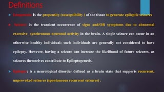
Astrocytes play a critical role in maintaining
neuronal homeostasis by buffering extracellular
potassium (K+) through the highly permeable
potassium channel KIR4.1.
Neurons extrude K+ with each action potential
repolarization and during periods of high activity,
K+ concentration in the extracellular space can be
rapidly elevated. Thus, the tight regulation of
[K+] via astrocytic uptake is critical for maintaining
neuronal homeostasis.
For instance, conditional knock-out of the KIR 4.1
channel in mice results in seizures resulting from
deficient K+ uptake in astrocytes.](https://image.slidesharecdn.com/epileptogenesis-170317005142/85/Epileptogenesis-46-320.jpg)


![Positron Emission Tomography
• Positron emission tomography (PET) uses
radiotracers to visualize and quantify functional
processes in the brain.
• [18F]FDG–PET: is the glucose analogue
[18F]FDG–PET, a biomarker for glucose uptake
and brain metabolism.
• A significant relationship has been reported
between the degree of hypometabolism on
[18F]FDG–PET in the entorhinal cortex early
following SE in the rat and the chance of later
developing spontaneous recurrent seizures,
suggesting that this may be a biomarker of early
epileptogenesis following a brain insult](https://image.slidesharecdn.com/epileptogenesis-170317005142/85/Epileptogenesis-66-320.jpg)
![a-[11C]Methyl-L-tryptophan “AMT PET”:
• AMT PET measures brain serotonin
synthesis.
• (AMT) is able to pinpoint the epileptic
focus itself in the interictal state, by
revealing a focus of increased AMT
uptake, even when an MRI or glucose
metabolism PET demonstrates normal
findings.
• AMT PET appears to be particularly
useful in patients with tuberous sclerosis
complex and in patients with cortical
developmental malformations.](https://image.slidesharecdn.com/epileptogenesis-170317005142/85/Epileptogenesis-67-320.jpg)


![Molecule Biomaterial Remarks [reference]
Neuron-specific
enolase
Serum Levels correlate with neurologic outcome after trauma and
reflect neuronal damage after SE
Myelin basic protein Serum Released in the course of brain trauma.
Ubiquitin carboxyl-
terminal
Hydrolase L1
CSF Increase in CSF correlates with epileptogenesis.
S100 calcium binding
protein B;
Serum/CSF Levels correlate with injury severity after trauma and have
predictive value on neurological outcome
Glial fibrillary acidic
protein
Serum/CSF Levels correlate with injury severity after trauma and have
predictive value on neurologic outcome; increase in CSF
correlates with Epileptogenesis stage in the kainic acid SE
model
Tau Serum/CSF Released in the course of brain trauma; predictive value for
Epileptogenesis to be determined
MicroRNA-9 Serum/Brain Increased after traumatic injury
Prolactin Serum/CSF Transiently increased in SE animal model in early
Epileptogenesis.](https://image.slidesharecdn.com/epileptogenesis-170317005142/85/Epileptogenesis-72-320.jpg)


