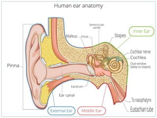
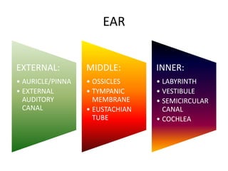
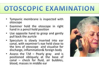
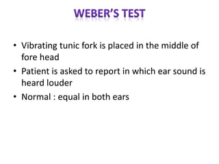
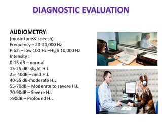
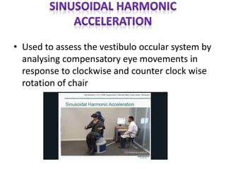
ENT
Mr. Manikandan.T,
RN., RM., M.Sc(N)., D.C.A .,(Ph.D)
Assistant Professor,
Dept. of Medical Surgical Nursing,
VMCON, Puducherry.
EXTERNAL
EAR
MIDDLE
EAR
INNER
EAR
EAR
EXTERNAL:
• AURICLE/PINNA
• EXTERNAL
AUDITORY
CANAL
MIDDLE:
• OSSICLES
• TYMPANIC
MEMBRANE
• EUSTACHIAN
TUBE
INNER:
• LABYRINTH
• VESTIBULE
• SEMICIRCULAR
CANAL
• COCHLEA
• The visible portion that is
commonly referred to as
"the ear"
• Helps localize sound sources
• Directs sound into the ear
• Each individual's pinna
creates a distinctive imprint
on the acoustic wave
traveling into the auditory
canal
• Extends from the pinna to the tympanic membrane
– About 26 millimeters (mm) in length and 7 mm in diameter in
adult ear.
– Size and shape vary among individuals.
– It contains hair, sebaceous land, ceruminous lands(wax)
• The purpose of wax:
– Repel water
– Trap dust, sand particles, micro-
organisms, and other debris
– Moisturize epithelium in ear
canal
– Odor discourages insects
– Antibiotic, antiviral, antifungal
properties
– Cleanse ear canal
• The eardrum separates the outer
ear from the middle ear
• Creates a barrier that protects
the middle and inner areas from
foreign objects
• Cone-shaped in appearance
– about 17.5 mm in diameter
• The eardrum vibrates in
response to sound pressure
waves.
• The eustachian tube (1MM WIDE 35 MM LONG)
connects the middle ear with the nasopharynx
• The eustachian tube normally it is closed, which
opens during swallowing and yawning
– This equalizes the pressure on either side of the
eardrum, which is necessary for optimal hearing.
• Malleus
(hammer)
• Incus (anvil)
• Stapes
(stirrup)
smallest
bone of the
body
• The cochlea resembles a
snail shell and spirals for
about 2 3/4 turns around
a bony column
• Within the cochlea are
three canals:
– Scala Vestibuli
– Scala Tympani
– Scala Media
FUNCTIONS
• Hearing
• Balance & equilibrium
• History of present illness
• General – onset, chronology, current situation,
location, radiation, quality, timing, factors,
associated symptoms, previous treatments
• Cardinal signs&symptoms:
EAR: Recent changes in hearing, itching,
earache, discharge, tinnitus, vertigo,ear
trauma, Q-TIP Use
• NOSE&SINUSES: Rhinorrhea, epistaxis,
obstruction of airflow, sinus pain & localised
headache, itching, anosomia, nasal trauma.
Sneezing
• MOUTH& THROAT: Hoarseness, dental chanes,
oral lesions, bleeding gums, sore throat,
dysphagia
• NECK: Pain, swelling, enlarged glands
• OTHERS: Fever, malaise, N/V
• Medical conditions& surgeries
• Allergies(season)
• Medications
• Herbal preparations
MEDICAL HISTORY RELATED TO ENT
• Frequent ear or throat infections
• Sinusitis
• Trauma to head, ENT
• ENT Surgery
• Seasonal allergies
• Asthma
• Hearing loss
• Meniere’s disease
• ENT Cancer
PERSONAL & SOCIAL HISTORY
• Smoking
• Frequent exposure to water
• Use of foreign object to ear
• Over crowding
• Use of ear protection
• Recent air travels
• Occupational exposure to toxins or loud
noises
• Inspection: auricle & surrounding tissue
should be inspected for deformities, lesions &
discharge
• Palpation: palpate the auricle – if pain – A/C
External otitis, tenderness on mastoid – A/C
Mastoiditis
• Tympanic membrane is inspected with
otoscope
• Examiner hold the otoscope in right
hand in a pencil hold position
• Use opposite hand to grasp and gently
pull back the auricle
• Speculum is slowly inserted into ear
canal, with examiner’s eye held close to
the lens of otoscope and visualise for
discharge, inflammation& foreign body
• Assess the T.M – Pearly gray and is
positioned obliquely at the base of
canal – check for fluid, air bubbles,
blood, masses in middle ear
• Place the base of vibrating tunic fork on
mastoid process
• When sound is no longer heard, the fork is
placed just outside (2 Inch)the ear
• Normal : 20 sec bone conduction, 30-40 sec
air conduction
• Vibrating tunic fork is placed in the middle of
fore head
• Patient is asked to report in which ear sound is
heard louder
• Normal : equal in both ears
HEARING STATUS WEBER RINNE
Normal Equal AC>BC
Conductive Sound is heard best in
affected ear
Sound is heard as long
or longer in affected
ear
Sensori neural Sound is heard best in
normal hearing ear
Air conduction is
audible longer than
bone conduction in
affected ear
WHISPER TEST
• Examiner cover the untested ear with palm of
the hand
• Then the examiner whispers softly from a
distance of 1 or 2 feet from unoccluded ear
and out of the patients sight
• The patient with normal acuity repeat what
was whispered.
AUDIOMETRY:
(music tone& speech)
Frequency – 20-20,000 Hz
Pitch – low 100 Hz –High 10,000 Hz
Intensity :
0-15 dB – normal
15-25 dB- slight H.L
25- 40dB – mild H.L
40-55 dB-moderate H.L
55-70dB – Moderate to severe H.L
70-90dB – Severe H.L
>90dB – Profound H.L
• Measure middle ear muscle reflex to sound
stimulation and compliance of tympanic
membrane by changing air pressure in a
sealed ear canal
• Electrodes are placed on the patients scalp &
an each ear lobe – connected to computer
• They record brain wave activity in response to
sounds you hear through earphones.
• Measurement of graphic recording of the changes in
electrical potential created by eye movements during
spontaneous, positionals(nystagmus)
• It is used to assess the occulomotor and vestibular
system and their corresponding interactions
• Used to assess the vestibulo occular system by
analysing compensatory eye movements in
response to clockwise and counter clock wise
rotation of chair
ENT ASSESSMENT

ENT ASSESSMENT

  • 2.
    ENT Mr. Manikandan.T, RN., RM.,M.Sc(N)., D.C.A .,(Ph.D) Assistant Professor, Dept. of Medical Surgical Nursing, VMCON, Puducherry.
  • 3.
  • 5.
    EAR EXTERNAL: • AURICLE/PINNA • EXTERNAL AUDITORY CANAL MIDDLE: •OSSICLES • TYMPANIC MEMBRANE • EUSTACHIAN TUBE INNER: • LABYRINTH • VESTIBULE • SEMICIRCULAR CANAL • COCHLEA
  • 6.
    • The visibleportion that is commonly referred to as "the ear" • Helps localize sound sources • Directs sound into the ear • Each individual's pinna creates a distinctive imprint on the acoustic wave traveling into the auditory canal
  • 7.
    • Extends fromthe pinna to the tympanic membrane – About 26 millimeters (mm) in length and 7 mm in diameter in adult ear. – Size and shape vary among individuals. – It contains hair, sebaceous land, ceruminous lands(wax)
  • 8.
    • The purposeof wax: – Repel water – Trap dust, sand particles, micro- organisms, and other debris – Moisturize epithelium in ear canal – Odor discourages insects – Antibiotic, antiviral, antifungal properties – Cleanse ear canal
  • 9.
    • The eardrumseparates the outer ear from the middle ear • Creates a barrier that protects the middle and inner areas from foreign objects • Cone-shaped in appearance – about 17.5 mm in diameter • The eardrum vibrates in response to sound pressure waves.
  • 10.
    • The eustachiantube (1MM WIDE 35 MM LONG) connects the middle ear with the nasopharynx • The eustachian tube normally it is closed, which opens during swallowing and yawning – This equalizes the pressure on either side of the eardrum, which is necessary for optimal hearing.
  • 11.
    • Malleus (hammer) • Incus(anvil) • Stapes (stirrup) smallest bone of the body
  • 12.
    • The cochlearesembles a snail shell and spirals for about 2 3/4 turns around a bony column • Within the cochlea are three canals: – Scala Vestibuli – Scala Tympani – Scala Media
  • 13.
  • 14.
    • History ofpresent illness • General – onset, chronology, current situation, location, radiation, quality, timing, factors, associated symptoms, previous treatments • Cardinal signs&symptoms: EAR: Recent changes in hearing, itching, earache, discharge, tinnitus, vertigo,ear trauma, Q-TIP Use
  • 15.
    • NOSE&SINUSES: Rhinorrhea,epistaxis, obstruction of airflow, sinus pain & localised headache, itching, anosomia, nasal trauma. Sneezing • MOUTH& THROAT: Hoarseness, dental chanes, oral lesions, bleeding gums, sore throat, dysphagia • NECK: Pain, swelling, enlarged glands • OTHERS: Fever, malaise, N/V
  • 16.
    • Medical conditions&surgeries • Allergies(season) • Medications • Herbal preparations
  • 17.
    MEDICAL HISTORY RELATEDTO ENT • Frequent ear or throat infections • Sinusitis • Trauma to head, ENT • ENT Surgery • Seasonal allergies • Asthma • Hearing loss • Meniere’s disease • ENT Cancer
  • 18.
    PERSONAL & SOCIALHISTORY • Smoking • Frequent exposure to water • Use of foreign object to ear • Over crowding • Use of ear protection • Recent air travels • Occupational exposure to toxins or loud noises
  • 19.
    • Inspection: auricle& surrounding tissue should be inspected for deformities, lesions & discharge • Palpation: palpate the auricle – if pain – A/C External otitis, tenderness on mastoid – A/C Mastoiditis
  • 20.
    • Tympanic membraneis inspected with otoscope • Examiner hold the otoscope in right hand in a pencil hold position • Use opposite hand to grasp and gently pull back the auricle • Speculum is slowly inserted into ear canal, with examiner’s eye held close to the lens of otoscope and visualise for discharge, inflammation& foreign body • Assess the T.M – Pearly gray and is positioned obliquely at the base of canal – check for fluid, air bubbles, blood, masses in middle ear
  • 21.
    • Place thebase of vibrating tunic fork on mastoid process • When sound is no longer heard, the fork is placed just outside (2 Inch)the ear • Normal : 20 sec bone conduction, 30-40 sec air conduction
  • 22.
    • Vibrating tunicfork is placed in the middle of fore head • Patient is asked to report in which ear sound is heard louder • Normal : equal in both ears
  • 23.
    HEARING STATUS WEBERRINNE Normal Equal AC>BC Conductive Sound is heard best in affected ear Sound is heard as long or longer in affected ear Sensori neural Sound is heard best in normal hearing ear Air conduction is audible longer than bone conduction in affected ear
  • 24.
    WHISPER TEST • Examinercover the untested ear with palm of the hand • Then the examiner whispers softly from a distance of 1 or 2 feet from unoccluded ear and out of the patients sight • The patient with normal acuity repeat what was whispered.
  • 25.
    AUDIOMETRY: (music tone& speech) Frequency– 20-20,000 Hz Pitch – low 100 Hz –High 10,000 Hz Intensity : 0-15 dB – normal 15-25 dB- slight H.L 25- 40dB – mild H.L 40-55 dB-moderate H.L 55-70dB – Moderate to severe H.L 70-90dB – Severe H.L >90dB – Profound H.L
  • 26.
    • Measure middleear muscle reflex to sound stimulation and compliance of tympanic membrane by changing air pressure in a sealed ear canal
  • 27.
    • Electrodes areplaced on the patients scalp & an each ear lobe – connected to computer • They record brain wave activity in response to sounds you hear through earphones.
  • 28.
    • Measurement ofgraphic recording of the changes in electrical potential created by eye movements during spontaneous, positionals(nystagmus) • It is used to assess the occulomotor and vestibular system and their corresponding interactions
  • 29.
    • Used toassess the vestibulo occular system by analysing compensatory eye movements in response to clockwise and counter clock wise rotation of chair