EAR
ANATOMY & PHYSIOLOGY
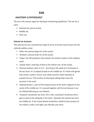
Theear is the sensory organ for hearing & maintaining equilibrium. The ear has 3
parts;
External ear/ pinna/ auricle.
Middle ear.
Inner ear.
External ear structures
The external ear has a characteristic shape & serves to funnel sound waves into the
external auditory canal.
Helix- the external large rim of the auricle.
Antihelix- external small rim of the auricle.
Tragus- the stiff projection that protects the anterior meatus of the auditory
canal.
Lobule/ lobus- small flap of flesh at the inferior end of the auricle.
External auditory canal- its 2.5 – 3cm long in the adults & it terminates at
the ear drum. It is S-shaped & leads to the middle ear. It is lined with glands
that secrete a yellow- brown wax called cerumen which lubricates &
protects the ear. The function of chewing & talking helps move the
cerumen in the canal.
Mastoid process- is part of the temporal bone of the skull is adjacent to the
cavity of the middle ear. It is assessed together with the ear because it can
be infected following an ear infection.
Tympanic membrane/ ear drum- this is thin, translucent membrane that is
grey in color & lies obliquely in the canal. Separates the external ear from
the middle ear. It has several distinct landmarks; handle & short process of
the malleus; umbo; cone light; pars flaccida; pars tensa.
2.
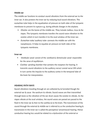
Middle ear
The middleear functions to conduct sound vibrations from the external ear to the
inner ear. It also protects the inner ear by reducing loud sound vibrations. The
eustachian tube helps in the equalization of pressure on both sides of the tympanic
membrane to prevent its rupture e.g. during altitude changes in the airplane.
Ossicles- are the bones of the middle ear. They include malleus, incus & the
stapes. The tympanic membrane transfers the sound wave vibration to the
ossicles which in turn transfers it to the oval window of the inner ear.
Eustachian tube/ auditory tube- connects the middle ear with the
nasopharynx. It helps to equalize air pressure on both sides of the
tympanic membrane.
Inner ear
Vestibular canal- consist of the vestibule & Semicircular canal- responsible
for the sense of equilibrium.
Cochlea- spiraling chamber that contains the receptors for hearing. It
transmits sound vibrations to the auditory nerve/ cranial nerve VIII which
in turn carries the impulse to the auditory cortex in the temporal lobe of
the brain for interpretation.
HEARING PATH WAYS
Sound vibrations traveling through air are collected by & funneled though the
external ear & cause the eardrum to vibrate. Sound waves are then transmitted
through bone as the vibration of the ear drum causes the ossicles to vibrate. As the
stapes vibrate at the oval window, the sound waves are passed are passed to the
fluid in the inner ear & then to the cochlea & to the brain. The transmission of the
sound through the external & middle ear is referred to as the conductive hearing &
transmission in the inner ear is called the perceptive/ sensorineural hearing. Hence
conductive hearing loss would be related to a dysfunction of the external or
3.
middle ear e.g.impacted ear wax. A sensorineural hearing loss is related to the
dysfunction of the inner ear.
The bones of the skull also conduct sound waves. This bone serves to augment the
usual pathway of sound waves through the air to the bone & finally to the fluid.
DATA COLLECTION
SUBJECTIVE DATA
Current symptoms
Earaches/otalgia.
Infection.
Discharge.
Hearing loss.
Tinnitus/ ringing sensations.
Dizziness/ loss of balance.
Trauma to the ear.
Past history
History of trauma or infections of the ear.
Past treatments for ear problems e.g. ototoxic drugs like antibiotics, aspirin,
quinine.
Ear surgery.
Family history
History of hearing loss in the family.
Lifestyle & health practices
Working or living in areas that have continuous loud noise- machinery,
music, explosives.
Use of ear guards in noisy environment.
Ear care/ ear cleaning.
4.
Spending lotsof time swimming or in water- swimmers ear i.e. infection
due to contaminated water being left in the ear therefore recommend use
of era plugs to keep water out of the ears.
OBJECTIVE DATA
Client preparation
Seated position.
Explain the tests to enhance client participation.
Equipments
Watch with a second hand.
Turning fork.
Otoscope.
EXTERNAL EAR STRUCTURES
The external ear & the tympanic membrane can be assessed by direct inspection &
otoscope. However, the middle & inner ear cannot be directly inspected but are
assessed by testing hearing acuity & conduction of sound.
Inspect the auricle, Tragus, & the lobule noting the size, shape & position.
Ears are equal in size bilaterally; the auricle aligns with the corner of each
eye. Abnormally. Low set ears or very small ear < 4cm or very large ears >
10cm.
Inspect for lesions, discolorations & discharge.
Palpate the auricle & mastoid process. Usually non- tender. Tenderness
indicates infection.
Otoscopic examination
- Used for inspection of the external auditory canal. Note the
following traits; lesions; foreign bodies; swellings; color &
consistency-should be pink & smooth without nodules; any
discharge- usually none; color & consistency of the cerumen- may
5.
be yellow, orange,red, brown. Gray or black,& is soft, moist, dry,
abnormally- foul smelling yellowish, purulent, bloody discharge;
- Inspect the tympanic membrane & note the color, shape,
consistency & landmarks = it is gray, shiny & translucent without
bulging or retraction; its concave (flat & slightly pulled at the
center), smooth & intact. A cone reflection of the otoscope light is
normally seen at 5 o’clock in the right ear & 7 o’clock in the left.
HEARING & EQUILIBRIUM TESTS
Whisper test
It tests the hearing acuity of high frequency sounds or gross hearing. Stand 1-2 ft
behind the client so that the/she cannot read your lips. Ask them to occlude on
ear with one finger on the tragus. Whisper a simple phrase & ask the client to
repeat the phrase. The client should be able to repeat the phrase correctly.
Inability to do so may indicate a loss of ability to hear high frequency sounds.
Weber’s test
Is valuable when a person reports hearing better with one ear than the other.
Place a vibrating tuning fork in the midline of the person’s skull & ask if the tone
sounds the same in both ears or better in one ear. It assesses sound conduction
via bone. Normally the person should hear the tone by bone conduction through
the skull & it should sound equally loud in both ears i.e. no lateralization of the
sound.
With conductive hearing loss there is lateralization of sound in the poorer ear
while in sensorineural hearing loss there is lateralization of sound to the better/
good ear.
Rinne test
It compares air conduction (AC) & bone conduction(BC).
6.
Place the stemof the vibrating tuning fork on the person’s mastoid process & ask
him/ her to signal when the sound goes away. Quickly invert the fork so that the
vibrating end is near the ear canal. The person should still hear the sound.
Normally the sound is heard twice as long by AC (next to the ear canal) as by BC(
through the mastoid process). I.e. AC> BC in a normal situation.
In conductive hearing loss BC>/= AC while in sensorineural loss AC> BC.
Romberg test
Done to test the client’s equilibrium or balance.
Ask the client to stand with feet together & arms at the sides &eyes closed. Client
maintains position for 20secs without swaying or with minimal swaying.
Abnormally, the client moves feet apart to prevent falls or starts to fall from loss
of balance & this may indicate a vestibular disorder.