Downloaded 101 times


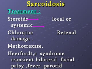


The document discusses several types of nasal inflammations and their causes, symptoms, diagnosis and treatment. It covers acute conditions like furunclosis, common cold and influenza rhinitis. It also discusses chronic conditions like chronic rhinitis, atrophic rhinitis, rhinoscleroma, syphilis, lupus, leprosy, Wegener's granulomatosis and sarcoidosis. For each condition, it provides details on etiology, clinical presentation, investigations and management approaches.


