
Recommended
Recommended
More Related Content
What's hot
What's hot (20)
Similar to Priniciples of Canine Endoscopic Surgery Prof.Dr. Awad Rizk.pdf
Similar to Priniciples of Canine Endoscopic Surgery Prof.Dr. Awad Rizk.pdf (20)
Recently uploaded
Recently uploaded (20)
Priniciples of Canine Endoscopic Surgery Prof.Dr. Awad Rizk.pdf
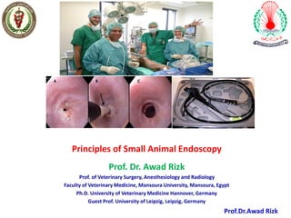
- 1. Principles of Small Animal Endoscopy Prof. Dr. Awad Rizk Prof. of Veterinary Surgery, Anesthesiology and Radiology Faculty of Veterinary Medicine, Mansoura University, Mansoura, Egypt Ph.D. University of Veterinary Medicine Hannover, Germany Guest Prof. University of Leipzig, Leipzig, Germany Prof.Dr.Awad Rizk
- 3. ❑Endoscopy is the use of specialized video cameras to evaluate areas within the body in a minimally invasive manner. ❑ In most instances, endoscopy is performed for diagnostic purposes allowing visualization and sampling of abnormalities. ❑Endoscopy can also be used for therapeutic purposes as well, termed interventional endoscopy.
- 4. Advantage of endoscopy Diagnostic ✓ Evaluating the digestive system is that it is nonsurgical. ✓ The technique allows for visualization of the lining of the digestive system ✓ Direct Biopsy samples can take from organs. ✓ Many foreign bodies in the esophagus and stomach may be removed via endoscopy. ✓ Specialized video camera with high resolution to evaluate is now available in market for better and accurate diagnosis
- 5. In surgical procedure (interventional endoscopy) ✓Endoscopies are almost painless although they may still cause some discomfort. ✓Compared with stress experienced by the body in a full surgical procedure ✓an endoscopy is simple, low risk, and cost effective. Other advantages include: ✓no scar – as a natural body opening is used ✓ quick recovery time ✓ less time in hospital, often, no time in hospital is required as the procedure is performed in the doctor’s rooms, and early detection of postoperative re-occurrence
- 6. other advantages of endoscopy ✓less in morbidity and mortality ✓minimally invasive ✓can be inserted into the natural openings of the body such as the mouth or anus. ✓ Also, they can be inserted into small incisions. ✓Its application in veterinary medicine has great role for the diagnosis and they can be used to examine visually or assist in surgery such as an arthroscopy and in therapeutic procedure.
- 7. Disadvantage of endoscopy ✓ Necessity to give general anesthesia to the patient ✓ Adequate laboratory testing and radiology is required before an endoscopy ✓ Evaluation of blood test before giving anesthesia is important for confirmation of patient is ready to take anesthetic risk or not ✓ Fasting of animals are at least 12 hours before an elective endoscopy ✓ A mouth gag is used to prevent damage to the endoscope. ✓ If lower part is to be examine it requires fasting more than 24 hrs to 48 hrs. ✓ Enemas are important to clean the intestines ✓ Care should be required at the time of endoscopy like tearing of intestine; perforation wound otherwise immediate surgery is required to correct the problem.
- 9. To avoid damage of endoscope 1. Mouth gag 2. Never force scope –avoid sharp bend 3. Inadequate anesthesia
- 10. Limitation of endoscopy in veterinary medicine ✓ Cannot assess functional disease luminal diameter, wall thickness ✓ Cannot identify disease in most of the small intestine ✓ Cannot detect disease in the deep submucosa, muscularis or serosa ✓ Not appropriate if bowel perforation is suspected ✓ Not ideal if pet not adequately prepared, ✓ Cannot assess or biopsy lymph nodes, Biopsy samples are very small, need multiple ✓ It requires experience and knowledge ✓ Requires regular cleaning
- 11. ➢ Endoscopy has become very important just as radiology or ultrasonography for veterinary specialist. ➢ Endoscopy is a valuable tool to peer view into interiors of the body. ➢ A basic knowledge of the normal appearance of the various luminal surfaces, and the underlying anatomy is essential. ➢ The use of endoscopy requires proficiency in anesthetic techniques, patient preparation, and in-depth knowledge of luminal affections in canines. ➢ Many gastrointestinal lesions have a polymorphic appearance, so biopsy is mandatory for definitive diagnosis. ➢ Endoscopic surgery results in less surgical morbidity, ➢ less postoperative pain and faster recovery.
- 12. History of endoscopy ✓ 1902: Georg Kelling of Dresden: performed the first laparoscopic procedure on dogs. ✓ 1910: Hans Christian Jacobaeus of Sweden: the first laparoscopic operation on humans. ✓ 1980: Patrick Steptoe, performed laparoscopic procedures in the operating room under sterile conditions. ✓ 1982: The first solid state camera was introduced and this was the start of video laparoscopy. ✓ 1983- First laparoscopic appendicectomy Semm, a German gynecologist. ✓ 1985- First Lap Cholecystectomy Erich Muhe a German surgeon. • 1987- First Laparoscopic repair of inguinal hernia by Ger. ✓ 1989- First lap hysterectomy, Reich
- 13. Types of Endoscopy Flexible endoscopy: • Bronchoscopy: an exam of the lower airways. • Colonoscopy:an exam of the transverse colon, ascending colon, cecum, large bowel, and rectum. • Endoscopy: an exam of the esophagus, stomach, and upper intestines. Rigid endoscopy: • Arthroscopy: an exam of soft tissue structures and joint cartilage, which is not visible on radiographs. Decreased damage to the joint and shortened recovery times are two advantages of arthroscopy over arthrotomy (surgical exam of the joint). Disadvantages include its limitation during diagnostic and corrective surgical procedures in small patients. • Cystoscopy: an examination of the vagina, urethral opening, urethra, bladder, and ureteral openings. • Laparoscopy: an exam of the abdominal cavity performed through a small incision in the wall of the abdomen or through the navel. It is done in veterinary medicine to obtain hepatic (liver) and renal (kidney) biopsy samples. • Proctoscopy: An exam of the large bowel and rectum. • Rhinoscopy: an exam of the nasal cavity and nasopharynx (junction between the nasal area and the back of the throat). • Thoracoscopy: an examination of the chest cavity. This is currently not performed frequently in veterinary medicine.
- 16. • Endoscopy is performed with either a rigid or flexible fiber optic instrument.
- 17. • ENDOSCOPY EQUIPMENT BASICS The basic endoscope consists of the: Insertion tube—depend on the mechanism for image transmission: Fiberoptic glass bundles (fiberscope) or charge-coupled device (CCD) chip (videoendoscope) Biopsy/suction channel Irrigation/insufflation channel Deflection control cables Handpiece—Includes the: Deflection control knobs Accessory channel entrance Irrigation/insufflation Suction valves Umbilical cord—Responsible for light transmission
- 19. FLEXIBLE ENDOSCOPES • There are 2 main types of flexible endoscopes: the fiberscope and videoendoscope. • The key differentiating feature is the mechanism by which an image is sensed and transmitted for viewing. • Flexible endoscopes are used predominately for navigating the complex anatomy of the GI tract.
- 20. • Flexible endoscopes such as those used in the examination of the stomach consist of a long, flexible insertion tube with a bending tip at the end that enters the body, an eyepiece, and a control section.
- 21. • The tip of the endoscope is manipulated using a control knob in the hand piece. • In addition to the fiber bundles which provide the light source, two channels are present within the endoscope.
- 22. ❖One channel permits various endoscopic tools to be passed and fluids to be suctioned or samples taken. ❖The other allows air or water to be passed into the stomach/intestine to insufflate (inject air into the area), or wash away mucus from the viewing port. ❖Special video cameras can be attached to the endoscopes which allow viewing of the exam on a television screen, as well as recording the exam on video.
- 26. • The rigid endoscope cannot be used in some areas, such as the stomach because it does not have the bending tip, so it cannot be flexed to allow examination of all parts of the stomach.
- 27. The Equipment 1. Laparoscope/video system 2. Light source 3. Insufflator 4. Diathermy /coagulation:cutting system 5. Suction irrigation system 6. Specialized hand instruments
- 29. 2- Monitor 1-Trolley 3-Endoscopic Camera 4-Light Source 6-Suction-Irrigation 5-Insufflator 7-High Frequency Unit
- 30. The Equipment 1. Laparoscope/video system There are two types: • Telescopic rod lens system, that is connected to a video camera (single chip or three chip) or • A digital laparoscope where the charge-coupled device[CCD] is placed at the end of the laparoscope, eliminateing the rod lens system.
- 31. Telescopic rod lens system There are three important structural differences in telescope available in the market. • 6 to18 rod lens system telescopes • 0 to 120 degree telescopes • 1.5 mm to 15 mm of telescopes
- 32. Telescopic rod lens system
- 33. Video camera Single chip VS three chip Three primary colours (Red,Blue, Green). In single chip camera all these 3 primary colours are sensed by single chip. In three chip camera there are 3 CCD- Chips for separate capture and processing of 3 primary colours—High resolution
- 34. Video camera
- 36. Monitor • No different from the T.V. • Basic principle of image reproduction is horizontal beam scanning on the face of the picture tube.
- 37. The existing television systems in use differ according to the country. • The U.S.A uses the NTSC (National Television System Committee) system. • In European countries the PAL (Phase Alternation b Line) system is in use. • French system called SECAM (Sequential color and memory).
- 38. Light source A fiber optic cable system connected to a 'cold' light source (halogen or xenon), to illuminate the operative field,
- 39. Light source
- 41. Insufflator The abdomen is insufflated with carbon dioxide gas [pneumoperitomeum] to create a working and viewing space. Elevates the abdominal wall above the internal organs like a dome. Gasless surgery –with mechanical wall elevators
- 43. Specialized hand instruments 5-10mm diameter instruments • Trocars & Ports---access devices • Graspers • Scissors • Dissectors • Clip applier,Knotting devices,Staplers • Cutting /coagulation – hooks,spatulas,balls,forceps • Irrigation suction tubes • Retrieval instruments
- 44. Disposable vs Reusable instruments Conventional vs. Needle scopic /miniaturized instruments—2mm size
- 45. Veress Needles
- 47. Trocars
- 49. Scissors
- 50. Dissectors
- 51. Graspers
- 52. Hook & spatula
- 54. Clip applicator
- 55. • Staplers • Knotting devices • Suturing devices
- 64. • The equipment used during laparoscopy includes: an optical system • consists of an endoscope, a light source, and a light cable. an insufflation system • Automatic, high-flow insufflators (10 L/min) usually are used to insufflate the abdomen. adapted surgical instruments
- 65. Diagnostic laparoscope (A), light cable (B), and video camera (C) used for laparoscopy.
- 66. • Carbon dioxide (CO2) is the most widely used gas for insufflation because: ❑ it is inexpensive ❑ is the least likely to cause gaseous emboli compared with nitrous oxide, air, and helium. • The primary disadvantage of CO2 ; ✓ is the production of postoperative discomfort when it turns into carbonic acid on the moist peritoneal surfaces. • This complication is reported frequently in people and has been observed in horses
- 67. Trocar-cannula system ❑ A trocar-cannula system is used to penetrate the body wall and allow introduction of the laparoscope and surgical instruments into the abdominal cavity ❑ Cannulas have a lateral Luer-Lok connector for the attachment of the insufflation tubing system ❑ They also contain a one-way valve that allows for entry and exit of instruments with maintenance of the pneumoperitoneum
- 68. Disposable cannula (A) and trocar (B) used for laparoscopy. Nondisposable cannula (A) and trocar (B) used for laparoscopy
- 69. Disposable laparoscopic Kelly forceps (A), Babcock forceps (B), and Metzenbaum scissors (C).
- 70. Non-disposable disassembled laparoscopic claw forceps
- 71. Veress needle (A) connected to an insufflator by a tubing system (B) is inserted through the body wall (C) to create a pneumoperitoneum before the laparoscopic cannula is inserted
- 72. During a laparoscopic procedure, laparoscopic retractors (A) and bowel clamps (B) are used to manipulate the surrounding tissue and organs away from the surgical site.
- 73. • When the laparoscopic examination is complete, the endoscope is removed, and the surgeon releases the CO2 gas/air via the open cannulas • The portal sites are not sutured until the end of the procedure, after all gas has been evacuated; this limits subcutaneous emphysema around the surgical sites and allows the maximal amount of CO2 to escape and helps prevent postoperative discomfort.
- 85. Laparoscopy
- 86. Surgical operations performed with laparoscopic methods are characterized by: ✓Their low invasiveness, ✓Small percentage of complications ✓ Low mortality. ✓To shorten significantly patients’ hospitalization time ✓ Faster recovery and entailing a higher level of satisfaction in their owners.
- 87. • The most common indications to laparoscopy in animals are: ▪ Biopsy of abdominal organs or abdominal tissue masses ▪ Surgical operations such as: ✓ Ovariohysterectomy ✓ Vasoligation ✓ nephrectomy, ✓ Cholecystectomy ✓ Cystotomy ✓ Thoracotomy
- 88. • Although laparoscopy is a low-invasive procedure, not every patient can be subjected to it. • Laparoscopic operations should not also be performed in obese animals, ones with abdominal dropsy, or in general bad condition • Pyometra is also listed as a contraindication to laparoscopy
- 90. ✓ Laparoscopic unit. From top to bottom: monitor ✓ Printer ✓ Camera ✓ insufflator ✓ DVD recorder ✓ source of light Basic elements of equipment necessary to perform laparoscopic operations include: ❖ a laparoscope (a telescope) ❖ a trocar ❖ a source of light ❖ a Veress needle ❖ an insufflator (a laparofflator) ❖ and a video camera
- 92. Rigid endoscopy: • Laparoscopy • Ovariectomy • Cystoscopy • Arthroscopy
- 93. Laparoscopy
- 94. Origins • First laparoscopic procedure performed in 1902 by Georg Kelling of Dresden, Germany in a dog. • 1910-First laparoscopic procedure performed in a human by Hans Christian Jacobaues of Sweden. Hans Christian Jacobaeus
- 95. • The surface between the pubic bone and 5 cm cranially to the umbilicus was shaved and scrubbed including 10 cm to each side and Lidocaine 2% was infiltrated at the sites of trocars placement.
- 96. Carbon Dioxide Gas and Laparoscopy • Before the laparoscopic procedure is performed, the abdomen is insufflated or blown up like a balloon with carbon dioxide gas. • This causes elevation of the abdominal wall above the internal organs like a dome to create a working and viewing space.
- 97. Three portal sites were used: • portal 1(primary port) for the laparoscope • portals 2-3 (secondary portals) for surgical instruments
- 98. • Portal 1 was located 2-3 cm cranial to the umblicus. • Portals 2 and 3 were distal to the laparoscope insertion site and 4-6 cm bilaterally from the ventral midline. • A 10mm skin incision was made at portal site 1, and then a Veress needle inserted perpendicular to the abdominal wall. • After an aspiration and injection test using a 5mL syringe containing 5mL sterile saline solution to verify that the needle was within the peritoneal cavity • An automatic high-flow CO2 insufflator was connected to the Veress needle and used to insufflate the abdominal cavity at the rate of 1L/min with a pressure gradient of 12– 15 mmHg according to the dogs' size.
- 99. • After complete pneumoperitoneum has been established, the Veress needle was replaced with a 10/11mm trocar-cannula unit • The trocar removed and the Co2 gas tube connected to the lure lock of the cannula and the insufflators was set to maintain an intra-abdominal pressure of 12-15mmHg during laparoscopy. • A rigid type telescope connected to a light source. • a high resolution one chip digital camera was then introduced through the cannula to transmit the scene to the video monitor.
- 105. Laparoscopic ovariectomy and OHE • Advantages of laparoscopy over conventional surgery include: ➢ Reduced pain from the surgical wounds – the pet is more comfortable post-operatively ➢ Smaller surgical wounds ➢ Fewer stitches ➢ A faster return to normal activity, due to improved patient comfort and reduced scar tissue formation
- 108. • All patients are positioned in dorsal recumbency and clipped from xiphoid to pubis and dorsally past the level of the 13th rib. • A wide clip is necessary to accommodate placement of the trans-abdominal suspension suture or weighted hook. • The abdomen is aseptically prepared as for traditional laparotomy.
- 109. • A skin incision no larger than the size of the intended cannula is made using a surgical blade. • The subcutaneous tissues are dissected down to the linea.
- 110. • The Hasson technique is performed by creating a mini- laparotomy. • A 5.5-mm cannula with blunt trocar is preferred. • The size of the skin and linea incision is no larger than the intended camera port (usually 5 mm). • Stay sutures are placed on both sides of the linea incision to allow traction during insertion of the blunt trocar.
- 111. • The trocar is inserted at an approximate 30-degree angle, towards the right caudal abdomen, to avoid iatrogenic injury to the spleen. • Once the cannula is inserted, the obturator is removed and the CO2 tubing attached. • The abdomen is insufflated to a pressure of 10 mm Hg. • All subsequent instrument trocars are placed under direct laparoscopic visualization to avoid intra-abdominal visceral injury. • A two-port laparoscopic OVE/OHE technique will be used.
- 112. • Dorsal recumbency is most commonly performed by ovariectomy, because both ovarian pedicles can be visualized without moving the patient. • The table can also be tilted from side to side to help manipulation and visualization • Mechanical ventilation is suggested because of the intra- abdominal insufflation pressure and the weight of the organs on the diaphragm • Vascular stapler is utilized if the ovarian pedicle is extremely wide.
- 113. • If the ovarian artery and vein are larger than 3 mm, endoscopic clips can be used. • A Babcock forceps was inserted into the 12 mm cannula to grasp the right ovary. • The ovary was lifted from the surrounding tissue and brought to the abdominal wall, which allowed percutaneous advancement of a transabdominal suspension suture with a round needle to maintain exposure of the ovarian pedicle.
- 114. Laparoscopic graspers. From the top to bottom: Babcock forceps Reddic-Olsen forceps Kelly forceps.
- 115. Laparoscopic Assisted OHE Laparoscopic-assisted OHE is performed similarly to laparoscopic OVE with the exception that the caudal cannula be placed in a more caudal location (over the uterine body) and the uterine body is ligated extracorporeally via the caudal port site. Because the uterine body is ligated extracorporeally, we prefers to use an 11.5-mm cannula in the caudal location for larger (>10 kg) dogs.
- 117. • The general location for the caudal cannula should be approximately 1/3 of the distance between the umbilicus and the pubis. • The cranial cannula can be placed 2–3 cm cranial to the umbilicus as for laparoscopic OVE. • Once the cannulas have been placed, the dog is repositioned into lateral-oblique recumbency and both ovarian pedicles ligated and divided as described for laparoscopic OVE.
- 118. • Dissection of the broad ligament to approximately midway between the ovarian pedicle and uterine body is all that is typically required prior to elevation from the caudal cannula site. • Once both ovarian pedicles have been divided and the broad ligament dissected, the dog is positioned in dorsal recumbency and the left proper ligament is grasped with the Babcock forceps and brought to the base of the caudal cannula. • If needed, the caudal port incision is lengthened directly over the cannula while maintaining the grasp of the proper ligament.
- 119. • The cannula is then removed over the Babcock forceps and the ovary and uterine horn extracted from the abdomen. • The contralateral uterine horn and ovary is easily traced and extracted similarly to the left. • Once the uterine body has been well exposed the uterine body can be ligated in standard fashion. • The uterine stump is inspected for bleeding and returned to the abdominal cavity. • The port sites are closed according to the guidelines described for laparoscopic OVE.
- 120. Complications • Complications from laparoscopic ovariectomy and ovariohysterectomy are rare but can include: ➢ Hemorrhage ➢ subcutaneous emphysema ➢ visceral organ injury (e.g., spleen ➢ inability to complete the procedure requiring conversion to laparotomy, pain, seroma formation, and rarely infection.
- 121. Canine Cystoscopy
- 122. 3.5mm with cystoscope-urethroscope sheath and telescope bridge
- 123. 2.7mm Telescope with operating sheath and forceps
- 124. Fiberscope
- 125. What is cystoscopy? • Cystoscopy is a procedure that allows specially trained veterinarians to look inside the urinary bladder and the urethra (tube connecting bladder to outside body) using a special instrument. • Cystoscopy is endoscopy of the bladder and urethra performed with a thin lighted tube called a cystoscope. • This procedure is performed in a sterile manner under anesthesia in dogs and cats. • This procedure does not require an incision as the camera, scope and instruments are passed through the penis in males or the vulva in females.
- 126. When would cystoscopy be needed? Cystoscopy could be helpful to determine the cause of: ✓Blood in the urine ✓Urine leakage (urinary incontinence) ✓Urinary blockages ✓Complicated urinary tract infections ✓When abnormalities in the bladder have been noted on radiographs (x-ray) or ultrasound
- 127. • Patient positioning in female dogs varies with the clinician's preference. • Most endoscopists favor right or left lateral recumbency with the hind end as close to the end of the table as possible. • Ventral-dorsal can also be utilized. • Male dogs are always in lateral recumbency.
- 128. Cystoscopy Procedure in Dogs • The dog is anesthetized intravenously. • The bladder is filled with sterile saline and distended, allowing the scope to enter. • A flexible optic instrument will be inserted in the urinary tract for male dogs, a rigid optical instrument will be used on female dogs(no incision is required). • The scope is placed through the urethra to obtain a visual of the bladder and urethra. • A video recording is often taken. • Biopsies of tissues can also be taken. • If bladder stones are found, they can then be removed with grasping forceps.
- 129. • Cystourethroscopy is a technique used to gain access to the lower genitourinary tract (urethra, urinary bladder, ureteral orifices, vagina). In most cases, cystourethroscopy is used as a diagnostic tool to visually assess the lower urinary tract if routine diagnostic evaluation (blood work, urinalysis, urine culture, radiography, ultrasonography) does not yield a definitive diagnosis
- 131. Indications for cystoscopy Dog with urinary tract infection and urinary bladder wall pustules.
- 132. Dog with struvite and calcium phosphate carbonate uroliths.
- 133. Cystoscopic image with a transitional cell carcinoma at the trigone.
- 134. Cystoscopic image from a 10-year-old female spayed dog with polypoid cystitis.
- 135. Cystoscopic image from a female spayed dog having urethrolith retrieved by graspers.
- 136. ➢ A variety of cystoscopes is needed to perform transurethral cystoscopy in both male and female dogs and cats because of anatomic variations and marked variability in urethral diameter. ➢ A rigid cystoscope can be used in female dogs and cats. ➢ Flexible cystoscopes are required in male dogs and cats for complete evaluation of the urethra and urinary bladder.
- 137. • What is lithotripsy? • Lithotripsy is the physical breaking of stones formed by the body within the urinary tract of cats and dogs. • Lithotripsy is usually performed within the body using a laser fiber via surgery or cystoscopy with a shock wave applied to the stones. • When would lithotripsy be needed? Lithotripsy can be helpful at treating: ✓Bladder stones (in certain cases) ✓Problematic kidney stones (causing infection, kidney damage, obstruction of urine flow) ✓Urethral stones
- 138. what happens once the stones are broken? • In the bladder, the stones are broken and removed with a small basket. • Once kidney stones have been broken, a specialized wand is used to suction the large stone fragments. A stent (plastic tube) is then placed in the ureter to ensure flow of urine from kidney to bladder and avoid obstruction by small pieces of stone.
- 140. Arthroscopy
- 141. • Arthroscopy is a technique for examining the inside of a joint using a tiny camera • To allow a detailed assessment of the joint in a minimally invasive fashion
- 142. ➢Arthroscopy has numerous advantages over arthrotomy for diagnosis and treatment of joint disease. ➢Arthroscopy entails less disruption of the periarticular soft tissue. Typically, two or three one-centimeter long skin incisions are required in conjunction with a 5-millimeter diameter tunnel through the underlying soft tissue and into the joint. ➢Decreased soft tissue disruption leads to less pain and less chance for infection.
- 143. ➢In most cases, return to use of the limb is quicker because of less surgically induced pain. ➢This is especially true when multiple joints are involved and are operated arthroscopically under the same anesthetic episode. ➢Examples of disease conditions where multiple joints are commonly treated under the same anesthetic episode include fragmented medial coronoid process of the elbow and OCD of the shoulder.
- 144. • Arthroscopy may be employed in a diagnostic, therapeutic or combined modality. • Using arthroscopy as an exploratory procedure may prevent the necessity for an arthrotomy and is an important advantage in cases where a surgically treatable lesion is not found. • Visualization of the joint typically is better with arthroscopy than with an arthrotomy. • In joints like the shoulder and elbow, arthroscopy allows inspection of areas of the joint that is not visible without performing multiple arthrotomies. `
- 145. • In addition, the magnification that occurs with arthroscopy in combination with the fluid medium within the joint allows one to see joint pathology that cannot be appreciated with an arthrotomy. • Visualization of synovial membrane and cartilage pathology, in particular, is better appreciated with arthroscopy than with arthrotomy. • With practice and development of proficiency, the length of an arthroscopic procedure is often less than an arthrotomy procedure. • Cosmetic appearance of the dog is typically better after arthroscopy compared to arthrotomy. For some owners, cosmetic appearance is very important.
- 152. Thank you for attention !!!!!!!!