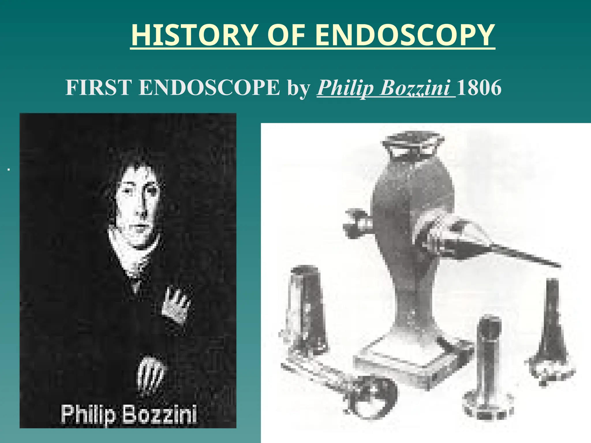
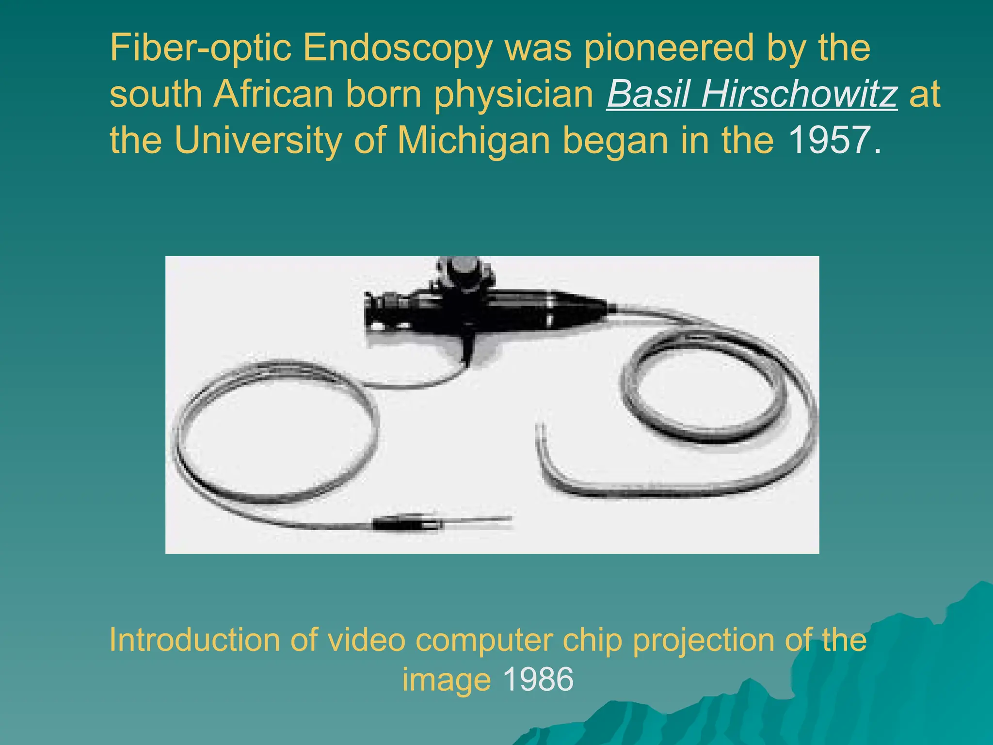
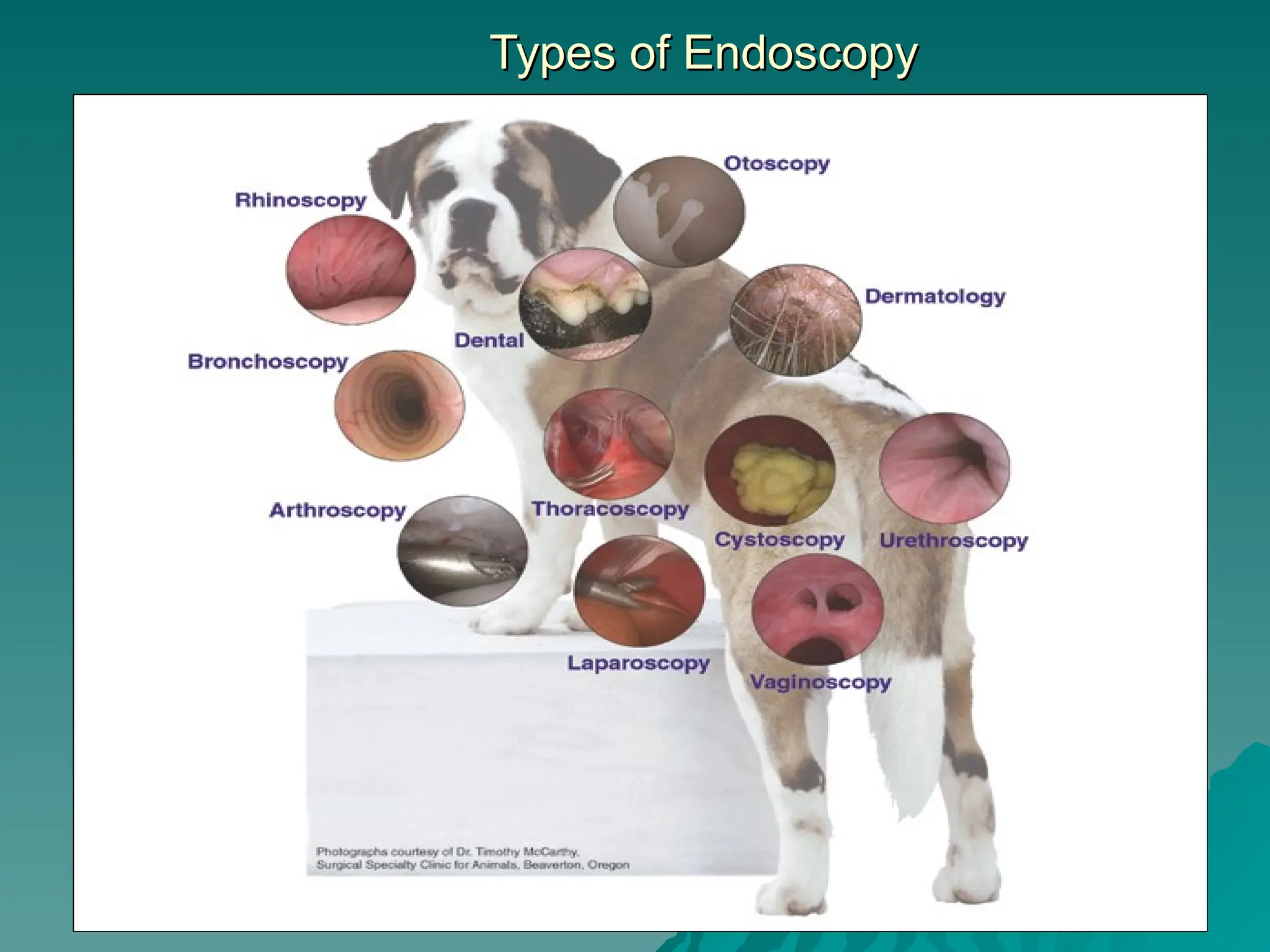
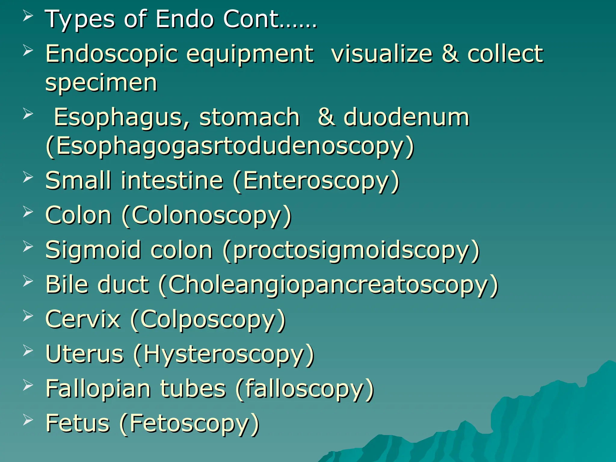
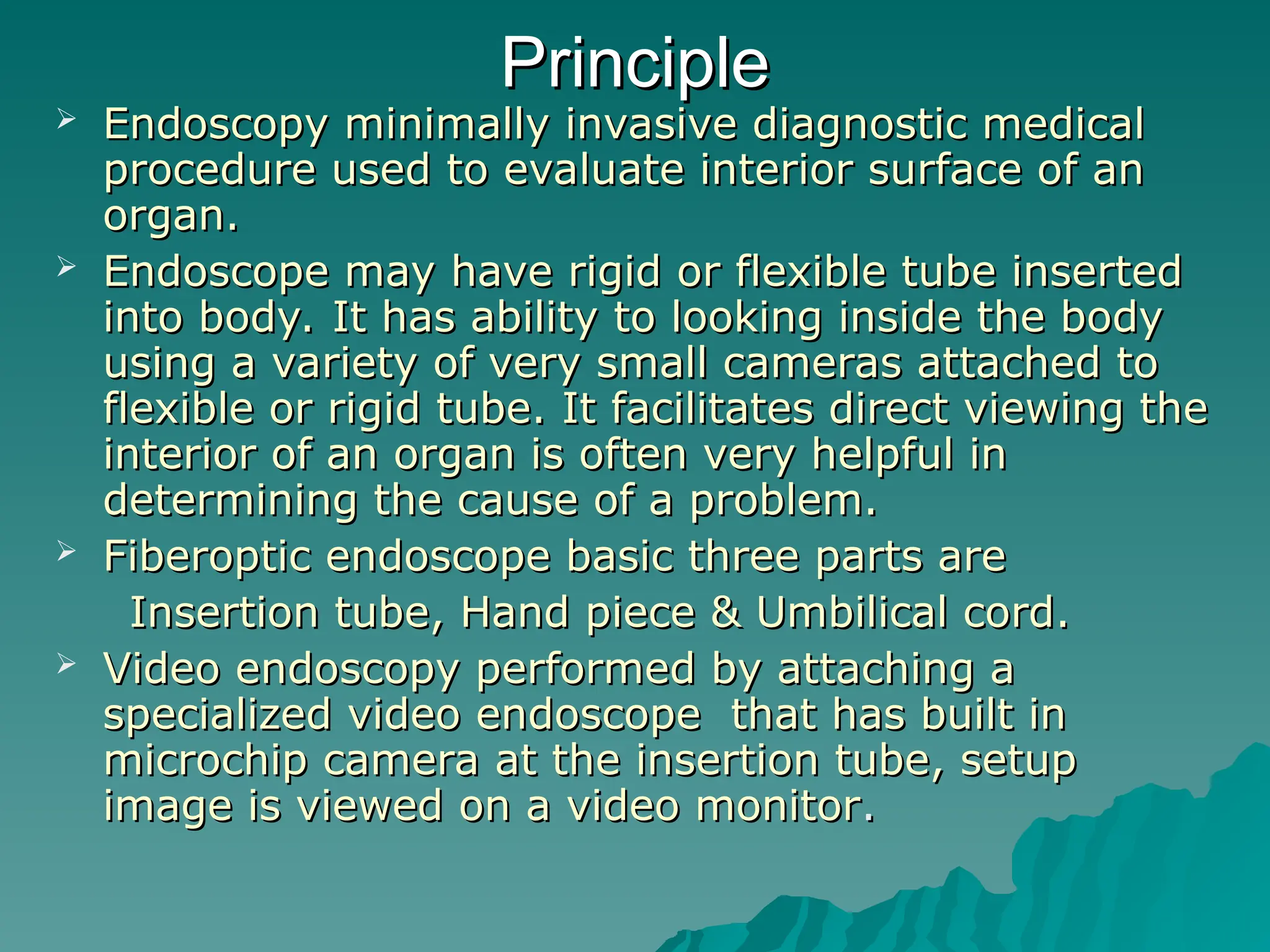
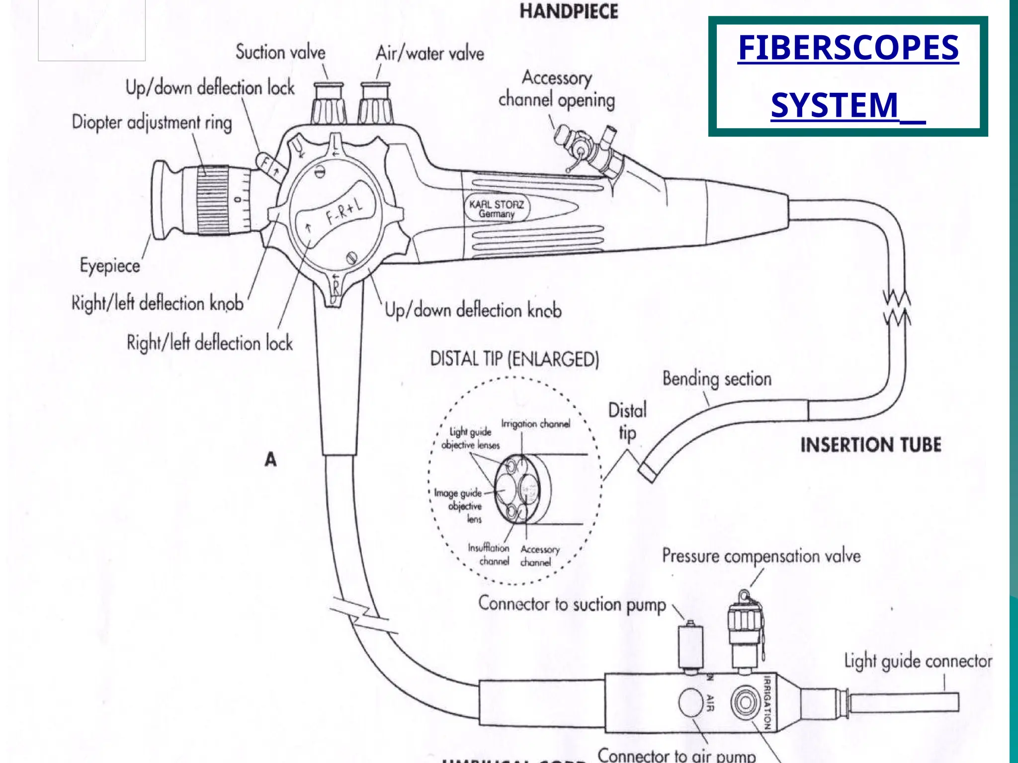
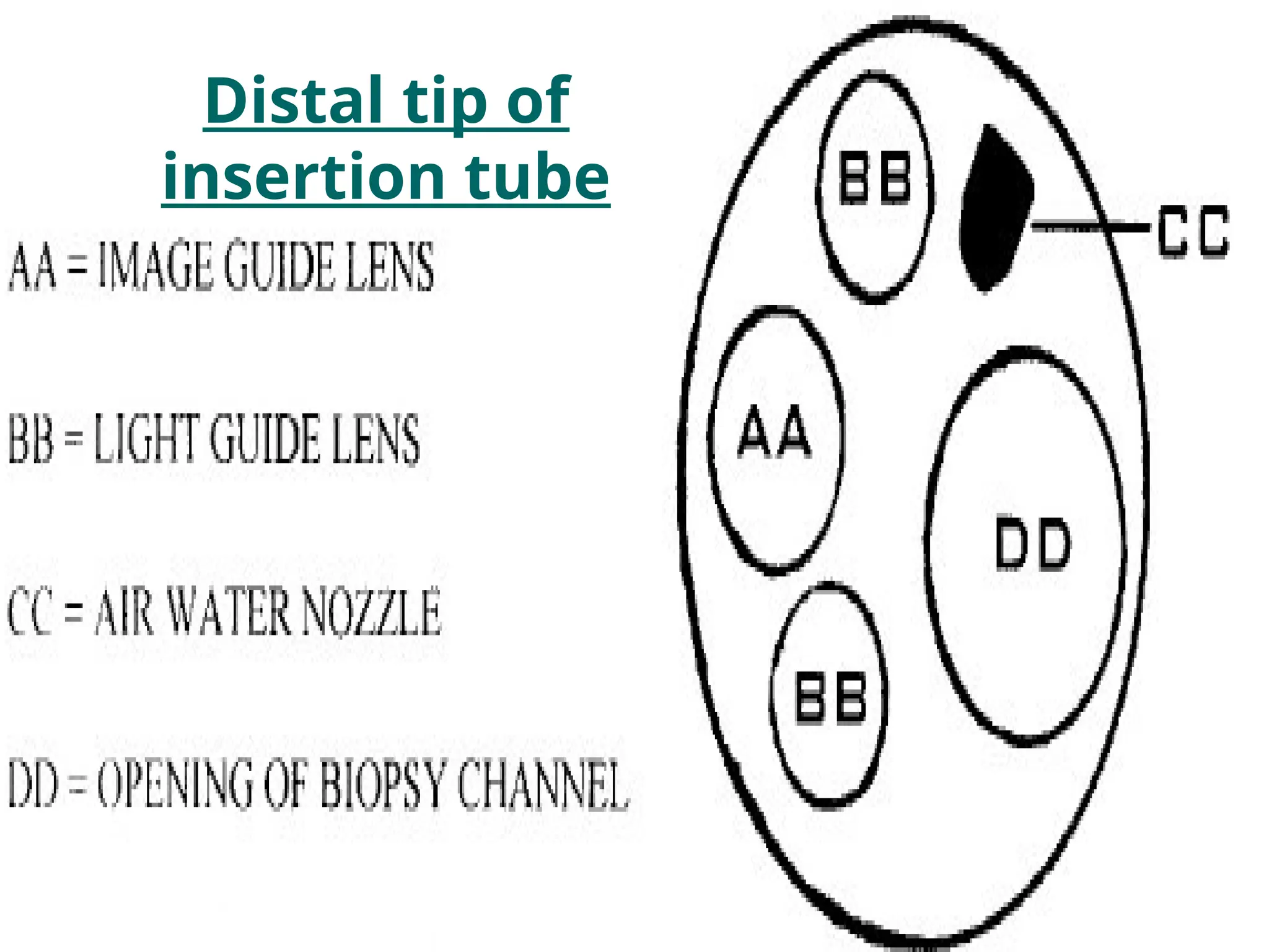
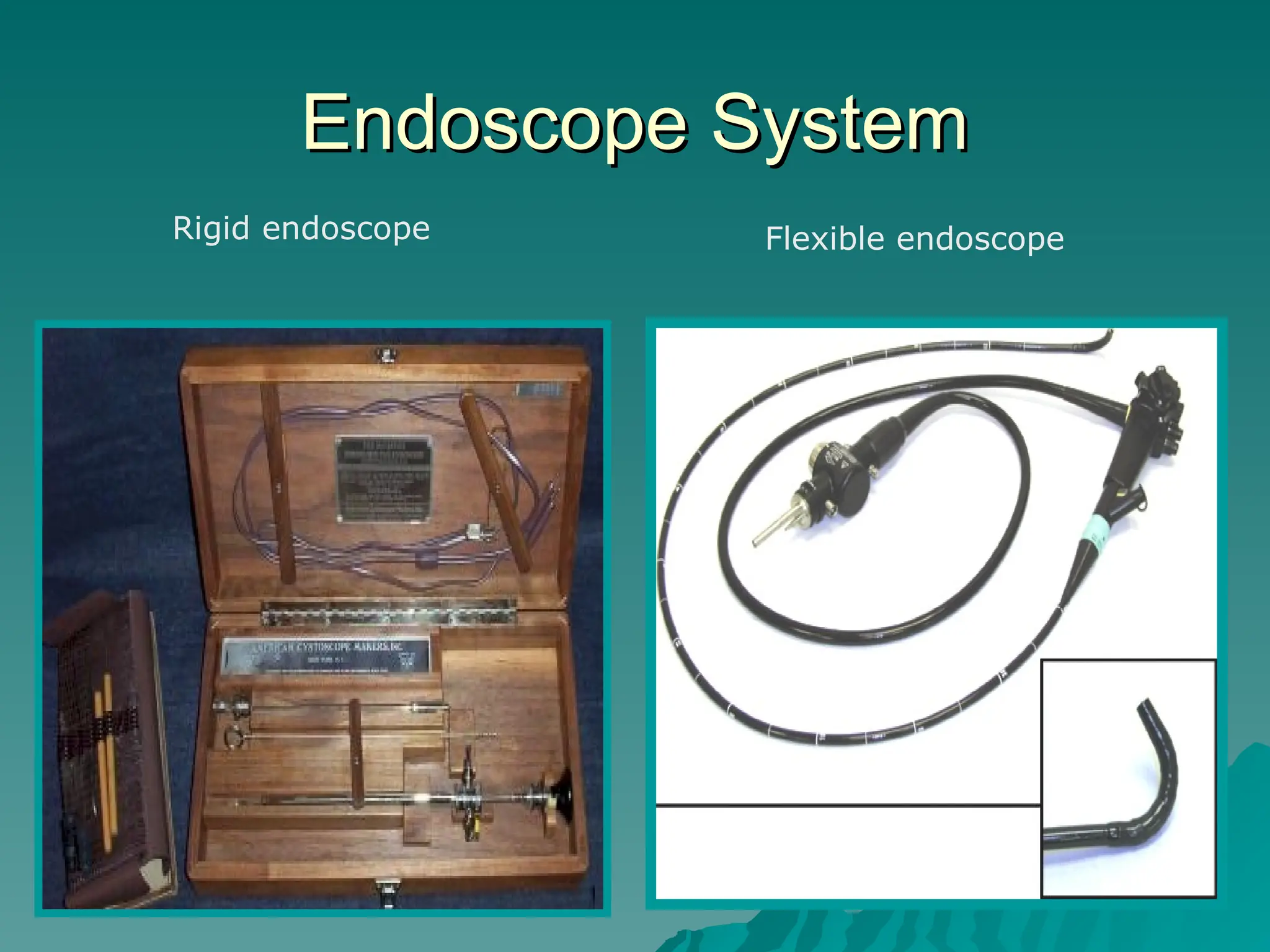
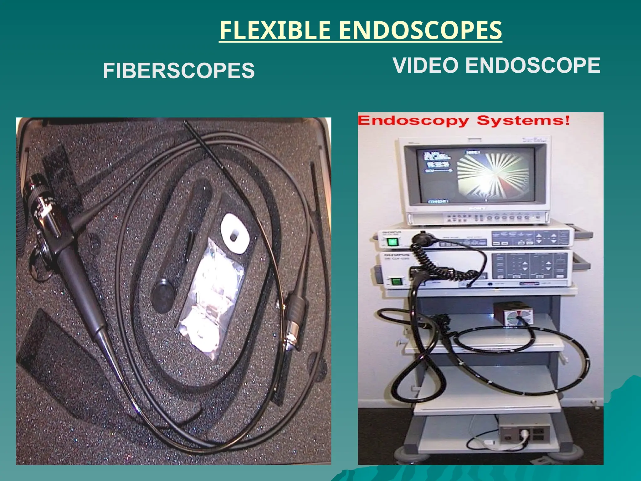
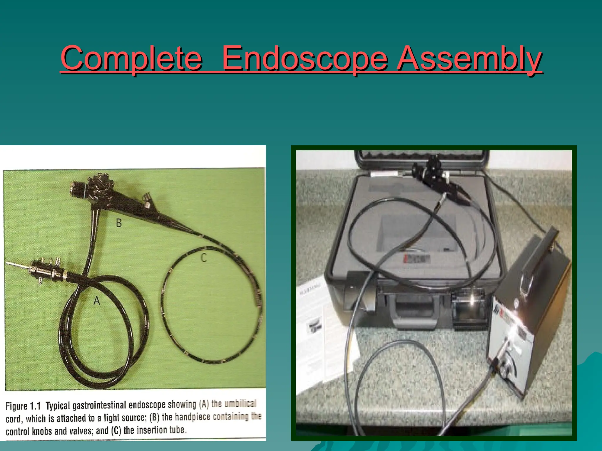
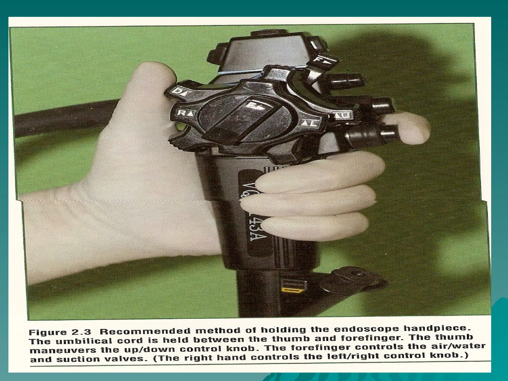
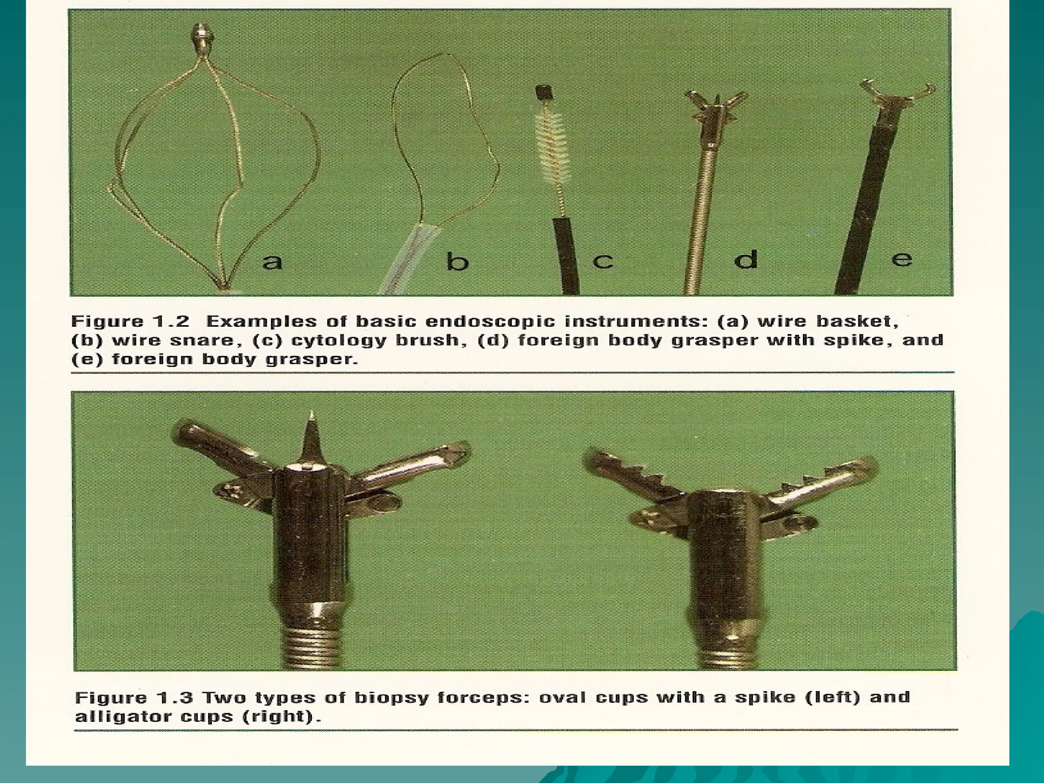
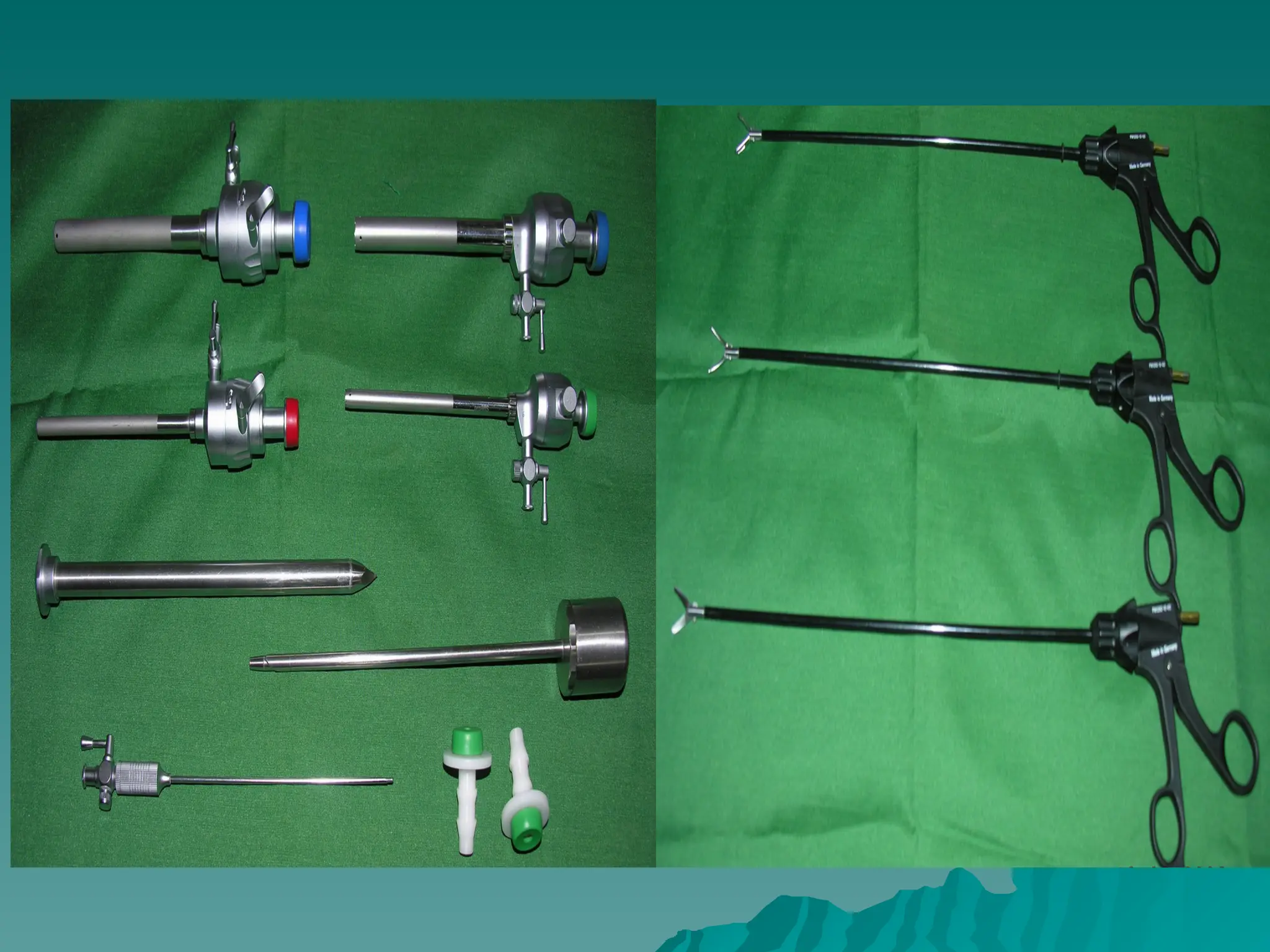
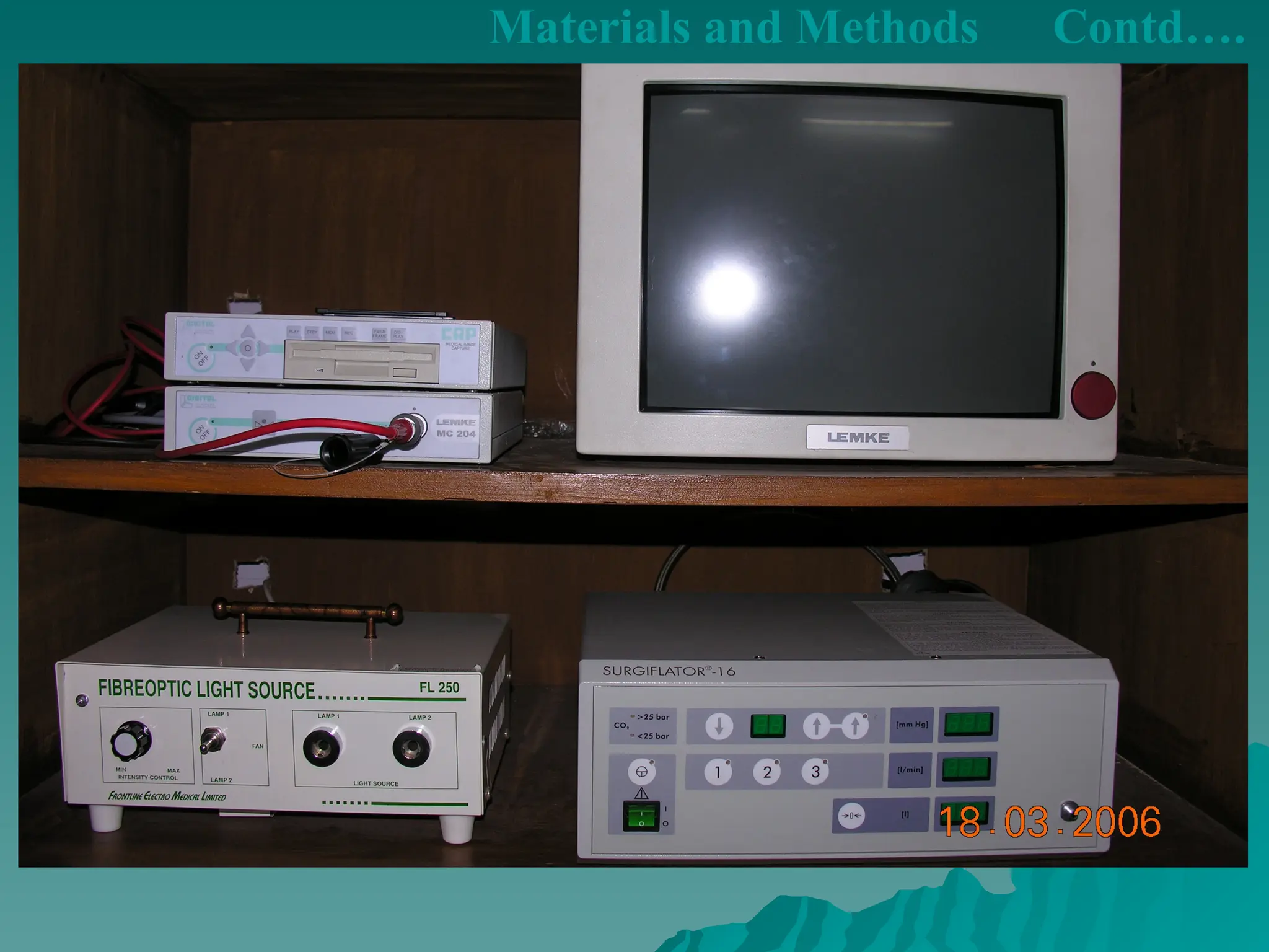
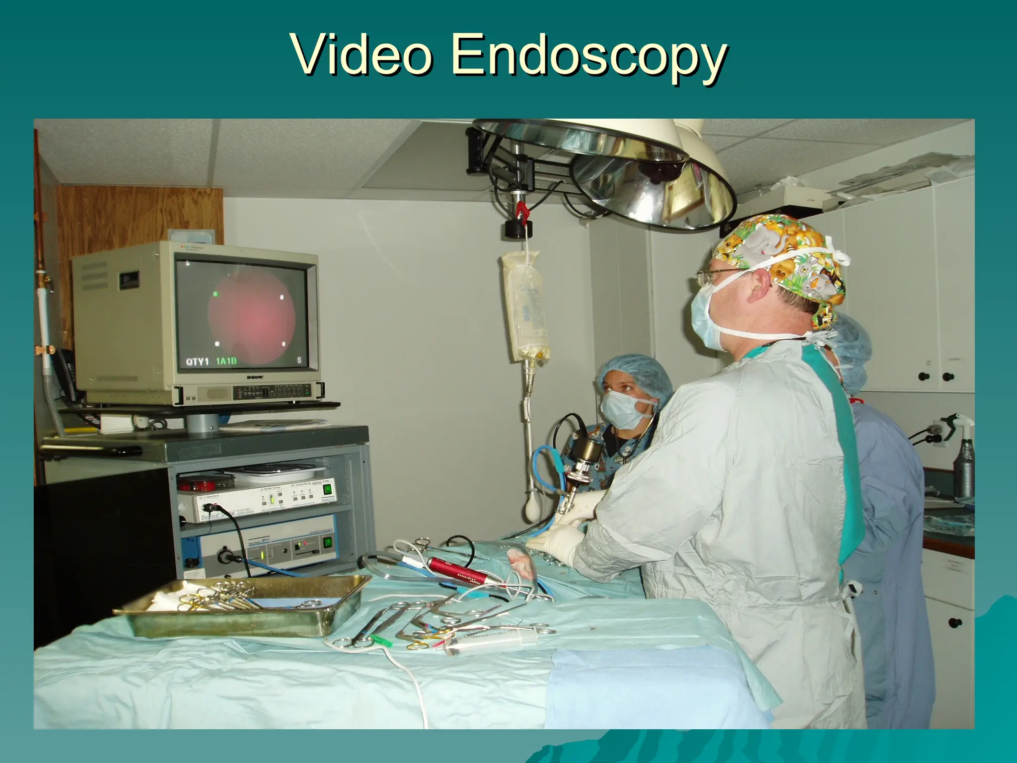
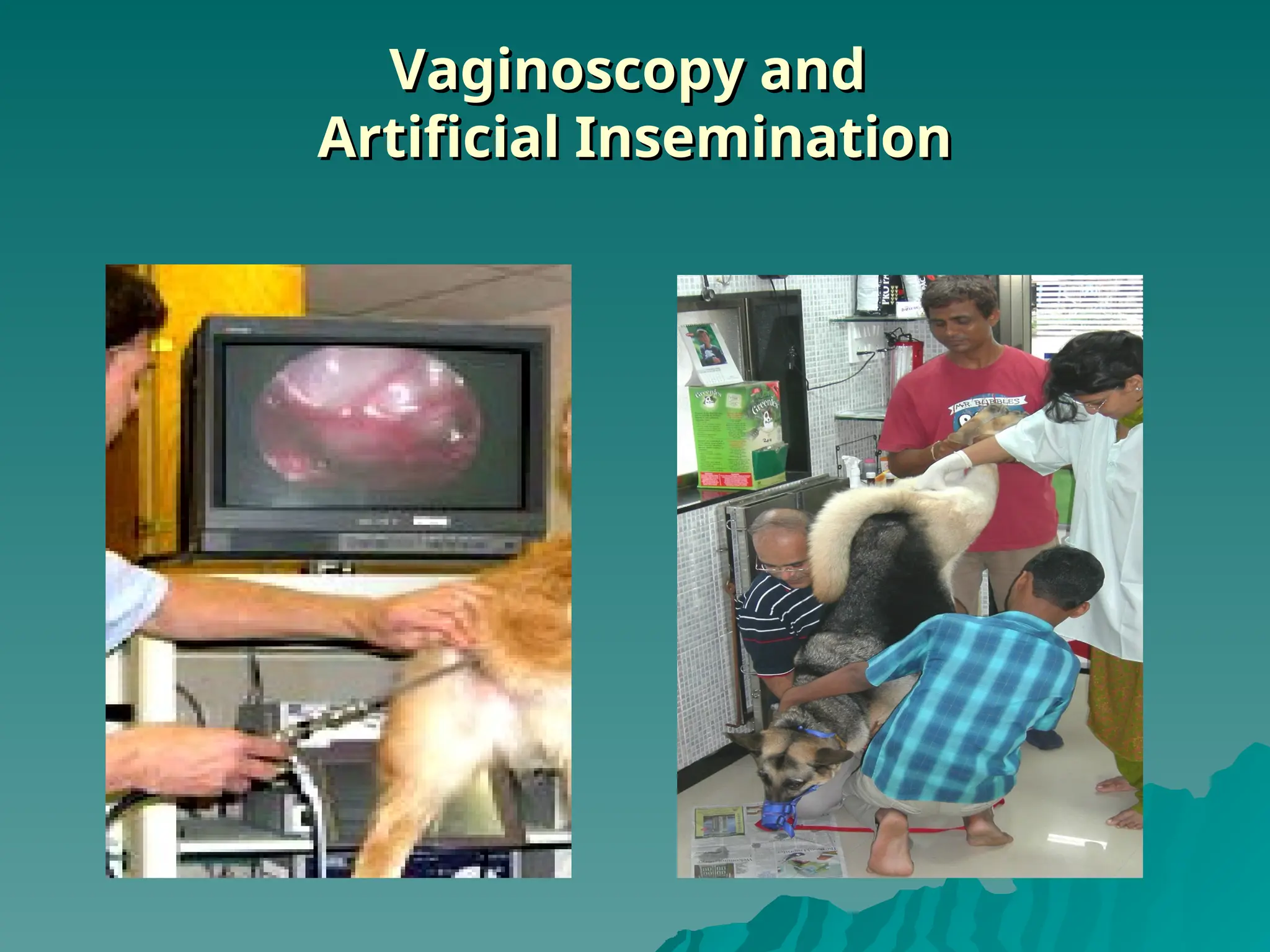
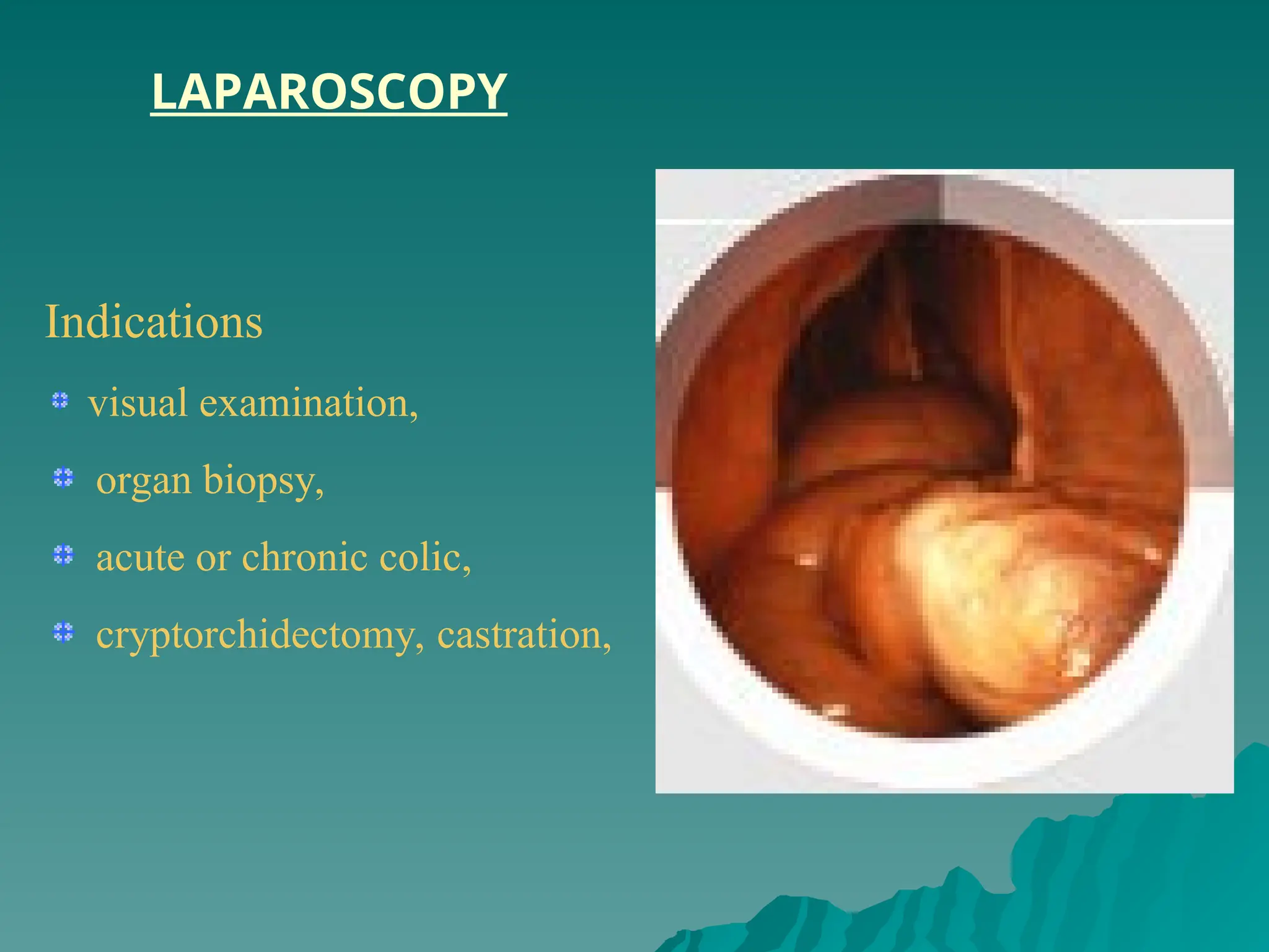
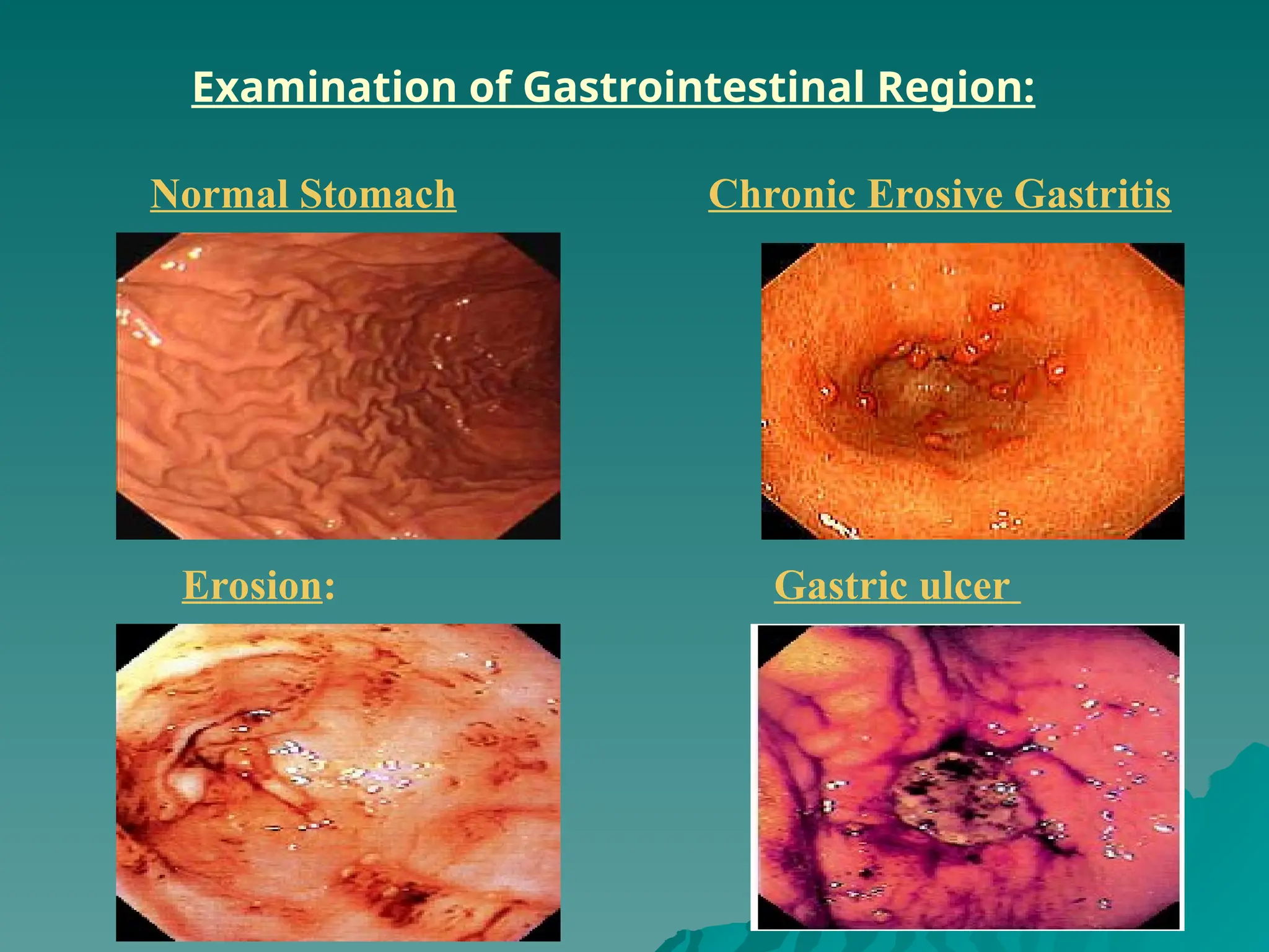
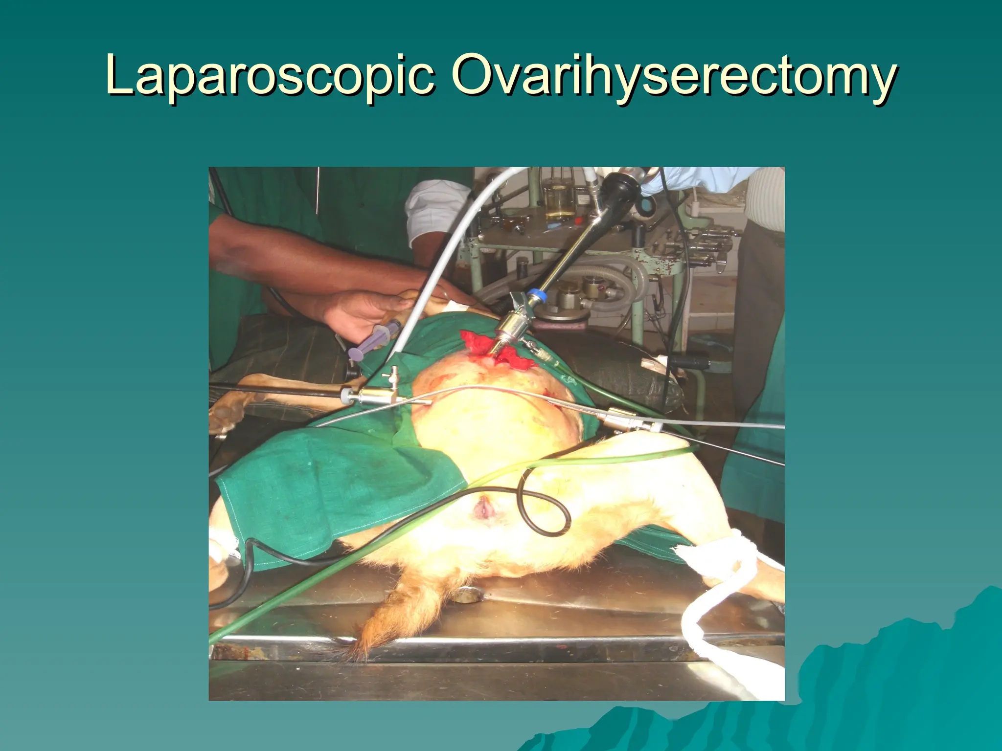
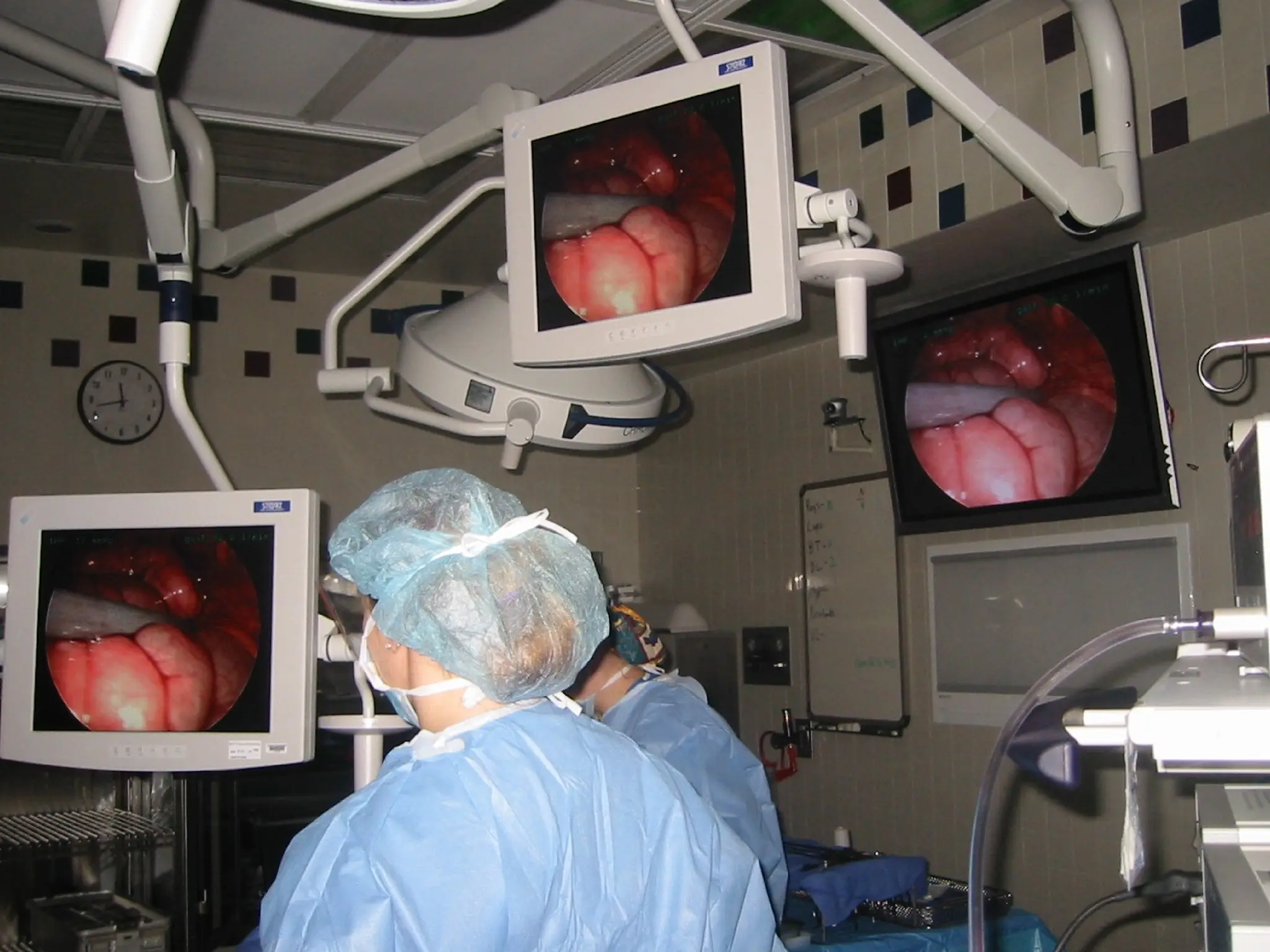
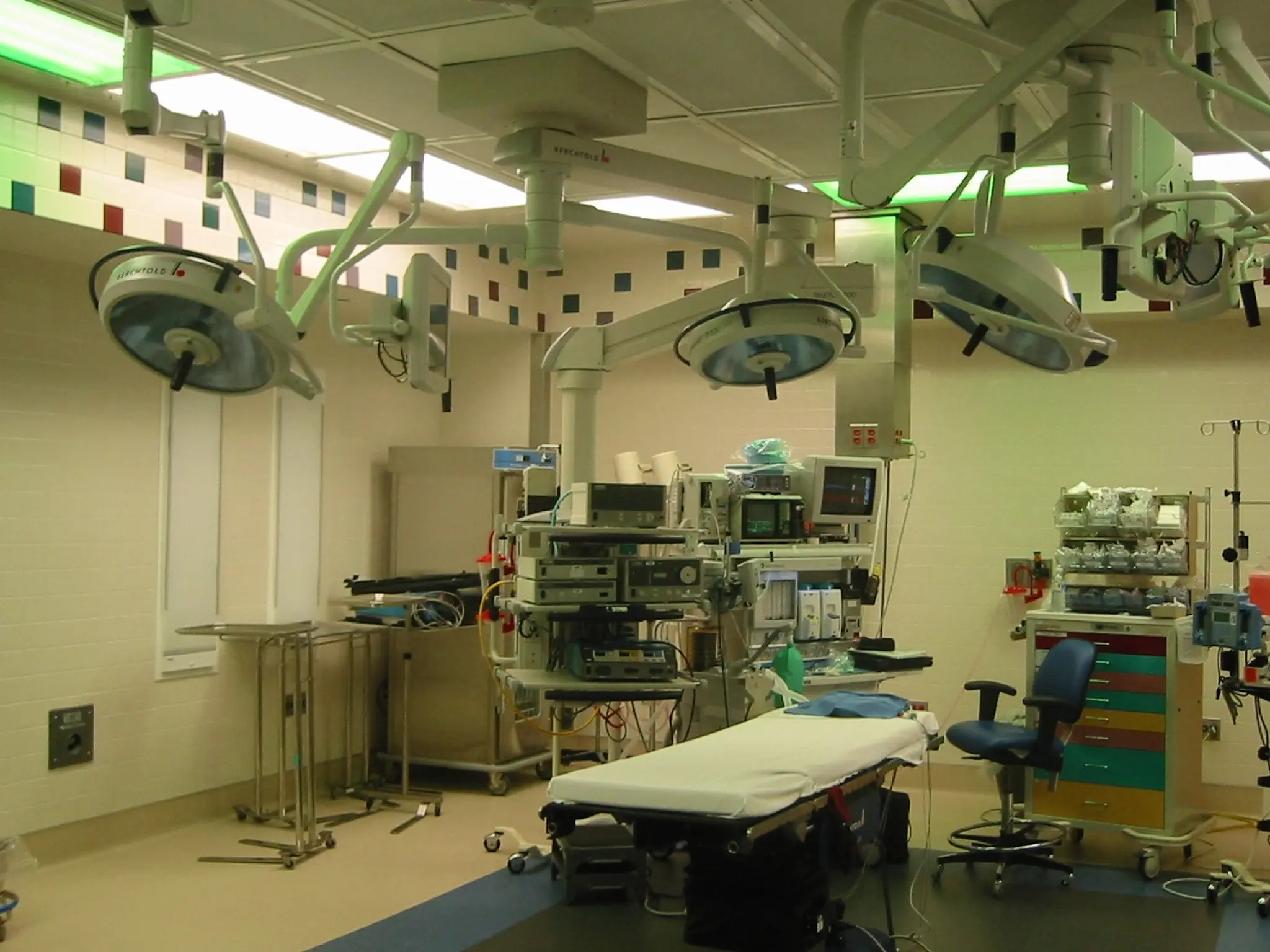
ENDOSCOPY
PRESENTED BY
DR. DHIREN B. BHOI
M. V. Sc.
VETERINARY GYNAECOLOGY AND OBSTETRICS
COLLEGE OF VETERINARY SCI. & ANIMAL HUSBANDRY
ANAND
Definition
Definition
 Endoscopy Greek Word “Endo”means “Inside”
“Skopeein ”means “To See”
 Means the ability to look inside the body
Means the ability to look inside the body
using a variety of very small cameras
attached to either a flexible or rigid tube.
 Direct viewing interior of an organ is often
Direct viewing interior of an organ is often
very helpful in determining the cause of a
problem & helpful in establishing a diagnosis.
 Endoscopy exceptionally valuable tool for
Endoscopy exceptionally valuable tool for
the diagnosis of many small animal
gastrointestinal disorders
gastrointestinal disorders.
.
History
History
 1806 Philp Bozzini developed first kind endoscope,
1806 Philp Bozzini developed first kind endoscope,
examination of canals & cavities of human body.
 1822 William Beaumont ,first introduced into human
1822 William Beaumont ,first introduced into human
being.
being.
 Jacobeus, developed endoscopic exploration,
Jacobeus, developed endoscopic exploration,
1910 – Thoracoscopy,
1912 - Laparoscopy
1912 - Laparoscopy
 1937 Hope reported laparoscopy to diagnose ectopic
1937 Hope reported laparoscopy to diagnose ectopic
pregnancy.
pregnancy.
 1944 Raoul Palmer performed gynecological
1944 Raoul Palmer performed gynecological
laparoscopy
laparoscopy
 1984 first laparoscopic cholecsytectomy performed
1984 first laparoscopic cholecsytectomy performed
 1987 first video-laparoscopic cholecystectmoy.
1987 first video-laparoscopic cholecystectmoy.
Kelling in 1902 – 1st
reported
cystoscopy in dogs & later was know
as “father of veterinary laparoscopy”.
Lettow in 1960 performed laparoscopy
in dogs involving ovarian function
studies & in 1972 1st
laparoscopic liver
biopsy procedure in dog.
HISTORY OF ENDOSCOPY
.
FIRST ENDOSCOPE by Philip Bozzini 1806
Fiber-optic Endoscopy was pioneered by the
south African born physician Basil Hirschowitz at
the University of Michigan began in the 1957.
Introduction of video computer chip projection of the
image 1986
Indications
Indications
 Role of evaluation gastrointestinal motility
Role of evaluation gastrointestinal motility
disorders, gastric adenocarcinoma, remove
foreign bodies, submucosal lesion, duodenal
or colonic abnormalities, stomach ulcers
&colon polyps.
&colon polyps.
 Perform therapeutic procedures esophageal
Perform therapeutic procedures esophageal
stricture dilation.
stricture dilation.
 Enabling visual examination of mucosal
Enabling visual examination of mucosal
surface, ability to mucosal biopsy for
histopathological examination or fluid
aspiration for analysis.
 Used Placement of gastrotomy tube in
Used Placement of gastrotomy tube in
stomach.
stomach.
Types of Endoscopy
Types of Endoscopy
 Types of Endo Cont……
Types of Endo Cont……
 Endoscopic equipment visualize & collect
Endoscopic equipment visualize & collect
specimen
specimen
 Esophagus, stomach & duodenum
Esophagus, stomach & duodenum
(Esophagogasrtodudenoscopy)
 Small intestine (Enteroscopy)
Small intestine (Enteroscopy)
 Colon (Colonoscopy)
Colon (Colonoscopy)
 Sigmoid colon (proctosigmoidscopy)
Sigmoid colon (proctosigmoidscopy)
 Bile duct (Choleangiopancreatoscopy)
Bile duct (Choleangiopancreatoscopy)
 Cervix (Colposcopy)
Cervix (Colposcopy)
 Uterus (Hysteroscopy)
Uterus (Hysteroscopy)
 Fallopian tubes (falloscopy)
Fallopian tubes (falloscopy)
 Fetus (Fetoscopy)
Fetus (Fetoscopy)
Principle
Principle
 Endoscopy minimally invasive diagnostic medical
Endoscopy minimally invasive diagnostic medical
procedure used to evaluate interior surface of an
organ.
organ.
 Endoscope may have rigid or flexible tube inserted
Endoscope may have rigid or flexible tube inserted
into body. It has ability to looking inside the body
using a variety of very small cameras attached to
flexible or rigid tube. It facilitates direct viewing the
interior of an organ is often very helpful in
determining the cause of a problem.
 Fiberoptic endoscope basic three parts are
Fiberoptic endoscope basic three parts are
Insertion tube, Hand piece & Umbilical cord.
 Video endoscopy performed by attaching a
Video endoscopy performed by attaching a
specialized video endoscope that has built in
microchip camera at the insertion tube, setup
image is viewed on a video monitor
image is viewed on a video monitor.
.
FIBERSCOPES
SYSTEM
Distal tip of
insertion tube
Endoscope System
Endoscope System
Rigid endoscope Flexible endoscope
FLEXIBLE ENDOSCOPES
FIBERSCOPES VIDEO ENDOSCOPE
Complete Endoscope Assembly
Materials and Methods Contd….
Advantages
Advantages
 Diagnostic as well as therapeutics solution.
Diagnostic as well as therapeutics solution.
 Minimum stress & mortality.
Minimum stress & mortality.
 Less traumatic surgery than std surgery.
Less traumatic surgery than std surgery.
 Faster recovery .
Faster recovery .
 Amount & duration of anesthesia much less than as
Amount & duration of anesthesia much less than as
compare to std surgical procedure.
 Minimal suture used, hence less body reaction.
Minimal suture used, hence less body reaction.
 Less adhesion after surgery.
Less adhesion after surgery.
 Can detect gastric parasite otherwise difficult to
Can detect gastric parasite otherwise difficult to
find.
find.
 Also used to place long term feeding tubes into the
Also used to place long term feeding tubes into the
stomachs of animals that cant eat.
 “
“According to an old adage in medicine-If you don’t
According to an old adage in medicine-If you don’t
look for it ,you will never diagnose it”.
Disadvantages
Disadvantages
 Requires expensive, flexible equipment,& specialized O.T
setup.
 Biopsies usually adequate but not as good as full thick-
ness.
 Cannot examine entire intestinal tract; lesion may be
beyond reach of scope.
 Cannot determine if an ulcer is going to rupture or
bleed.
 There are few chances herniation of abdominal contents
after laparoscopy.
 Always possibility of introduction unsertilized instrument
Always possibility of introduction unsertilized instrument
into the body
into the body
 Inexpert handling of the endoscope may lead to
Inexpert handling of the endoscope may lead to
punctured organ with variable effects.
PATIENT PREPARATION
ANESTHESIA PATIENT POSITIONING
Insertion of Endoscopy
Video Endoscopy
Video Endoscopy
Vaginoscopy and
Vaginoscopy and
Artificial Insemination
Indications
visual examination,
organ biopsy,
acute or chronic colic,
cryptorchidectomy, castration,
LAPAROSCOPY
Examination of Gastrointestinal Region:
Gastric ulcer
Erosion:
Normal Stomach Chronic Erosive Gastritis
Bleeding in Stomach Linear hemorrhage
Gastric Polyps Snare Polypectomy
Intestinal intussusception in a pup
Duodenal ulcer
Duodenitis
Duodenal Affections
EXAMINATION OF OESOPHAGUS
Normal oesophagus Oesophagitis
Spleen abscess in a dog
THORACOSCOPY
• Distending the Joint With Fluid or Gas.
Location and Extent of Articular Trauma
ARTHROSCOPY
 It is used to examine &
resect the bovine teat, either
via the streak canal or with a
lateral, single puncture
approach.
 Used for removal of
obstruction, neoplasm's,
adhesions, polyps, ect.
Under visual aid.
THELORESECTOSCOPY
Cystoscopy / Urethroscopy
Urinary
calculi
Neoplasms
Bladder
worms
Rupture of
bladder
BIOPSY OF THE ORGANS
LIVER BIOPSY
View Of The Lower Respiratory Tract Through An
Endoscope
Gastric Parasite
Gastric Parasite
Removal of foreign body
Foreign Body
Dog’s esophagus with a
chicken
bone lodged in the lumen.
Laparoscopic Castration (Vasectomy)
Laparoscopic Ovarihyserectomy
Thanking you

endoscope-higher advanced technology.ppt

  • 1.
    ENDOSCOPY PRESENTED BY DR. DHIRENB. BHOI M. V. Sc. VETERINARY GYNAECOLOGY AND OBSTETRICS COLLEGE OF VETERINARY SCI. & ANIMAL HUSBANDRY ANAND
  • 2.
    Definition Definition  Endoscopy GreekWord “Endo”means “Inside” “Skopeein ”means “To See”  Means the ability to look inside the body Means the ability to look inside the body using a variety of very small cameras using a variety of very small cameras attached to either a flexible or rigid tube. attached to either a flexible or rigid tube.  Direct viewing interior of an organ is often Direct viewing interior of an organ is often very helpful in determining the cause of a very helpful in determining the cause of a problem & helpful in establishing a diagnosis. problem & helpful in establishing a diagnosis.  Endoscopy exceptionally valuable tool for Endoscopy exceptionally valuable tool for the diagnosis of many small animal the diagnosis of many small animal gastrointestinal disorders gastrointestinal disorders. .
  • 3.
    History History  1806 PhilpBozzini developed first kind endoscope, 1806 Philp Bozzini developed first kind endoscope, examination of canals & cavities of human body. examination of canals & cavities of human body.  1822 William Beaumont ,first introduced into human 1822 William Beaumont ,first introduced into human being. being.  Jacobeus, developed endoscopic exploration, Jacobeus, developed endoscopic exploration, 1910 – Thoracoscopy, 1910 – Thoracoscopy, 1912 - Laparoscopy 1912 - Laparoscopy  1937 Hope reported laparoscopy to diagnose ectopic 1937 Hope reported laparoscopy to diagnose ectopic pregnancy. pregnancy.  1944 Raoul Palmer performed gynecological 1944 Raoul Palmer performed gynecological laparoscopy laparoscopy  1984 first laparoscopic cholecsytectomy performed 1984 first laparoscopic cholecsytectomy performed  1987 first video-laparoscopic cholecystectmoy. 1987 first video-laparoscopic cholecystectmoy.
  • 4.
    Kelling in 1902– 1st reported cystoscopy in dogs & later was know as “father of veterinary laparoscopy”. Lettow in 1960 performed laparoscopy in dogs involving ovarian function studies & in 1972 1st laparoscopic liver biopsy procedure in dog.
  • 5.
    HISTORY OF ENDOSCOPY . FIRSTENDOSCOPE by Philip Bozzini 1806
  • 6.
    Fiber-optic Endoscopy waspioneered by the south African born physician Basil Hirschowitz at the University of Michigan began in the 1957. Introduction of video computer chip projection of the image 1986
  • 7.
    Indications Indications  Role ofevaluation gastrointestinal motility Role of evaluation gastrointestinal motility disorders, gastric adenocarcinoma, remove disorders, gastric adenocarcinoma, remove foreign bodies, submucosal lesion, duodenal foreign bodies, submucosal lesion, duodenal or colonic abnormalities, stomach ulcers or colonic abnormalities, stomach ulcers &colon polyps. &colon polyps.  Perform therapeutic procedures esophageal Perform therapeutic procedures esophageal stricture dilation. stricture dilation.  Enabling visual examination of mucosal Enabling visual examination of mucosal surface, ability to mucosal biopsy for surface, ability to mucosal biopsy for histopathological examination or fluid histopathological examination or fluid aspiration for analysis. aspiration for analysis.  Used Placement of gastrotomy tube in Used Placement of gastrotomy tube in stomach. stomach.
  • 8.
  • 9.
     Types ofEndo Cont…… Types of Endo Cont……  Endoscopic equipment visualize & collect Endoscopic equipment visualize & collect specimen specimen  Esophagus, stomach & duodenum Esophagus, stomach & duodenum (Esophagogasrtodudenoscopy) (Esophagogasrtodudenoscopy)  Small intestine (Enteroscopy) Small intestine (Enteroscopy)  Colon (Colonoscopy) Colon (Colonoscopy)  Sigmoid colon (proctosigmoidscopy) Sigmoid colon (proctosigmoidscopy)  Bile duct (Choleangiopancreatoscopy) Bile duct (Choleangiopancreatoscopy)  Cervix (Colposcopy) Cervix (Colposcopy)  Uterus (Hysteroscopy) Uterus (Hysteroscopy)  Fallopian tubes (falloscopy) Fallopian tubes (falloscopy)  Fetus (Fetoscopy) Fetus (Fetoscopy)
  • 10.
    Principle Principle  Endoscopy minimallyinvasive diagnostic medical Endoscopy minimally invasive diagnostic medical procedure used to evaluate interior surface of an procedure used to evaluate interior surface of an organ. organ.  Endoscope may have rigid or flexible tube inserted Endoscope may have rigid or flexible tube inserted into body. It has ability to looking inside the body into body. It has ability to looking inside the body using a variety of very small cameras attached to using a variety of very small cameras attached to flexible or rigid tube. It facilitates direct viewing the flexible or rigid tube. It facilitates direct viewing the interior of an organ is often very helpful in interior of an organ is often very helpful in determining the cause of a problem. determining the cause of a problem.  Fiberoptic endoscope basic three parts are Fiberoptic endoscope basic three parts are Insertion tube, Hand piece & Umbilical cord. Insertion tube, Hand piece & Umbilical cord.  Video endoscopy performed by attaching a Video endoscopy performed by attaching a specialized video endoscope that has built in specialized video endoscope that has built in microchip camera at the insertion tube, setup microchip camera at the insertion tube, setup image is viewed on a video monitor image is viewed on a video monitor. .
  • 11.
  • 12.
  • 13.
    Endoscope System Endoscope System Rigidendoscope Flexible endoscope
  • 14.
  • 15.
  • 19.
  • 20.
    Advantages Advantages  Diagnostic aswell as therapeutics solution. Diagnostic as well as therapeutics solution.  Minimum stress & mortality. Minimum stress & mortality.  Less traumatic surgery than std surgery. Less traumatic surgery than std surgery.  Faster recovery . Faster recovery .  Amount & duration of anesthesia much less than as Amount & duration of anesthesia much less than as compare to std surgical procedure. compare to std surgical procedure.  Minimal suture used, hence less body reaction. Minimal suture used, hence less body reaction.  Less adhesion after surgery. Less adhesion after surgery.  Can detect gastric parasite otherwise difficult to Can detect gastric parasite otherwise difficult to find. find.  Also used to place long term feeding tubes into the Also used to place long term feeding tubes into the stomachs of animals that cant eat. stomachs of animals that cant eat.  “ “According to an old adage in medicine-If you don’t According to an old adage in medicine-If you don’t look for it ,you will never diagnose it”. look for it ,you will never diagnose it”.
  • 21.
    Disadvantages Disadvantages  Requires expensive,flexible equipment,& specialized O.T setup.  Biopsies usually adequate but not as good as full thick- ness.  Cannot examine entire intestinal tract; lesion may be beyond reach of scope.  Cannot determine if an ulcer is going to rupture or bleed.  There are few chances herniation of abdominal contents after laparoscopy.  Always possibility of introduction unsertilized instrument Always possibility of introduction unsertilized instrument into the body into the body  Inexpert handling of the endoscope may lead to Inexpert handling of the endoscope may lead to punctured organ with variable effects. punctured organ with variable effects.
  • 22.
  • 23.
  • 25.
  • 26.
    Vaginoscopy and Vaginoscopy and ArtificialInsemination Artificial Insemination
  • 27.
    Indications visual examination, organ biopsy, acuteor chronic colic, cryptorchidectomy, castration, LAPAROSCOPY
  • 28.
    Examination of GastrointestinalRegion: Gastric ulcer Erosion: Normal Stomach Chronic Erosive Gastritis
  • 29.
    Bleeding in StomachLinear hemorrhage Gastric Polyps Snare Polypectomy
  • 30.
    Intestinal intussusception ina pup Intestinal intussusception in a pup
  • 31.
  • 32.
    EXAMINATION OF OESOPHAGUS Normaloesophagus Oesophagitis
  • 33.
    Spleen abscess ina dog Spleen abscess in a dog
  • 34.
  • 35.
    • Distending theJoint With Fluid or Gas. Location and Extent of Articular Trauma ARTHROSCOPY
  • 36.
     It isused to examine & resect the bovine teat, either via the streak canal or with a lateral, single puncture approach.  Used for removal of obstruction, neoplasm's, adhesions, polyps, ect. Under visual aid. THELORESECTOSCOPY
  • 37.
  • 38.
    BIOPSY OF THEORGANS LIVER BIOPSY
  • 40.
    View Of TheLower Respiratory Tract Through An Endoscope
  • 42.
  • 46.
    Removal of foreignbody Removal of foreign body
  • 47.
    Foreign Body Dog’s esophaguswith a chicken bone lodged in the lumen.
  • 48.
  • 50.
  • 53.

Editor's Notes

  • #51 We’re finishing an appy here. In the background is the 3 x4 LCD flat screen wall mounted monitor that can also display MRI, xray, CT images, etc. Oh, and whatever the hottest DVD of the day is- Ice Age seemed to be very popular last week.
  • #52 This demonstrates how serious SLCH is about minimally invasive procedures. This our brand new, state-of-the-art endoscopic suite. Last I heard, the bill for it was over a million dollars and climbing. The green lighting allows the main overheads to be turned off to improve the surgeon’s view while allowing the other staff visibility as well. The laparoscopy equipment is on it’s own power boom and has a direct CO2 supply. There are multiple monitors, again mounted on booms from the ceiling, along with a touch screen monitor for the equipment tower. There’s also a boom mounted camera at this end of the room that can take video and still shots to be burned onto a dvd at the nurse’s command post, or shown on the wall monitor