Thyroid surgery involves rendering patients euthyroid with antithyroid drugs before operation and using potassium iodide to reduce thyroid size and vascularity. A subtotal thyroidectomy is performed, leaving a portion of one lobe. While complications are rare, 15% of patients become permanently hypothyroid and 5% remain thyrotoxic. Radioactive iodine treatment with 131I is also described, being effective in 75% of patients within 4-12 weeks but sometimes resulting in hypothyroidism. Hypothyroidism is then discussed, including its causes, features, and treatment with levothyroxine replacement.
in this presentation lecture we gone take a hypo and hyper thyrodism that affect the human cell because both situation may increase or decrease the basal metabolic rate.
Graves' disease, also known as toxic diffuse goiter, is the most common cause of hyperthyroidism in the United States.
Hyperthyroidism is a disorder that occurs when the thyroid gland makes more thyroid hormone than the body needs.
Toxic shock syndrome is a serious, life threatening illness caused by toxins released by two specific bacteria Streptococcus pyogenes or Staphylococcus aureus
It is a medical emergency requiring prompt care
As a board-certified otolaryngologist, Dr. Frank Brettschneider treats a variety of disorders of the ear, nose, throat, head, and neck. Dr. Frank Brettscheider draws on an in-depth knowledge of both hypothyroidism and hyperthyroidism.
Hypothyroidism Diagnosis, Etiopathogenesis and TreatmentPranatiChavan
Hypothyroidism is a condition in which the thyroid gland doesn't produce enough thyroid hormone.
Hypothyroidism's deficiency of thyroid hormones can disrupt such things as heart rate, body temperature and all aspects of metabolism. Hypothyroidism is most prevalent in older women.
Major symptoms include fatigue, cold sensitivity, constipation, dry skin and unexplained weight gain.
Treatment consists of thyroid hormone replacement.
thyrotoxicosis in special situation the let.pptHamedRashad1
how thyroid hyperfunction affects children and elderly , when to suspect and how to treat Summary of clinical manifestations and how to peck and diagnose and methods of treatment
Seminar of U.V. Spectroscopy by SAMIR PANDASAMIR PANDA
Spectroscopy is a branch of science dealing the study of interaction of electromagnetic radiation with matter.
Ultraviolet-visible spectroscopy refers to absorption spectroscopy or reflect spectroscopy in the UV-VIS spectral region.
Ultraviolet-visible spectroscopy is an analytical method that can measure the amount of light received by the analyte.
This pdf is about the Schizophrenia.
For more details visit on YouTube; @SELF-EXPLANATORY;
https://www.youtube.com/channel/UCAiarMZDNhe1A3Rnpr_WkzA/videos
Thanks...!
Earliest Galaxies in the JADES Origins Field: Luminosity Function and Cosmic ...Sérgio Sacani
We characterize the earliest galaxy population in the JADES Origins Field (JOF), the deepest
imaging field observed with JWST. We make use of the ancillary Hubble optical images (5 filters
spanning 0.4−0.9µm) and novel JWST images with 14 filters spanning 0.8−5µm, including 7 mediumband filters, and reaching total exposure times of up to 46 hours per filter. We combine all our data
at > 2.3µm to construct an ultradeep image, reaching as deep as ≈ 31.4 AB mag in the stack and
30.3-31.0 AB mag (5σ, r = 0.1” circular aperture) in individual filters. We measure photometric
redshifts and use robust selection criteria to identify a sample of eight galaxy candidates at redshifts
z = 11.5 − 15. These objects show compact half-light radii of R1/2 ∼ 50 − 200pc, stellar masses of
M⋆ ∼ 107−108M⊙, and star-formation rates of SFR ∼ 0.1−1 M⊙ yr−1
. Our search finds no candidates
at 15 < z < 20, placing upper limits at these redshifts. We develop a forward modeling approach to
infer the properties of the evolving luminosity function without binning in redshift or luminosity that
marginalizes over the photometric redshift uncertainty of our candidate galaxies and incorporates the
impact of non-detections. We find a z = 12 luminosity function in good agreement with prior results,
and that the luminosity function normalization and UV luminosity density decline by a factor of ∼ 2.5
from z = 12 to z = 14. We discuss the possible implications of our results in the context of theoretical
models for evolution of the dark matter halo mass function.
Richard's aventures in two entangled wonderlandsRichard Gill
Since the loophole-free Bell experiments of 2020 and the Nobel prizes in physics of 2022, critics of Bell's work have retreated to the fortress of super-determinism. Now, super-determinism is a derogatory word - it just means "determinism". Palmer, Hance and Hossenfelder argue that quantum mechanics and determinism are not incompatible, using a sophisticated mathematical construction based on a subtle thinning of allowed states and measurements in quantum mechanics, such that what is left appears to make Bell's argument fail, without altering the empirical predictions of quantum mechanics. I think however that it is a smoke screen, and the slogan "lost in math" comes to my mind. I will discuss some other recent disproofs of Bell's theorem using the language of causality based on causal graphs. Causal thinking is also central to law and justice. I will mention surprising connections to my work on serial killer nurse cases, in particular the Dutch case of Lucia de Berk and the current UK case of Lucy Letby.
Richard's entangled aventures in wonderlandRichard Gill
Since the loophole-free Bell experiments of 2020 and the Nobel prizes in physics of 2022, critics of Bell's work have retreated to the fortress of super-determinism. Now, super-determinism is a derogatory word - it just means "determinism". Palmer, Hance and Hossenfelder argue that quantum mechanics and determinism are not incompatible, using a sophisticated mathematical construction based on a subtle thinning of allowed states and measurements in quantum mechanics, such that what is left appears to make Bell's argument fail, without altering the empirical predictions of quantum mechanics. I think however that it is a smoke screen, and the slogan "lost in math" comes to my mind. I will discuss some other recent disproofs of Bell's theorem using the language of causality based on causal graphs. Causal thinking is also central to law and justice. I will mention surprising connections to my work on serial killer nurse cases, in particular the Dutch case of Lucia de Berk and the current UK case of Lucy Letby.
A brief information about the SCOP protein database used in bioinformatics.
The Structural Classification of Proteins (SCOP) database is a comprehensive and authoritative resource for the structural and evolutionary relationships of proteins. It provides a detailed and curated classification of protein structures, grouping them into families, superfamilies, and folds based on their structural and sequence similarities.
THE IMPORTANCE OF MARTIAN ATMOSPHERE SAMPLE RETURN.Sérgio Sacani
The return of a sample of near-surface atmosphere from Mars would facilitate answers to several first-order science questions surrounding the formation and evolution of the planet. One of the important aspects of terrestrial planet formation in general is the role that primary atmospheres played in influencing the chemistry and structure of the planets and their antecedents. Studies of the martian atmosphere can be used to investigate the role of a primary atmosphere in its history. Atmosphere samples would also inform our understanding of the near-surface chemistry of the planet, and ultimately the prospects for life. High-precision isotopic analyses of constituent gases are needed to address these questions, requiring that the analyses are made on returned samples rather than in situ.
Observation of Io’s Resurfacing via Plume Deposition Using Ground-based Adapt...Sérgio Sacani
Since volcanic activity was first discovered on Io from Voyager images in 1979, changes
on Io’s surface have been monitored from both spacecraft and ground-based telescopes.
Here, we present the highest spatial resolution images of Io ever obtained from a groundbased telescope. These images, acquired by the SHARK-VIS instrument on the Large
Binocular Telescope, show evidence of a major resurfacing event on Io’s trailing hemisphere. When compared to the most recent spacecraft images, the SHARK-VIS images
show that a plume deposit from a powerful eruption at Pillan Patera has covered part
of the long-lived Pele plume deposit. Although this type of resurfacing event may be common on Io, few have been detected due to the rarity of spacecraft visits and the previously low spatial resolution available from Earth-based telescopes. The SHARK-VIS instrument ushers in a new era of high resolution imaging of Io’s surface using adaptive
optics at visible wavelengths.
Observation of Io’s Resurfacing via Plume Deposition Using Ground-based Adapt...
Endo crine 2 dr saad تكملة
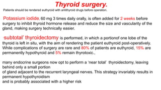
1. Thyroid surgery.
Patients should be rendered euthyroid with antithyroid drugs before operation.
Potassium iodide, 60 mg 3 times daily orally, is often added for 2 weeks before
surgery to inhibit thyroid hormone release and reduce the size and vascularity of the
gland, making surgery technically easier.
‘subtotal’ thyroidectomy is performed, in which a portionof one lobe of the
thyroid is left in situ, with the aim of rendering the patient euthyroid post-operatively.
While complications of surgery are rare and 80% of patients are euthyroid, 15% are
permanently hypothyroid and 5% remain thyrotoxic.,
many endocrine surgeons now opt to perform a ‘near total’ thyroidectomy, leaving
behind only a small portion
of gland adjacent to the recurrent laryngeal nerves. This strategy invariably results in
permanent hypothyroidism
and is probably associated with a higher risk
2. Radioactive iodine. 131I
131I is administered orally as a single dose400 MBq (10 mCi) is given orally.
, and is trapped and organified in the thyroid Although 131I decays within a few weeks, it has long-lasting
inhibitory effects on survival and replication of follicular cells. This regimen is effective in 75% of patients within
4–12 weeks.
During the lag period, symptoms can be controlled by a β-blocker or, in more severe cases, by
carbimazole. However, reduces the efficacy of 131I therapy because it prevents organification of 131I in the gland,
and so should be carbimazole avoided until 48 hours after radio-iodine administration.
If thyrotoxicosis persists after 6 months, a further dose of 131I can be given.
The disadvantage of 131I treatment
1 hypothyroidism.
2 131I isusually avoided in patients with Graves’ ophthalmopathy It can be administered with caution in those with mild
or ‘burnt-out’ eye disease, when it is customary to cover the treatment with a 6-week tapering course of oral
prednisolone. 3 In women of reproductive age, pregnancy must be excluded before administration of 131I and avoided
for 6 months there after; men are also advised against fathering children for 6 months after receiving 131I
3.
4.
5. The majority of patients require no treatment other than reassurance.
Smoking cessation should be actively
Methylcellulose eye drops and gel counter
the gritty discomfort of dry eyes, and tinted glasses or
side shields attached to spectacle frames reduce the
excessive lacrimation triggered by sun or wind. In
patients with mild Graves’ ophthalmopathy,
oral selenium
(100 μg twice daily for 6 months) improves
quality of life, reduces ocular involvement and slows
progression of disease; the mechanism of action is not
known but may relate to an antioxidant effect More severe inflammatory episodes are treated
with glucocorticoids (e.g. daily oral prednisolone or
pulsed IV methylprednisolone) and sometimes
orbitalradiotherapy
. There is also an increasing trend to use
immunosuppressant therapies, such as ciclosporin, in
combination with glucocorticoids.
6.
7. Pretibial myxoedema
This infiltrative dermopathy occurs in fewer than 10% of patients
with Graves’ disease and has similar pathological
features as occur in the orbit. It takes the form of raised pink-
coloured or purplish plaques on the anterior
aspect of the leg, extending on to the dorsum of the foot The
lesions may be itchy and the skin may have
a ‘peau d’orange’ appearance with growth of coarse hair; less
commonly, the face and arms are affected.
Treatment is rarely required, but in severe cases topical
glucocorticoids may be helpful.
8.
9.
10.
11.
12. Thyrotoxicosis in pregnancy
The coexistence of pregnancy and thyrotoxicosis is unusual, as anovulatory cycles are common in thyrotoxic
patients and autoimmune disease tends to remitduring pregnancy, when the maternal immune responseis
suppressed. Thyroid function tests must be interpretedin the knowledge that thyroid-binding globulin, and hence total
T4 and T3 levels, are increased in pregnancyand that TSH reference ranges may be lower (see
a fully suppressed TSH with elevatedfree thyroid hormone levels indicates thyrotoxicosis.
The thyrotoxicosis is almost always caused by Graves’ disease. Both mother and fetus must be considered,
since maternal thyroid hormones, TRAb and antithyroiddrugs can all cross the placenta to some degree, exposing
the fetus to the risks of thyrotoxicosis, iatrogenic hypothyroidism and goitre. Poorly controlled maternal
thyrotoxicosis can result in fetal tachycardia, retardation, prematurity, stillbirth and possibly even congenital
malformations. Antithyroid drugs are the treatment of choice for thyrotoxicosis in pregnancy. Carbimazole has been
associated with rare cases of embryopathy, particularly skin defect known as aplasia cutis, and should be
a
13. Hypothyroidism
Hypothyroidism is a common condition with various
causes (but autoimmune disease(Hashimoto’s
thyroiditis) and thyroid failure following131I or
surgical treatment of thyrotoxicosis account for
over 90% of cases, except in areas where iodine
deficiency is endemic. Women are affected
approximately six times more frequently than men
14.
15. Features of hypothyroidism
consequence of prolonged hypothyroidism is the infiltration of many body tissuesby
the mucopolysaccharides, hyaluronic acid and chondroitin sulphate, resulting in a low-
pitched voice, poorhearing, slurred speech due to a large tongue, and compressionof
the median nerve at the wrist (carpal tunnel syndrome).
Infiltration of the dermis gives rise to nonpitting oedema (myxoedema), which is most
marked inthe skin of the hands, feet and eyelidsThe resultantperiorbital puffiness is
often striking and may be combined with facial pallor due to vasoconstriction
andanaemia, or a lemon-yellow tint to the skin caused bycarotenaemia, along with
purplish lips and malar flush. Most cases of hypothyroidism are not clinically obvious,
. maintained so that the diagnosis is not overlooked inindividuals complaining of non-
specific symptoms suchas tiredness, weight gain, depression or carpal tunnel
16.
17.
18. Hashimoto’s thyroiditis
Hashimoto’s thyroiditis is characterised by destructive lymphoid infiltration of the thyroid, leading to a varying
degree of fibrosis and thyroid enlargement.
There is an increased risk of thyroid lymphoma although this is exceedingly rare. The
autoimmune hypothyroidism is confusing. Some authorities reserve the term ‘Hashimoto’s
thyroiditis’ for patients with positive antithyroid peroxidase autoantibodies and a firm goitre who may or may
not
be hypothyroid, and use the term ‘spontaneous atrophic hypothyroidism’ for hypothyroid patients without a
goitre in whom TSH receptor-blocking antibodies may be more important than antiperoxidase antibodies.
However, these syndromes can both be considered asvariants of the same underlying disease process.
Hashimoto’s thyroiditis increases in incidence with age and affects approximately 3.5 per 1000 women
and 0.8 per 1000 men each year
. Many present with a small or moderately sized diffuse goitre, firm or rubbery in consistency
. The goitre may be soft, however, and impossible to differentiate from
. Around 25%of patients are hypothyroid at presentation. In the
Antithyroid peroxidase antibodies are present in the serum in more than 90% of
patients with Hashimoto’s thyroiditis. In those under
19. Sub acute(de Quervain’s)
thyroiditis
In its classical painful form, sub acute thyroiditis is a transient inflammation of the
thyroid gland occurring There is pain in the region of the thyroid that may radiate to the
angle of the jaw and the ears, and is made worse by swallowing, coughing and
movement of the neck. The thyroid is usually palpably enlarged and tender. Systemic upset is
common. Affected patients are usually females aged 20–40 years.. The condition can also be
precipitated by drugs, including interferon-α and lithium.
also after infection with. with damage to follicular cells and impaired synthesisCoxsackie, mumps
or adenoviruses
of new thyroid hormones. As a result, T4 and T3 levels are raised for 4–6 weeks until the pre-formed
colloid is depleted. Thereafter, there is usually a period of hypothyroidism of variable severity before the
follicular cells recover and normal thyroid function is restored within 4–6 is suppressed. Low-titre thyroid
autoantibodies appear transiently in the serum, and the erythrocyte sedimentation
rate (ESR) is usually raised.
High-titre autoantibodie ssuggest an underlying autoimmune pathology and greater
risk of recurrence and ultimate progression to hypothyroidism.
20.
21. Management
Treatment is with levothyroxine replacement. It is customary
to start with a low dose of 50 μg per day for3 weeks, increasing thereafter
to 100 μg per day for afurther 3 weeks and finally to a maintenance dose
of100–150 μg per day.
In younger patients, it is safe toinitiate levothyroxine at a higher dose
to allow a more rapid normalisation of thyroid hormone levels.
Levothyroxine has a half-life of 7 days so it should always be taken as a
single daily dose and at least 6 weeks should pass before repeating
thyroid function tests and adjusting the dose, usually
by 25 μg per day. Patients feel better within 2–3 weeks.
Reduction in weight and periorbital puffiness occurs
quickly, but the restoration of skin and hair texture may take 3–6 months It is
important to measure thyroid function every1–2 years
22. Levothyroxine replacement in ischaemic heart disease
exacerbation of myocardial ischaemia, infarction and
sudden death are recognised complications of levothyroxine
replacement, even using doses as low as 25 μgper day. In
replacement should be introduced at low dose and increased
very slowly under specialistsupervision. It has been suggested
that T3 has an advantage over T4, since T3 has a shorter half-
life and anyadverse effect will reverse more quickly, but the
moredistinct peak in hormone levels after each dose of T3 isa
disadvantage. Coronary angioplasty or bypass surgerymay be
required if angina is exacerbated by levothyroxine
23. Myxoedema coma
depressed level of consciousness,
usually in an elderly patient who appears myxoedematous. Body temperature may be as low as 25°C, convulsions
are not uncommon and cerebrospinal fluid (CSF) pressure and protein content are raised. The mortalityrate is 50%
and survival depends on early recognitionand treatment of hypothyroidism and other factors
contributing to the altered consciousness level, as medication, cardiac failure, pneumonia, dilutional
hyponatraemia and respiratory failure.
Myxoedema coma is a medical emergency and treatmentmust begin before biochemical confirmation of the
diagnosis. Suspected cases should be treated with anintravenous injection of 20 μg triiodothyronine, followed
by further injections of 20 μg 3 times daily unti lthere is sustained clinical improvement. In survivors,
there is a rise in body temperature within 24 hours
after 48–72 hours, it is usually possible to switch patientsto oral levothyroxine in a dose of 50 μg daily. Unlessit is
apparent that the patient has primary hypothyroidism, the thyroid failure should also be assumed to be secondary to
hypothalamic or pituitary disease
and treatment given with hydrocortisone 100 mg IM
3 times daily, pending the results of T4, TSH and cortisol
measurement (p. 787). Other measures include slow
rewarming (p. 105), cautious use of intravenous fluids,
broad-spectrum antibiotics and high-flow oxygen. Occasionally,
assisted ventilation may be necessary
24. Hypothyroidism in pregnancy
Most pregnant women with primary hypothyroidismrequire an increase in the dose of
levothyroxine of
approximately 25–50 μg daily to maintain normal TSH levels. This may reflect increased
metabolism of thyroxine
by the placenta and increased serum thyroxinebindingglobulin during pregnancy,
resulting in an increase in the total thyroid hormone pool to maintain the same free T4
and T3 concentrations
. Inadequate maternal T4 therapy may be associated with impaired cognitive
development in an unborn child and so women are usually advised to increase their
daily levothyroxine dose by 25 μg when pregnancy is confirmed. Serum TSH
and free T4 should be measured during each trimester and the dose of
levothyroxine adjusted to maintain a normal TSH
25. Subclinical hypothyroidism
Serum TSH is raised, and serum T3 and T4 lower end of the
reference range.
This may persist for many years, although there is a risk of
progression to overt thyroid failure, particularly if
antibodies to thyroid peroxidase are present or if the
TSH rises above 10 mU/L.
Levothyroxine should be given in a dose sufficient to
restore the serum TSH concentration
26. Subclinical thyrotoxicosis
Serum TSH is decrease , and serum T3 and T4
are at the upper end of the reference range.
This combination
in older patients with multinodular goitre.
increased risk atrial fibrillation and osteoporosis,
mild thyrotoxicosis and require therapy, usually
with 131I. Otherwise, annual review is essential, as
the conversion rate to overt thyrotoxicosis
5% each year.
27. Non-thyroidal illness (‘sick euthyroidism’)
This typically presents with a low serum TSH, raised T4and normal or low T3
,.
These abnormalities are caused by
1 decreased peripheral conversion of T4 to T3,
2 altered levels of binding proteins and their affinity for thyroid
hormones
, 3 reduced secretion of TSH. During convalescence, serum
28. Symptoms of hypothyroidism with normal thyroid function tests
The classic symptoms of hypothyroidism are, by theirvery nature, non-specific There is a wide
differential diagnosis for symptoms such as ‘fatigue’, weight gain’ and ‘low mood’. As has been noted,
outside the context of pituitary and hypothalamicdisease, serum TSH is an excellent measure of an individual’s
thyroid hormone status. However, some individualsbelieve that they have hypothyroidism despite
normal serum TSH concentrations. There are a large number of websites which claim that serum TSH is not
a good measure of thyroid hormone status and suggestthat other factors, such as abnormalities of T4 to T3 conversion,
may lead to low tissue levels of active thyroidhormones. Such websites often advocate a variety of
tests of thyroid function of dubious scientific validity, including measurement of serum reverse T3, 24-hour
urine T3, basal body temperature, skin iodine absorption, and levels of selenium in blood and urine. Individuals
who believe they have hypothyroidism, despitenormal conventional tests of thyroid function, can be
difficult to manage. They require reassurance that theirsymptoms are being taken seriously and that organic
disease has been carefully considered; if their symptomspersist, then referral to a team specialising in medically
unexplained symptoms should be considered.
‘
29. Iodine-induced thyroid dysfunction
Iodine has complex effects on thyroid function. Ver yhigh concentrations of iodine inhibit thyroid
hormone release and this forms the rationale for iodine treatmen tof thyroid storm and prior to
thyroid surgery for thyrotoxicosis . Iodine administration initially enhances, but then inhibits,
iodination of tyrosine and thyroid hormone synthesis
The resulting effect of iodine on thyroid function varies according to whether the patient has an
iodine-deficient diet or underlying thyroid disease. In iodine-deficient parts of the
world, transient thyrotoxicosis may be precipitated by prophylactic iodinisation programmes.
In iodine-sufficient areas, thyrotoxicosis can be precipitated by radiographic
contrast medium or expectorants in individuals who have underlying thyroid disease
predisposing to thyrotoxicosis, such as multinodular goiter or Graves’ disease in remission.
Induction of thyrotoxicosis by iodine is called the Jod–Basedow effect.
Chronic excess iodine administration can, however, result in hypothyroidism. Increased iodine
within the thyroid gland down-regulates iodine trapping,
30. Amiodarone
The anti-arrhythmic agent amiodarone has a structurethat is analogous to that of T4 and contains
huge amounts of iodine; a 200 mg dose contains 75 mg iodine, compared with a daily dietary
requirement of
just 125 μg. Amiodarone also has a cytotoxic effect on thyroid follicular cells and inhibits conversion of T4
toT3. Most patients receiving amiodarone have normal thyroid function, but up to 20% develop hypothyroidism
or thyrotoxicosis and so thyroid function should be monitored regularly. The ratio of T4:T3 is elevated and
TSH provides the best indicator of thyroid function.
The thyrotoxicosis can be classified as either:
• type I: iodine-induced excess thyroid hormone synthesis in patients with an underlying thyroid
disorder, such as nodular goitre or latent Graves’ disease treatment Antithyroid drugs may be effective in
patients with the type I form, but are ineffective in type
II thyrotoxicosis.
• type II: thyroiditis due to a direct cytotoxic effect if amiodarone administration results in a transient
Thyrotoxicosis treatment
Prednisolone is beneficial in the type II
42. More than 99% of total body calcium is in
bone. Prolonged exposure of bone to high
levels of PTH is associated with increased
osteoclastic activity and new bone
formation, but the net effect is to cause bone
loss with mobilisation of calcium into the
extracellular
43. 50% of total calcium is bound to organic ions, such as citrate or
phosphate, and to proteins, especially albumin. 50% of total calcium is
free
Accordingly, if the serum albumin level is reduced, total calcium
concentrations should be ‘corrected’ by adjusting
the value for calcium upwards by 0.02 mmol/L
(0.4 mg/dL) for each 1 g/L reduction in albumin below
40 g/L. If albumin concentrations are significantly low, as in severe
acute illness and other chronic illness suchas liver cirrhosis, this
correction is less accurate and measurement of ionised calcium is
needed.
Calcitoninis secreted from the parafollicular C cells of the thyroid gland.
Although it is a useful tumour marker in medullary carcinoma of thyroid and
can be given therapeutically in Paget’s disease of bone
44.
45. Hypercalcaemia
Hypercalcaemia is one of the most common biochemical abnormalities and is often detected during routine
biochemical analysis in asymptomatic patients. However, it can present with chronic symptoms, as
described below,
and occasionally as an acute emergency with severe hypercalcaemia and dehydration.
Causes of hypercalcaemia are listed in Of these, primary hyperparathyroidism and malignant
hypercalcaemia are by far the most common. Familial hypocalciuric hypercalcaemia (FHH) is a
rare but important
cause that needs differentiation from primary hyperparathyroidism (HPT). Lithium may cause
hyperparathyroidism
by reducing the sensitivity of the calcium-sensing receptor.
46.
47. Clinical assessment
50% of patients with primary hyperparathyroidismare
asymptomatic while others have nonspecific
symptoms such as fatigue, depression and generalised aches
and pains. Some present with renal calculi 5% of first stone
formers, 15% of recurrent stone formers
have
Hypertension is a common feature of hyperparathyroidism.
Parathyroid
tumours are almost never palpable.
48. 1 High plasmaphosphate and alkaline phosphatase accompanied by renal impairment
suggest tertiary hyperparathyroidism.
2nephrocalcinosis and renaltubular impairment
3 Patients with FHH can present with a similar biochemical picture to primary
hyperparathyroidism but typically have low urinary calcium excretion (a ratio of urinary calcium
clearance to creatinine clearance of < 0.01). The diagnosis of FHH can be confirmed by screening
family members for hypercalcaemia and/or a mutation in the gene encoding the calcium-sensing
receptor.
4 If PTH is low and no other cause is apparent, then malignancy with or without bony
metastases is likely PTH-related peptide
49. management
1 Patients should initially be treated with intravenous 0.9% saline to improve renal function and increase
urinary calcium excretion. This alone often results in clinical improvement
2 Intravenous bisphosphonates should be given to inhibit bone resorption. serum calcium levels to
normal within 5 days The duration of action is up to 4 weeks and repeated therapy can be given at 3–4-weekly
intervals
3Calcitonin acts rapidly to increase calcium excretion and to reduce bone resorption and can be combined
with fluid and bisphosphonate therapy for the first 24–48 hours in patients with life-threatening hypercalcaemia.
50.
51. The prevalence of primary hyperparathyroidism is about 1 in 800 and it is 2–3 times more common in
women than men; 90% of patients are over 50 years of age. It also occurs in the familial MEN syndromes
Clinical and radiological features
The clinical presentation of primary hyperparathyroidism is bone disease is now rare due to earlier diagnosis and treatment.
Osteitis fibrosa results from increased bone resorption by osteoclasts with fibrous replacement in the
lacunae. This may present as bone pain and tenderness, fracture and deformity.
Chondrocalcinosis can occur due to deposition of calcium pyrophosphate crystals within articular cartilage. It
typically affects the menisci at the knees and can result in secondary degenerative arthritis or predispose to attacks of acute
pseudogout
Skeletal X-rays are usually normal in mild primary hyperparathyroidism, but in patients with advanced disease
characteristic changes are observed. In the early stages there is demineralisation, with subperiosteal
erosions and terminal resorption in the phalanges.
A ‘pepper-pot’ appearance may be seen on lateral X-rays of the skull.
In nephrocalcinosis, scattered opacities may be visible within the renal outline.
There may be soft tissue calcification in arterial walls and hands and in the cornea
.
52. Investigations
The diagnosis can be confirmed by finding a raised PTH level in the presence of hypercalcaemia, provided that
FHH is excluded .
Parathyroid scanning by 99mTcsestamibi scintigraphy and/or ultrasound
, in an attempt to localise an adenoma and allow a targetedresection. However, negative imaging does not exclude
the diagnosis.
53. Surgery is usually
indicated for individuals aged less than 50 years, with clear-cut symptoms or documented complications (such
as peptic ulceration, renal stones, renal impairment or osteoporosis), and (in asymptomatic
patients) significant hypercalcaemia (corrected serum calcium > 2.85 mmol/L (> 11.4 mg/dL)).
Patients who are treated conservatively without surgery should have calcium biochemistry and renal function checked
annually and bone density monitored periodically. They should be encouraged to maintain a high oral fluid
intake to avoid renal stones
.
54. Familial hypocalciuric hypercalcaemia
This autosomal dominant disorder is caused by an inactivating mutation in one of the alleles of the calcium sensing
receptor gene which reduces the ability of the parathyroid gland to ‘sense’ ionised calcium concentrations.
As a result, higher than normal calcium levels are required to suppress PTH secretion. The typical presentation
is with mild hypercalcaemia with PTH concentrations that are ‘inappropriately’ at the upper end of the reference range
or are slightly elevated
. Calcium-sensing receptors in the renal tubules are also affected and thisleads to increased renal tubular
reabsorption of calcium and hypocalciuria
.
The hypercalcaemia of FHH is always asymptomatic and complications do not occur. The main risk of FHH is of
the patient being subjected to an unnecessary(and ineffective) parathyroidectomy if misdiagnosed as having primary
hyperparathyroidism.
Testing of family members for hypercalcaemia is helpful in confirming the diagnosis and it is also possible to
perform
genetic testing. No treatment is necessary.
55.
56. Hypocalcaemia
may also develop as a result of magnesium depletion and should be considered in patients
with malabsorption, on diuretic or proton pump inhibitor therapy, and/or with a history of alcohol excess.
Magnesium deficiency causes hypocalcaemia by impairing the ability of the parathyroid glands to secrete
PTH
(resulting in PTH concentrations that are low or inappropriately in the reference range) and may also impair
the actions of PTH on bone and kidney
57. Clinical assessment
Mild hypocalcaemia is often asymptomatic but, with more profound reductions in serum
calcium, tetany canoccur.
This is characterised by muscle spasms due to increased excitability of peripheral
nerves.
Children are more liable to develop tetany than adults and present with a characteristic
triad of carpopedal spasm {the hands adopt a characteristic position with flexion of
the metacarpophalangeal joints}
, stridor caused by spasm of the glottis.
and convulsions, although one or more of these may be found independently of the
others.
frequent. Stridor is Adults can also develop carpopedal spasm in association with tingling
of the hands and feet and around the
Trousseau’s sign; inflation of a sphygmomanometer cuff on the upper arm to
more than the systolic blood pressurefollowed by carpal spasm within 3 minutes.
is Less specificis Chvostek’s sign, tapping over the branches of the facial nerve
as they emerge from the parotid gland produces twitching of the facial muscles.
58. Hypocalcaemia can cause papilloedema and
prolongation of the ECG QT interval, which may
predisposeto ventricular arrhythmias. Prolonged
hypocalcaemia and hyperphosphataemia (as in
hypoparathyroidism)
may cause calcification of the basal ganglia,
grand mal epilepsy, psychosis and cataracts.
Hypocalcaemia associated
with hypophosphataemia, as in vitamin D deficiency,
causes rickets in children and osteomalacia in
Adults
59. Pseudohypoparathyroidism
In this disorder, the individual is functionally hypoparathyroid, but instead of PTH deficiency there is tissue resistance to the
effects of PTH, such that PTH concentrations are markedly elevated. The PTH receptor itself is normal, but there are
defective post-receptor mechanisms due to mutations at the GNAS1 locus. There are several subtypes, but in the most
common form (type 1a) features include short stature, short 4th metacarpals and metatarsals, rounded face, obesity and
subcutaneous calcification (Albright's hereditary osteodystrophy, AHO).
The term 'pseudo-pseudohypoparathyroidism' is used to describe patients with AHO in whom
serum calcium and PTH concentrations are normal. The inheritance of these disorders is an example of genetic imprinting (p.
49); inheritance of the defective allele from a mother with pseudohypoparathyroidism results in pseudohypoparathyroidism
in the offspring, but inheritance from the father results in pseudo-pseudohypoparathyroidism.
60.
61.
62.
63.
64.
65.
66.
67. Some clinical features are more common in ectopic ACTH syndrome. Unlike pituitary
tumours secreting ACTH, ectopic tumours have no residual negative feedback sensitivity
to cortisol, and both ACTH and cortisol levels are usually higher than with other causes.
Very high ACTH levels are associated with marked pigmentation.
high cortisol levels overcome the barrier of 11β-HSD2 in the kidney and cause
hypokalaemic alkalosis. Hypokalaemia aggravates both myopathy
hyperglycaemia (by inhibiting insulin secretion). When the tumour secreting ACTH is
malignant, then the onset is usually rapid and may be associated with cachexia. For these
reasons, the classical features of Cushing's syndrome are less common in ectopic ACTH
syndrome, and if present suggest that a less aggressive tumour (e.g. bronchial carcinoid) is
responsible.
In Cushing's disease, the pituitary tumour is usually a microadenoma
(< 10 mm in diameter); hence other features of a pituitary
macroadenoma (hypopituitarism, visual failure or disconnection
68.
69.
70. Management
Untreated Cushing's syndrome has a 50% 5-year mortality.
Most patients are treated surgically with medical therapy given for a few weeks prior to
operation. A number of drugs are used to inhibit corticosteroid biosynthesis, including metyrapone and
ketoconazole. The dose of these agents is best titrated against 24-hour urine free cortisol.
Cushing's disease
Trans-sphenoidal surgery with selective removal of the adenoma is the treatment of choice. Experienced
surgeons can identify microadenomas which were not detected by MRI and cure about 80% of patients. If the
operation is unsuccessful, then bilateral adrenalectomy is an alternative.
If bilateral adrenalectomy is used in patients with pituitary-dependent Cushing's syndrome, then there is a risk
that the pituitary tumour will grow in the absence of the negative feedback suppression previously provided by elevated cortisol
levels. This can result in Nelson's syndrome, with an aggressive pituitary macroadenoma and very high ACTH
levels causing pigmentation. Nelson's syndrome can be prevented by pituitary irradiation.
71. Adrenal tumours
Adrenal adenomas are removed by laparoscopy or a loin incision. Surgery offers the only prospect of cure for
adrenocortical carcinomas, but in general prognosis is poor with high rates of
recurrence even in patients with localised disease at presentation.
Although often used, there is little evidence that radiotherapy, chemotherapy or the adrenolytic drug
mitotane improves recurrence rates or survival.
Ectopic ACTH syndrome
Localised tumours such as bronchial carcinoid should be removed surgically. In patients with incurable malignancy it is
important to reduce the severity of the Cushing's syndrome using medical therapy
72.
73. Management of glucocorticoid withdrawal
All glucocorticoid therapy, even if inhaled or applied topically, can suppress the HPA axis. In
practice, this is only likely to result in a crisis due to adrenal insufficiency on withdrawal of
treatment if glucocorticoids have been administered orally or systemically for longer than 3
weeks, if repeated courses have been prescribed within the previous year, or if the dose is
higher than the equivalent of 7.5 mg prednisolone per day.
Once the dose of glucocorticoid is reduced to a minimum (e.g. 4 mg prednisolone or 0.5 mg
dexamethasone per day), then measure plasma cortisol at 0900 hrs before the next dose. If this is
detectable, then perform an ACTH stimulation test to confirm that glucocorticoids can be
withdrawn completely.
Even once glucocorticoids have been successfully withdrawn, short-term replacement
therapy is often advised during significant intercurrent illness occurring in subsequent
months, as the HPA axis may not be able to respond fully to severe stress.
Adrenal insufficiency
74.
75.
76.
77. Assessment of glucocorticoids
Random plasma cortisol is usually low in patients
with adrenal insufficiency, but it may be within
the normal range, Random measurement of
plasma cortisol cannot therefore be used to
confirm or refute the diagnosis, unless the value
is high (> 460 nmol/L (> 17 μg/dL)).
78. Assessment of mineralocorticoids
Assessment of mineralocorticoids
Mineralocorticoid secretion in patients with suspected
Addison's disease cannot be adequately assessed by
electrolyte measurements since hyponatraemia occurs in
both aldosterone and cortisol deficiency (see Box 20.45 and
p. 435). Hyperkalaemia is common, but not universal, in
aldosterone deficiency. Plasma renin activity
and aldosterone should be measured in the supine
position. In mineralocorticoid deficiency, plasma renin
activity is high, with plasma aldosterone being either low or
in the lower part of the normal range.
79. Assessment of adrenal androgens
Assessment of adrenal androgens
This is not necessary in men because testosteron
from the testes is the principal androgen. In wome
dehydroepiandrosterone (DHEA) and androstened
may be measured in a random specimen of blood
though levels are highest in the morning.
80.
81. Hydrocortisone (cortisol) is the drug of choice. In someone
who, hydrocortisone should be given by mouth,10- 15 mg on
waking and 5 mg at around 1800 hrs..
weight gain usually indicates over-replacement, whilst
persistent lethargy or hyperpigmentation may be due to an
inadequate dose. Measurement of plasma cortisol levels is
unhelpful,
These are physiological replacement doses which should not
cause Cushingoid side-effects.
82. Androgen replacement
DHEA (approximately 50 mg/day) is
sometimes given to women with primary
adrenal insufficiency. Some trials have
suggested that DHEA increases libido and
sense of well-being, but complications
include acne and hirsutism.
83.
84. incidental adrenal mass
It is not uncommon for a mass in the adrenal gland to be identified on a CT or MRI scan of the abdomen that has been
performed for another indication. Such lesions are known as adrenal 'incidentalomas'. They are
present in up to 10% of adults and the prevalence increases with age.
Eighty-five per cent of adrenal incidentalomas are non-functioning
adrenal adenomas. The remainder includes functional tumours of the adrenal cortex (secreting cortisol,
aldosterone or androgens), phaeochromocytomas, primary and secondary carcinomas, hamartomas and other rare disorders
including granulomatous infiltrations.
Clinical assessment and investigations
There are two key questions to be resolved: is the lesion
secreting hormones, and is it benign or malignant?
Patients with an adrenal incidentaloma are usually
asymptomatic.
85. Investigations should include a 24-hour urine collection for metanephrines and urine free cortisol
and,
in women, measurement of serum testosterone, DHEA and androstenedione
Patients with hypertension should be investigated for mineralocorticoid excess as described
below.
CT and MRI are equally effective in assessing the malignant potential of an adrenal mass, using
the following parameters:
Size. The larger the lesion, the greater the malignant potential. Around 90% of adrenocortical
carcinomas are > 4 cm in diameter, but specificity is poor since only approximately 25% of such
lesions are malignant.
Configuration. Homogeneous and smooth lesions are more likely to
be benign. Adenomas are usually lipid-rich
86. Management
Functional lesions and tumours > 5 cm in diameter should be removed by laparoscopic
adrenalectomy. In patients with radiologically benign, non-functioning lesions < 5 cm in diameter, surgery is
only required if serial imaging suggests tumour growth.
87.
88. Primary hyperaldosteronism
as 10% of people with hypertension. Indications to test for mineralocorticoid
excess in hypertensive patients include hypokalaemia (including hypokalaemia
induced by thiazide diuretics), poor control of blood pressure with conventional
therapy, a family history of early-onset hypertension, or presentation at a young
age.
It is important to differentiate primary hyperaldosteronism, caused by an intrinsic
abnormality of the adrenal glands resulting in aldosterone excess, from secondary
hyperaldosteronism, which is usually a consequence of enhanced activity of renin
in response to inadequate renal perfusion and hypotension. Most individuals with
primary hyperaldosteronism have bilateral adrenal hyperplasia
(idiopathic hyperaldosteronism), while only a minority have an aldosterone-
producing adenoma (APA; Conn's syndrome).
89. Clinical features
Individuals with primary hyperaldosteronism are usually asymptomatic,
but they may have features of sodium retention or potassium loss.
Sodium retention may cause oedema, while hypokalaemia may causes
muscle weakness (or even paralysis, especially in Chinese),
polyuria (secondary to renal tubular damage which produces
nephrogenic diabetes insipidus)
tetany (because of associated metabolic alkalosis and low ionised
calcium).
Blood pressure is elevated but accelerated phase hypertension is
rare.
90.
91. Management
Mineralocorticoid receptor antagonists are
valuable in treating both hypokalaemia and
hypertension in all forms of mineralocorticoid
excess. Up to 20% of males develop
gynaecomastia on spironolactone. Eplerenone
or amiloride (10-40 mg/day), which blocks the
epithelial sodium channel regulated by
aldosterone, is an alternative.
92. Phaeochromocytoma and paraganglioma
These are rare neuro-endocrine tumours that may secrete
catecholamines (adrenaline/epinephrine,
noradrenaline/norepinephrine) and are responsible for less than 0.1%
of cases of hypertension.
Approximately 80% of these tumours occur in the adrenal medulla
(phaeochromocytomas), while 20% arise elsewhere in the body in
sympathetic ganglia (paragangliomas). Most are benign but
approximately 15% show malignant . Around 25% of
phaeochromocytomas are associated with inherited disorders, including
neurofibromatosis , von Hippel-Lindau syndrome and MEN type 2
Paragangliomas are particularly associated with mutations in the
succinate dehydrogenase B, C and D genes.
93. Investigations
Excessive secretion of catecholamines can be confirmed by measuring metabolites in plasma and/or urine
(metanephrine and normetanephrine). There is a high 'false positive' rate as misleading metanephrine
concentrations may be seen in stressed patients (during acute illness or following vigorous exercise or severe
pain) and following ingestion of some drugs such as tricyclic antidepressants.
this reason, a repeat sample should usually be requested if elevated levels are found, although as
a rule the higher the concentration of metanephrines, the more likely the diagnosis of phaeochromocytoma/paraganglioma.
Serum chromogranin A is often elevated and may be a useful tumour marker in patients with non-secretory tumours
and/or metastatic disease.
Genetic testing should be considered in individuals with other features of a genetic syndrome, with a family history of
phaeochromocytoma/paraganglioma and in those presenting under the age of 50 years.
Localisation
Phaeochromocytomas are usually identified by abdominal CT or MRI . Localisation of paragangliomas may be more
difficult. Scintigraphy using meta-iodobenzyl guanidine (MIBG) can be useful, particularly if combined with CT.
94. Medical therapy
is required to prepare the patient for surgery, preferably for a minimum of 6 weeks
to allow restoration of normal plasma volume.
The most useful drug in the face of very high circulating catecholamines is the α-blocker phenoxybenzamine (10-20
mg orally 6-8-hourly) because it is a non-competitive antagonist, unlike prazosin or doxazosin. If α-blockade produces a marked
tachycardia, then a β-blocker (e.g. propranolol) or combined α- and β-antagonist (e.g. labetalol) can be added. On
no account should the β-antagonist be given before the α-antagonist, as it may cause a paradoxical rise in blood pressure due to
unopposed α-mediated vasoconstriction.
During surgery sodium nitroprusside and the short-acting α-antagonist
phentolamine are useful in controlling hypertensive episodes which may result from anaesthetic
induction or tumour mobilization
Post-operative hypotension may occur and require volume expansion and, very occasionally,
noradrenaline (norepinephrine) infusion. This is uncommon if the patient has been prepared adequately with
phenoxybenzamine.
95.
96.
97.
98.
99.
100.
101.
102.
103.
104.
105.
106.
107.
108.
109.
110.
111.
112.
113.
114.
115.
116.
117.
118.
119.
120.
121.
122.
123.
124.
125.
126.
127.
128.
129.
130.
131.
132.
133.
134.
135. Nutrients
the diet can be classified into ‘macronutrients’,
which are eaten in relatively large amounts to
provide fuel for energy, and ‘micronutrients’ (e.g.
vitaminsand minerals), which do not contribute to
energy balance but are required in small amounts
because they are not synthesised in the body.
136. Energy intake is determined by the ‘macronutrient’
content of food. Carbohydrates, fat, protein and alcohol
provide fuel for oxidation in the mitochondria to
generate
energy (as adenosine triphosphate (ATP). The
energy provided by each of these elements differs:
• carbohydrates (16 kJ/g)
• fat (37 kJ/g)
• protein (17 kJ/g)
• alcohol (29 kJ/g).
137.
138.
139.
140.
141.
142.
143.
144. Vitamins are organic substances with key
roles in certain
metabolic pathways, and are categorised into
those that
are fat-soluble (vitamins A, D, E and K) and those
that
are water-soluble (vitamins of the B complex
group and
vitamin C).
145.
146.
147.
148.
149. Vitamin A (retinol)
Pre-formed retinol is found only in foods of animalorigin. Vitamin A can also be derived from
carotenes,
which are present in green and coloured vegetables and some fruits. Carotenes provide most of
the total
vitamin A in the UK, and constitute the only supply invegans. Retinol is converted to several
other important
molecules:
• 11-cis retinaldehyde is part of the photoreceptor complex in rods of the retina
.
• Retinoic acid induces differentiation of epithelial
cells by binding to specific nuclear receptors, which
induce responsive genes. In vitamin A deficiency,
mucus-secreting cells are replaced by keratinproducing
cells.
• Retinoids are necessary for normal growth, fetal
development, fertility, haematopoiesis and immune
function.
150. Globally, the most important consequence of
vitamin A deficiency is irreversible blindness in young
children.
Asia is most notably affected and the problem
is being addressed through widespread vitamin Asupplementation
programmes. Adults are not usually at
risk because liver stores can supply vitamin A when
foods containing vitamin A are unavailable.
151. Early deficiency causes impaired adaptation to thedark (night blindness). Keratinisation of the cornea
(xerophthalmia) gives rise to characteristic Bitot’s spots, and progresses to keratomalacia, with corneal ulceration,
scarring and irreversible blindness ).
Incountries where vitamin A deficiency is endemic, pregnantwomen should be advised to eat dark-green, leafy
vegetables and yellow fruits (to build up stores of retinolin the fetal liver), and infants should be fed the same.
WHO is according high priority to prevention in communitieswhere xerophthalmia occurs, giving single
prophylactic oral doses of 60 mg retinyl palmitate (providing200 000 U retinol) to pre-school children.
Thisalso reduces mortality from gastroenteritis and respiratory
infections.
152.
153. Repeated moderate or high doses of retinol can cause
liver damage, hyperostosis and teratogenicity.
Women
in countries where deficiency is not endemic are therefore
advised not to take vitamin A supplements in pregnancy.
Retinol intake may also be restricted in those at
risk of osteoporosis. Acute overdose leads to nausea and
headache, increased intracranial pressure and skin
desquamation.
Excessive intake of carotene can cause pigmentation
of the skin (hypercarotenosis); this gradually
fades when intake is reduced.
154.
155. The natural form of vitamin D, cholecalciferol or vitaminD3, is formed in the skin by the action of UV light on
7-dehydrocholesterol, a metabolite of cholesterol. Fewfoods contain vitamin D naturally and skin exposure to
sunlight is the main source. Moving away from theequator, the intensity of UV light decreases, so that at
a latitude above 50° (including northern Europe),
vitamin D is not synthesised in winter, and even above
30° there is seasonal variation. The body store accumulated
during the summer is consumed during the winter. Vitamin D is converted in the liver to 25-hydroxy vitamin
D (25(OH)D), which is further hydroxylated in thekidneys to 1,25-dihydroxy-vitamin D (1,25 (OH)2D), the
active form of the vitamin
1,25(OH)2D activates specific intracellular receptorswhich influence calcium metabolism, bone mineralisation
and tissue differentiation. The synthetic form, ergocalciferol, or vitamin D2, is considered to be less potent
than the endogenous D3.
156.
157. The effects of vitamin D deficiency (calcium
deficiency,
rickets and osteomalacia) are described on An
analogue of vitamin D (calcipotriol) is used for
treatment of skin conditions such as psoriasis.
Excessive
doses of cholecalciferol, ergocalciferol or the
hydroxylated
metabolites cause hypercalcaemia
158. -Vitamihasn E
αtocopherol. many direct metabolic
actions:
• It prevents oxidation of polyunsaturated
fatty acids
in cell membranes by free radicals.
1 helps maintain cell membrane structure.
2 DNA synthesis and cell signalling.
3 anti-inflammatory and immunesystems.
159. Human deficiency is rare and has only been
described
in premature infants and in malabsorption. It can
cause a mild haemolytic anaemia, ataxia and
scotomas. visual
160. Vitamin K is supplied in the diet mainly as vitamin K1
Gla(phylloquinone) in the UK, or as vitamin K2 (menaquinone)
from fermented products in parts of Asia. Vitamin
K2 is also synthesised by bacteria in the colon. Vitamin
K is a co-factor for carboxylation reactions: in particular,
the production of γ-carboxyglutamate (gla).
residues are found in four of the coagulation factor proteins
(II, VII, IX and X; , conferring their capacity
to bind to phospholipid surfaces in the presence of
calcium. Other important gla proteins are osteocalcin
and matrix gla protein, which are important in bone
mineralisation
161. Vitamin K deficiency leads to delayed coagulation
and bleeding.
In obstructive jaundice, dietary vitamin K
is not absorbed and it is essential to administer the
vitamin in parenteral form before surgery.
Warfarin andrelated anticoagulants act by antagonising
vitamin K. Vitamin K is given routinely to newborn
babies to prevent haemorrhagic disease. Symptoms of
excess have been reported only in infants, with synthetic
preparations linked to haemolysis and liver damage
162. Water-soluble vitamins
Thiamin (vitamin B1 )
Thiamin is widely distributed in foods of both vegetableand
animal origin. Thiamin pyrophosphate (TPP) isa co-factor for
enzyme reactions involved in themetabolism of macronutrients
(carbohydrate, fat andalcohol), including
• decarboxylation of pyruvate to acetyl-co-enzyme A,
which bridges between glycolysis and the
tricarboxylic acid (Krebs) cycle
• transketolase activity in the hexose monophosphate
shunt pathway
• decarboxylation of α-ketoglutarate to succinate in
the Krebs cycle.
163. In thiamin deficiency, cells cannot metabolise
glucoseaerobically to generate energy as
ATP. Neuronal cellsare most vulnerable,
since they depend almost exclusivelyon
glucose for energy requirements. Impaired
glucose oxidation also causes an
accumulation ofpyruvic and lactic acids,
which produce vasodilatationand increased
cardiac output.
164. Deficiency – beri-beri
In the developed world, thiamin deficiency is
mainlyencountered in chronic alcoholics. Poor diet,
impairedabsorption, storage and phosphorylation of
thiamin inthe liver, and the increased requirements for
thiamin to metabolise ethanol all contribute. In the
developingworld, deficiency usually arises as a
consequence of adiet based on polished rice. The body
has very limitedstores of thiamin, so deficiency is manifest
after only 1month on a thiamin-free diet. There are two
forms of thedisease in adults:
165. • Dry (or neurological) beri-beri manifests with chronic
peripheral neuropathy and with wrist and/or foot
drop, and may cause Korsakoff’s psychosis and
Wernicke’s encephalopathy
• Wet (or cardiac) beri-beri causes generalised oedema
due to biventricular heart failure with pulmonary
congestion.
In dry beri-beri, response to thiamin administration
is not uniformly good. However, multivitamin therapy
seems to produce some improvement, suggesting
that other vitamin deficiencies may be involved.
Wernicke’s encephalopathy and wet beri-beri should
be treated without delay with intravenous vitamin B
and C mixture (‘Pabrinex’,
Korsakoff’s psychosis is irreversible and does not respond to thiamin
166. A rare but important effect of chronic alcohol misuse
is the Wernicke–Korsakoff syndrome. This organic brain
disorder results from damage to the mamillary bodies,
dorsomedial nuclei of the thalamus and adjacent areas
of periventricular grey matter caused by a deficiency of
thiamin (vitamin B1), which most commonly results
from long-standing heavy drinking and an inadequate
diet. It can also arise from malabsorption or even protracted
vomiting. Without prompt treatment (see below),
the acute presentation of Wernicke’s encephalopathy
(nystagmus, ophthalmoplegia, ataxia and confusion)
can progress to the irreversible deficits of Korsakoff’s
syndrome (severe short-term memory deficits and
confabulation, and also reduced red blood cell transketolase).
In those who die in the acute stage, microscopic
examination of the brain shows hyperaemia,
petechial haemorrhages and astrocytic prolifera
167. Riboflavin (vitamin B2 )
Riboflavin is required for the flavin co-factors
involved
in oxidation–reduction reactions. It is widely
distributed
in animal and vegetable foods. Levels are low in staple
cereals but germination increases its content. It is
destroyed under alkaline conditions by heat and by
exposure to sunlight.
Deficiency is rare in developed countries. It mainly
affects the tongue and lips and manifests as glossitis,
angular stomatitis and cheilosis. The genitals may be
affected, as well as the skin areas rich in sebaceous
glands, causing nasolabial or facial dyssebacea.
Rapid
168. Niacin (vitamin B3 )
Niacin encompasses nicotinic acid and nicotinamide. Nicotinamide is an essential part of the two
pyridinenucleotides, nicotinamide adenine dinucleotide (NAD) and
nicotinamide adenine dinucleotide phosphate
(NADP), which play a key role as hydrogen acceptors and donors for many enzymes. Niacin can be synthesised
in the body in limited amounts from the aminoacid tryptophan
.
Deficiency – pellagra
Pellagra was formerly endemic among poor people whosubsisted chiefly on maize, which contains niacytin, a
form of niacin that the body is unable to utilise. Pellagracan develop in only 8 weeks in individuals eating
diets
that are very deficient in niacin and tryptophanremains a problem in parts of Africa, and is occasionally
seen in alcoholics and in patients with chronic smallintestinal disease in developed countries.
Pellagra canoccur in Hartnup’s disease, a genetic disorder characterisedby impaired absorption of several
amino acids, including tryptophan.
It is also seen occasionally in carcinoid syndrome , when tryptophan is consumed
in the excessive production of 5-hydroxytryptamine(5-HT). Pellagra has been called the disease of the
three D
169.
170. • Dermatitis. Characteristically, there is erythemaresembling severe
sunburn, appearingsymmetrically over the parts of the body exposed to
sunlight, particularly the limbs and especiallyon the neck, but not the
face (Casal’s necklace,
The skin lesions may progress tovesiculation, cracking, exudation and
secondaryinfection.
• Diarrhoea. This is often associated with anorexia, nausea, glossitis
and dysphagia, reflecting thepresence of a non-infective inflammation
thatextends throughout the gastrointestinal tract.
• Dementia. In severe deficiency, delirium occursacutely and dementia
develops in chronic cases
171. Toxicity
Excessive intakes of niacin may lead to
reversible
hepatotoxicity.
Nicotinic acid is a lipid-lowering agent,
but at doses above 200 mg a day gives
rise to vasodilatorysymptoms (‘flushing’
and/orhypotension)
172. Pyridoxine (vitamin B6 )
Pyridoxine, pyridoxal and pyridoxamine are differentforms of vitamin B6 that undergo phosphorylation to
produce pyridoxal 5-phosphate (PLP). PLP is theco-factor for a large number of enzymes involved in the
metabolism of amino acids. Vitamin B6 is available inmost foods.
Deficiency is rare, although certain drugs, such asisoniazid and penicillamine, act as chemical antagonists
to pyridoxine.
Pyridoxine administration is effectivein isoniazid-induced peripheral neuropathy and some
cases of sideroblastic anaemia.
Large doses of vitaminB6 have an antiemetic effect in radiotherapy-induced nausea
. Although vitamin B6 supplements have become popular in the treatment of nausea in pregnancy, carpal
tunnel syndrome and premenstrual syndrome, there is no convincing evidence of benefit.
Very high doses ofvitamin B6 taken for several months can cause a sensorypolyneuropathy
.
173. Biotin
Biotin is a co-enzyme in the synthesis of fatty
acids, isoleucine and valine and is also involved
in gluconeogenesis.
Deficiency results from consuming very large
quantities of raw egg whites (> 30% energy
intake)
because the avidin they contain binds to and
inactivates
biotin in the intestine. It may also be seen after
long
periods of total parenteral nutrition. The clinical
174. Folate (folic acid)
Folates exist in many forms. The main circulating formis 5-
methyltetrahydrofolate. Folic acid is the stable synthetic form. Folate works
as a methyl donor for cellular methylationand protein synthesis. It is
directly involved in DNAand RNA synthesis, and requirements increase
during embryonic development
Folate deficiency may cause three major birth defects
(spina bifida, anencephaly and encephalocele) resulting
from imperfect closure of the neural tube, which takesplace 3–4 weeks after
conception.
Departmentof Health advises that women who have experienced a
pregnancy affected by a neural tube defect should take5 mg of folic acid daily
from before conception andthroughout the first trimester
All
175. women planning a pregnancy are advised to include good sources of
folate in their diet, and to take folate supplements throughout the first
trimester. Liver is the richest source of folate but an alternative source
(e.g. leafy vegetables) is advised in early pregnancy because of the
high vitamin A content of liver
Folate deficiency also has been associated with heart disease, dementia and
cancer.. There are now concerns that this may contribute to the increased
incidence of colon cancer through promotion of the growth of polyps.
176.
177. Hydroxycobalamin (vitamin B12 )
Vitamin B12 is a co-factor in folate co-enzyme recyclingand nerve myelination. Vitamin B12 and
folate are particularlyimportant in DNA synthesis in red blood cells
The haematological disorders (macrocytic ormegaloblastic anaemias) due to their deficiency
are discussedon . Vitamin B12, but not folate, is needed for the integrity of myelin, so that
vitamin B12deficiency is also associated with neurological disease
Neurological consequences of vitamin B12 deficiency
. In older people and chronic alcoholics, vitamin B12 deficiencyarises from
insufficient intake and/or frommalabsorption. Several drugs, including
neomycin, canrender vitamin B12 inactive.
Adequate intake of folatemaintains erythropoiesis and there is a concern
that fortification of foods with folate may mask underlyingvitamin B12
deficiency. In severe deficiency there is insidious, diffuse and uneven
demyelination. It may be clinically manifest as peripheral neuropathy or
spinalcord degeneration affecting both posterior and lateral columns
(‘subacute combined degeneration of the spinal
cord’ or there may be cerebral
manifestations(resembling dementia) or optic
atrophy. Vitamin B12therapy improves symptoms in most cases
178. The average daily diet contains 5–30 μg of vitamin B12,
mainly in meat, fish, eggs and milk – well in excess of
the 1 μg daily requirement. In the stomach, gastric
enzymes release vitamin B12 from food and at gastric pH
it binds to a carrier protein termed R protein. The gastric
parietal cells produce intrinsic factor, a vitamin B12-
binding protein which optimally binds vitamin B12 at pH
8. As gastric emptying occurs, pancreatic secretion raises
the pH and vitamin B12 released from the diet switches
from the R protein to intrinsic factor. Bile also contains
vitamin B12 which is available for reabsorption in the
intestine. The vitamin B12–intrinsic factor complex binds
to specific receptors in the terminal ileum, and vitamin
B12 is actively transported by the enterocytes to plasma
179.
180.
181. where it binds to transcobalamin II, a transport protein
produced by the liver, which carries it to the tissues for
utilisation.
The liver stores enough vitamin B12 for
3 years and this, together with the enterohepatic
circulation,
.
Blood levels of vitamin B12 provide a reasonable
indicationof tissue stores and are usually diagnostic of
deficiency. Levels of cobalamins fall in normal pregnancy.
Reference ranges vary between laboratories but levels
below 150 ng/L are common and, in the last trimester,
. Spuriously
low B12 values occur in women using the oral
contraceptive pill and in patients with myeloma,.