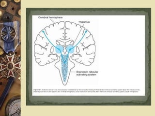
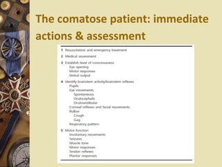
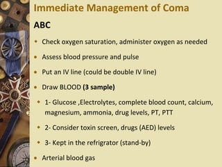
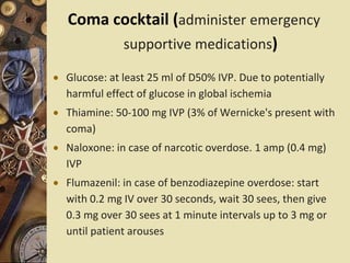
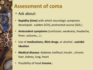
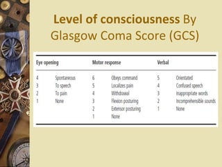
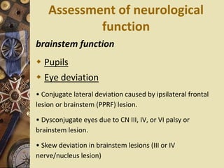
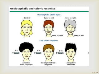
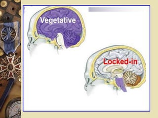
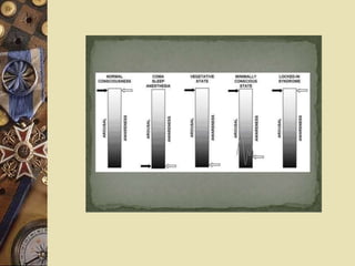
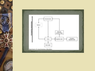
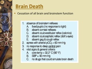
COMA
“9”
Presented By:
Dr. Raed Ahmed
MBChB , FIBMS
Neurologist
1
Neuroanatomy
 Consciousness, which is the state of awareness of self and
environment with the ability to respond appropriately to stimuli,
results from:
 • arousal (ascending reticular activating system (RAS));
 • awareness (cerebral cortex).
Neuropathology
Coma results from one or more of the
following:
 1) Dysfunction of RAS in the brainstem
 2) Bilateral diencephalic dysfunction
(thalamus)
 3) Diffuse lesions in both cerebral
hemispheres (cortical or subcortical white
matter).
 Disorders of consciousness can affact either
level (arousal ,awareness) or content.
COMA
• Coma A state of unrousable unresponsiveness in
which the subject lies with eyes closed.
• No understandable response to external stimuli
or inner need and the patient does not utter
understandable responses nor accurately
localizes noxious stimuli.
• Total absence of awareness of self and
environment even when the subject is externally
stimulated.
• No spontaneous eye opening, response to voice,
localization to painful stimuli or verbal output
DEFINITIONS
 Stupor: patient can be transiently awakened
only by vigorous stimuli, accompanied by
little motor or verbal activity once aroused
 Drowsiness: simulates light sleep and is
characterized by easy arousal and the
persistence of alertness for brief periods
 Ambiguous terms such as “ lethargy”,
“semicoma” , or “obtundation”
Acute confusional state
 Inability to maintain a coherent sequence of
thoughts
 Patients are drowsy, disorientated in time,
and have poor short-term memory and
comprehension
 Difficulty undertaking complex tasks and
show day–night reversal
 Delirium: disturbed consciousness,
disorientation, severe motor restlessness,
fear, irritability,consistent misperception of
sensory stimuli and visual hallucinations.
The comatose patient: immediate
actions & assessment
Immediate Management of Coma
ABC
 Check oxygen saturation, administer oxygen as needed
 Assess blood pressure and pulse
 Put an IV line (could be double IV line)
 Draw BLOOD (3 sample)
 1- Glucose ,Electrolytes, complete blood count, calcium,
magnesium, ammonia, drug levels, PT, PTT
 2- Consider toxin screen, drugs (AED) levels
 3- Kept in the refrigrator (stand-by)
 Arterial blood gas
Coma cocktail (administer emergency
supportive medications)
 Glucose: at least 25 ml of D50% IVP. Due to potentially
harmful effect of glucose in global ischemia
 Thiamine: 50-100 mg IVP (3% of Wernicke's present with
coma)
 Naloxone: in case of narcotic overdose. 1 amp (0.4 mg)
IVP
 Flumazenil: in case of benzodiazepine overdose: start
with 0.2 mg IV over 30 seconds, wait 30 sees, then give
0.3 mg over 30 sees at 1 minute intervals up to 3 mg or
until patient arouses
Assessment of coma
 Ask about:
 Rapidity (time) with which neurologic symptoms
developed; sudden (ICH), protracted course (SOL) ;
 Antecedent symptoms (confusion, weakness, headache,
fever, seizures,…) ;
 Use of medications, illicit drugs, or alcohol ; suicidal
ideation
 Medical disease: diabetes mellitus Insulin , chronic
liver, kidney, lung, heart
 Possibility of head trauma ;
GENERAL PHYSICAL EXAMINATION
 Breath odour (alcohol, ketones, hepatic, or renal failure).
 Mucous membranes may show evidence of cyanosis,
 Bruising in the mastoid or orbital regions
 Skin : hypodermic needle marks in opiate intoxication. A
purpuric petechial rash suggesting meningococcal
 Temperature
 Hyperthermia suggests a systemic infection, bacterial
meningitis, encephalitis, heat stroke, malignant
hyperthermia due to anesthetics or anticholinergic drug
intoxication;
 Hypothermia is observed with exposure; alcoholic,
barbiturate, sedative, or phenothiazine intoxication;
hypoglycemia; or extreme hypothyroidism.
Hypothermia itself causes coma only when the
temperature is <31°C
 BP
 Marked hypertension suggests hypertensive
encephalopathy, but it may also be secondary to a rapid
rise in intracranial pressure (ICP) (the Cushing response)
 Hypotension is characteristic of coma from alcohol or
barbiturate intoxication , internal hemorrhage,
myocardial infarction, sepsis , or Addisonian crisis.
 Otoscopic examination may reveal otorrhoea or
haemotympanum from a basal skull fracture.
Respiratory patterns
Cheyne-Stokes
breathing gradually crescendos in amplitude and then trails
off, followed by an expiratory pause, and then the pattern
repeats.
 Bilateral cerebral hemisphere dysfunction or metabolic
abnormality.
Cluster breathing periods of rapid irregular breathing
separated by apneic spells, High medulla or lower pons
lesion (an ominous sign)
 Apneustic (rare): a pause at full inspiration. Indicates
pontine lesion, e.g. with basilar artery occlusion
 Ataxic : no pattern in rate or depth of respirations.
Seen with medullary lesion. Usually preterminal
Level of consciousness By
Glasgow Coma Score (GCS)
Assessment of neurological
function
brainstem function
 Pupils
 Eye deviation
• Conjugate lateral deviation caused by ipsilateral frontal
lesion or brainstem (PPRF) lesion.
• Dysconjugate eyes due to CN III, IV, or VI palsy or
brainstem lesion.
• Skew deviation in brainstem lesions (III or IV
nerve/nucleus lesion)
 Spontaneous eye movements
 • Repetitive horizontal deviations (ping-pong gaze) =
brainstem lesion.
 • Downward ocular bobbing = pontine lesion
 Reflex eye movements
 Corneal reflex
 facial grimace to painful stimuli reflects VIIth
nerve function.
 Gag reflex
21 of 19
22
Assessment of neurological
function
Motor and sensory system
 Look for asymmetry, evidence of
significant cortical (decorticate) or
brainstem (decerebrate) damage.
 • Observe for seizure activity (focal or
general: implies cortical damage).
 • Tone • Posture • Reflexes, plantar
responses • Response to pain.
Decorticate posturing:
 Classically attributed to disinhibition by removal of
corticospinal pathways above the midbrain.
 It is characterized by abnormal flexion in UE and
extension in LE.
Decerebrate posturing:
 Usually caused by brainstem lesions, particularly of the
bilateral midbrain or pons, and carries a poor prognosis.
 It is characterized by bilateral abnormal extension in UE
and LE.
Coma without focal signs or meningism
 • Hypoxic–ischaemic injury
 • Toxins : Carbon monoxide, methanol, cyanide, Alcohol
 • Drugs overdose: e.g. barbiturates, opioids,
 • Seizures, epilepsy: CSE , NCSE
 • Organ failure
 • Electrolyte disturbances: Hyponatraemia,
hypernatraemia; hypocalcaemia, hypercalcaemia;
hypermagnesaemia
 • Diabetes mellitus: Hypoglycaemia, ketoacidosis, lactic
acidosis, hyperosmolar non-ketotic diabetic coma
 Coma with meningism
 • Meningoencephalitis • SAH
 Coma with focal signs
 • Vascular : infarction (Ischaemia ,Embolic) ,
Haemorrhage (EDH,SDH,SAH)
 • Traumatic brain injury
 • Infection : brain abscess, tuberculoma , subdural
empyema
 • Brain neoplasms (primary or metastatic)
 • Herniation syndromes
Toxic and metabolic coma from
structural coma
Indicators favour metabolic coma
• Preceding medical history may suggest a metabolic
abnormality
• Coma without lateralizing or brainstem signs
• Presence of involuntary limb movements (tremor,
myoclonus and asterixis)
• level of consciousness tends to fluctuate and be lighter
in patients with metabolic disorders
30
Investigations
 • Metabolic , toxicology screen
 • CT scan or MRI if possible, especially in
coma with meningism or focal signs.
 • Normal CT does not exclude increased
ICP
 • EEG
 • If no contraindications, consider LP
 PSEUDOCOMA (conditions mimic coma)
 1. Locked-in syndrome
 2. Psychiatric: catatonia, conversion
reaction, malingering
 3. Neuromuscular weakness: myasthenia
gravis, Guillain-Barre
Psychogenic unresponsiveness
 Obvious psychiatric precipitating factors
 Inconsistent volitional responses, particularly on
eyelid opening
 Pupillary constriction will occur on eye opening
 Oculo-vestibular stimulation with cold stimulus
will show preservation of the fast phase away
from the stimulated side (Normal)
 EEG will show responsive alpha rhythms
Catatonia
• Hypomobile and mute syndrome that
• Occurs as part of a major psychosis, usually
schizophrenia or major depression.
 No voluntary or responsive movements, although they
blink, swallow, and may not appear distressed.
 O/E Eyelid elevation is actively resisted, blinking occurs
in response to a visual threat, and the eyes move
concomitantly with head rotation
 In catatonia for the limbs to retain the postures in which
they have been placed by the examiner (“waxy
flexibility,” or catalepsy).
Locked-in syndrome
 No impairment of consciousness
 An awake patient has no means of producing speech or
volitional movement
 Communicate with eye movements (voluntary vertical
eye movements and lid elevation)
 The pupils are normally reactive
 The usual cause is an infarction or hemorrhage of the
ventral pons that transects all descending motor
(corticospinal and corticobulbar) pathways
Vegetative state
 Awake with their eyes open but show no evidence of
awareness of self or, are unable to interact with others
 Respiratory and autonomic functions are retained
 Sleep–wake cycles are preserved
 No responses to visual, auditory, tactile or noxious
stimuli
 No evidence of language comprehension or expression
 Patients are able to breathe spontaneously and the gag,
cough, sucking and swallowing reflexes are usually
present
 Vegetative state usually develops after a variable period
of coma; it may progress to a persistent vegetative state or
death.
Brain Death
 Cessation of all brain and brainstem function
9  coma

9 coma

  • 1.
    COMA “9” Presented By: Dr. RaedAhmed MBChB , FIBMS Neurologist 1
  • 2.
    Neuroanatomy  Consciousness, whichis the state of awareness of self and environment with the ability to respond appropriately to stimuli, results from:  • arousal (ascending reticular activating system (RAS));  • awareness (cerebral cortex).
  • 3.
    Neuropathology Coma results fromone or more of the following:  1) Dysfunction of RAS in the brainstem  2) Bilateral diencephalic dysfunction (thalamus)  3) Diffuse lesions in both cerebral hemispheres (cortical or subcortical white matter).  Disorders of consciousness can affact either level (arousal ,awareness) or content.
  • 6.
    COMA • Coma Astate of unrousable unresponsiveness in which the subject lies with eyes closed. • No understandable response to external stimuli or inner need and the patient does not utter understandable responses nor accurately localizes noxious stimuli. • Total absence of awareness of self and environment even when the subject is externally stimulated. • No spontaneous eye opening, response to voice, localization to painful stimuli or verbal output
  • 7.
    DEFINITIONS  Stupor: patientcan be transiently awakened only by vigorous stimuli, accompanied by little motor or verbal activity once aroused  Drowsiness: simulates light sleep and is characterized by easy arousal and the persistence of alertness for brief periods  Ambiguous terms such as “ lethargy”, “semicoma” , or “obtundation”
  • 8.
    Acute confusional state Inability to maintain a coherent sequence of thoughts  Patients are drowsy, disorientated in time, and have poor short-term memory and comprehension  Difficulty undertaking complex tasks and show day–night reversal  Delirium: disturbed consciousness, disorientation, severe motor restlessness, fear, irritability,consistent misperception of sensory stimuli and visual hallucinations.
  • 9.
    The comatose patient:immediate actions & assessment
  • 11.
    Immediate Management ofComa ABC  Check oxygen saturation, administer oxygen as needed  Assess blood pressure and pulse  Put an IV line (could be double IV line)  Draw BLOOD (3 sample)  1- Glucose ,Electrolytes, complete blood count, calcium, magnesium, ammonia, drug levels, PT, PTT  2- Consider toxin screen, drugs (AED) levels  3- Kept in the refrigrator (stand-by)  Arterial blood gas
  • 12.
    Coma cocktail (administeremergency supportive medications)  Glucose: at least 25 ml of D50% IVP. Due to potentially harmful effect of glucose in global ischemia  Thiamine: 50-100 mg IVP (3% of Wernicke's present with coma)  Naloxone: in case of narcotic overdose. 1 amp (0.4 mg) IVP  Flumazenil: in case of benzodiazepine overdose: start with 0.2 mg IV over 30 seconds, wait 30 sees, then give 0.3 mg over 30 sees at 1 minute intervals up to 3 mg or until patient arouses
  • 13.
    Assessment of coma Ask about:  Rapidity (time) with which neurologic symptoms developed; sudden (ICH), protracted course (SOL) ;  Antecedent symptoms (confusion, weakness, headache, fever, seizures,…) ;  Use of medications, illicit drugs, or alcohol ; suicidal ideation  Medical disease: diabetes mellitus Insulin , chronic liver, kidney, lung, heart  Possibility of head trauma ;
  • 14.
    GENERAL PHYSICAL EXAMINATION Breath odour (alcohol, ketones, hepatic, or renal failure).  Mucous membranes may show evidence of cyanosis,  Bruising in the mastoid or orbital regions  Skin : hypodermic needle marks in opiate intoxication. A purpuric petechial rash suggesting meningococcal  Temperature  Hyperthermia suggests a systemic infection, bacterial meningitis, encephalitis, heat stroke, malignant hyperthermia due to anesthetics or anticholinergic drug intoxication;
  • 15.
     Hypothermia isobserved with exposure; alcoholic, barbiturate, sedative, or phenothiazine intoxication; hypoglycemia; or extreme hypothyroidism. Hypothermia itself causes coma only when the temperature is <31°C  BP  Marked hypertension suggests hypertensive encephalopathy, but it may also be secondary to a rapid rise in intracranial pressure (ICP) (the Cushing response)  Hypotension is characteristic of coma from alcohol or barbiturate intoxication , internal hemorrhage, myocardial infarction, sepsis , or Addisonian crisis.  Otoscopic examination may reveal otorrhoea or haemotympanum from a basal skull fracture.
  • 16.
    Respiratory patterns Cheyne-Stokes breathing graduallycrescendos in amplitude and then trails off, followed by an expiratory pause, and then the pattern repeats.  Bilateral cerebral hemisphere dysfunction or metabolic abnormality. Cluster breathing periods of rapid irregular breathing separated by apneic spells, High medulla or lower pons lesion (an ominous sign)
  • 17.
     Apneustic (rare):a pause at full inspiration. Indicates pontine lesion, e.g. with basilar artery occlusion  Ataxic : no pattern in rate or depth of respirations. Seen with medullary lesion. Usually preterminal
  • 18.
    Level of consciousnessBy Glasgow Coma Score (GCS)
  • 19.
    Assessment of neurological function brainstemfunction  Pupils  Eye deviation • Conjugate lateral deviation caused by ipsilateral frontal lesion or brainstem (PPRF) lesion. • Dysconjugate eyes due to CN III, IV, or VI palsy or brainstem lesion. • Skew deviation in brainstem lesions (III or IV nerve/nucleus lesion)
  • 20.
     Spontaneous eyemovements  • Repetitive horizontal deviations (ping-pong gaze) = brainstem lesion.  • Downward ocular bobbing = pontine lesion  Reflex eye movements  Corneal reflex  facial grimace to painful stimuli reflects VIIth nerve function.  Gag reflex
  • 21.
  • 22.
  • 23.
    Assessment of neurological function Motorand sensory system  Look for asymmetry, evidence of significant cortical (decorticate) or brainstem (decerebrate) damage.  • Observe for seizure activity (focal or general: implies cortical damage).  • Tone • Posture • Reflexes, plantar responses • Response to pain.
  • 24.
    Decorticate posturing:  Classicallyattributed to disinhibition by removal of corticospinal pathways above the midbrain.  It is characterized by abnormal flexion in UE and extension in LE. Decerebrate posturing:  Usually caused by brainstem lesions, particularly of the bilateral midbrain or pons, and carries a poor prognosis.  It is characterized by bilateral abnormal extension in UE and LE.
  • 28.
    Coma without focalsigns or meningism  • Hypoxic–ischaemic injury  • Toxins : Carbon monoxide, methanol, cyanide, Alcohol  • Drugs overdose: e.g. barbiturates, opioids,  • Seizures, epilepsy: CSE , NCSE  • Organ failure  • Electrolyte disturbances: Hyponatraemia, hypernatraemia; hypocalcaemia, hypercalcaemia; hypermagnesaemia  • Diabetes mellitus: Hypoglycaemia, ketoacidosis, lactic acidosis, hyperosmolar non-ketotic diabetic coma
  • 29.
     Coma withmeningism  • Meningoencephalitis • SAH  Coma with focal signs  • Vascular : infarction (Ischaemia ,Embolic) , Haemorrhage (EDH,SDH,SAH)  • Traumatic brain injury  • Infection : brain abscess, tuberculoma , subdural empyema  • Brain neoplasms (primary or metastatic)  • Herniation syndromes
  • 30.
    Toxic and metaboliccoma from structural coma Indicators favour metabolic coma • Preceding medical history may suggest a metabolic abnormality • Coma without lateralizing or brainstem signs • Presence of involuntary limb movements (tremor, myoclonus and asterixis) • level of consciousness tends to fluctuate and be lighter in patients with metabolic disorders 30
  • 31.
    Investigations  • Metabolic, toxicology screen  • CT scan or MRI if possible, especially in coma with meningism or focal signs.  • Normal CT does not exclude increased ICP  • EEG  • If no contraindications, consider LP
  • 33.
     PSEUDOCOMA (conditionsmimic coma)  1. Locked-in syndrome  2. Psychiatric: catatonia, conversion reaction, malingering  3. Neuromuscular weakness: myasthenia gravis, Guillain-Barre
  • 34.
    Psychogenic unresponsiveness  Obviouspsychiatric precipitating factors  Inconsistent volitional responses, particularly on eyelid opening  Pupillary constriction will occur on eye opening  Oculo-vestibular stimulation with cold stimulus will show preservation of the fast phase away from the stimulated side (Normal)  EEG will show responsive alpha rhythms
  • 35.
    Catatonia • Hypomobile andmute syndrome that • Occurs as part of a major psychosis, usually schizophrenia or major depression.  No voluntary or responsive movements, although they blink, swallow, and may not appear distressed.  O/E Eyelid elevation is actively resisted, blinking occurs in response to a visual threat, and the eyes move concomitantly with head rotation  In catatonia for the limbs to retain the postures in which they have been placed by the examiner (“waxy flexibility,” or catalepsy).
  • 36.
    Locked-in syndrome  Noimpairment of consciousness  An awake patient has no means of producing speech or volitional movement  Communicate with eye movements (voluntary vertical eye movements and lid elevation)  The pupils are normally reactive  The usual cause is an infarction or hemorrhage of the ventral pons that transects all descending motor (corticospinal and corticobulbar) pathways
  • 37.
    Vegetative state  Awakewith their eyes open but show no evidence of awareness of self or, are unable to interact with others  Respiratory and autonomic functions are retained  Sleep–wake cycles are preserved  No responses to visual, auditory, tactile or noxious stimuli  No evidence of language comprehension or expression  Patients are able to breathe spontaneously and the gag, cough, sucking and swallowing reflexes are usually present  Vegetative state usually develops after a variable period of coma; it may progress to a persistent vegetative state or death.
  • 41.
    Brain Death  Cessationof all brain and brainstem function