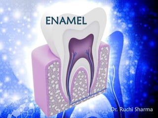
SlideShare a Scribd company logo
1 of 87
Dr. Ruchi Sharma
C TIP2
 Gene that controls tooth enamel production.
 This is transcription factor, which regulates skin and
nervous system.
 Discovered by Chrissa Kioussi; Oregon state
university.
 Enamel is hardest tissue of the body that forms
protective cover over the crown of teeth.
 It is hardest due to high mineralization.
COMPOSITION
Calcium phosphate
Hydroxyapatite
Fluoride
Strontium
Magnesium
Lead
Tyrosine rich
amelogenin
Non
amelogenins
PHYSICAL PROPERTIES
 I] Extremely hard like mild steel; so it can withstand
mechanical forces.
 II] Bulk: - Maximum bulk around 2 to 25 mm at
cuspal region and knife edge thickness at neck region.
 III] Specific gravity - 2.8
 IV] Permeability:-It acts as a semipermeable
membrane for: C-labeled urea, I etc.
 V] Colour: - Light yellow to grayish white. Depends
upon translucency of enamel that is attributable to
calcification and homogeneity.
STRUCTURE OF ENAMEL
Enamel rods
 The enamel is composed of elongated structures
called rods or prisms.
 The rods are roughly cylindrical and packed with
hydroxyapatite crystals.
 The crystals run along the longitudinal axis of rods
but the crystals that are away from central axis of
rods flare laterally.
ROD SHEATH
 The boundary where crystals
of rod meet the inter-rod
region at sharp angles.
 The average width of rods is 5
μm, but they vary in thickness
of enamel.
 From dentino-enamel junction
the rods run a tortuous course
towards surface of tooth.
 So the length of most rods is greater than thickness of
enamel. (Due to oblique direction wavy course).
 The diameter of rod is 2:1 more at surface than
dentino enamel protein than other regions, because
crystals meeting at different angles can not be packed
tightly. Hence in demineralised sections, it appears as
Fish scale.
 In longitudinal sectioning the section parses from
heads of rods of one row and tails of adjacent row
appearing like key hole pattern.
 The bodies of rods are towards occlusal and incisal
surface and tails towards cervical.
 In first 5 μm next to dentin, no rod structure is seen.
(Tomes’ Processes were not formed when this enamel
was laid down) similarly, rods are absent at enamel
surface.
 This rodless enamel is 30 μm of all primary teeth and
gingival third enamel of permanent teeth.
 The rods are perpendicular to the surface of dentin.
Near the cusp tips rods are somewhat vertical. In
cervical region they are horizontal except few which
tilt apically.
GNARLED ENAMEL
 In oblique section the enamel rods seen to be
intertwine at cuspal region near the dentin surface.
This optical phenomenon is termed as gnarled
enamel.
 Normally rods are arranged radially in horizontal
rows, surrounding long axis of tooth like washer.
There is back and forth undulation in rows. In cuspal
regions this undulation also seen in vertical direction.
CROSS STRIATIONS
 In human enamel forms (deposited) 4 μm/day. So in
ground section these periodic dark bands are seen at
interval of 4 μm in rods.
 This could also be due to structured interrelations of
group of rods than single rod.
 These are more prominent in less calcified enamel so
striae are considered due to daily variation in
secretary activity of ameloblasts.
INCREMENTAL LINES OF
RETZIUS
 These are brownish bands appearing in ground
section. They due to incremental deposition of
enamel during crown formation.
 In longitudinal section they surround tip of dentin.
Whereas in cross section they form concentric circles
like growth rings of tree. From dentino-enamel
junction they deviate occlusally. Prominent in
permanent teeth and rare in deciduous teeth.
 They form due to temporary constriction of Tomes’
Processes with secretary inter rod enamel.
 Sometimes they are also sent in fever that affect
amelogenesis.
 It is seen that crystals are decreased in striae and
enamel rods bend when they cross the striae.
NEONATAL LINE
 “Enlarged striae due to variation in physiologic
environment during tooth development (i.e.
intrauterine and postnatal stage).
 Fetus develops in well protected environment with
good nutritional supply. So, prenatal enamel is better
than postnatal. The striae indicate weekly variation in
the secretary activity of ameloblasts.
HUNTER SCHREGER BANDS
This is the optical
phenomenon produced
due to change in
direction of enamel rods.
They are clearly visible in
longitudinal sections by
reflected light. They
appear as dark and light
bands.
 Commonly seen near
dentino enamel junction
and move outward.
 According to some, it is
also formed due to
variation in calcification
with different permeability
and different organic
content.
ENAMEL TUFTS
 Enamel tufts arise at dentino – enamel junction. They
project towards enamel reaching 1/5th to 1/3rd
thickness of enamel.
 Tufts develop due to abrupt change in direction of
groups of rods. They are termed because of grass like
appearance, when viewed in ground section.
 They are best seen in transverse section. Tufts appear
like branches of grass filled with more concentration
of high molecular weight protein.
 Their presence shows adaptive nature of enamel in
various conditions.
 Due to less interprismatic substance and higher
protein concentration these are considered as
hypocalcified areas.
ENAMEL LAMELLAE
 These are thin leaf like structures extend form enamel
surface to dentino - enamel junction. Some times they
penetrate dentin.
 Lamellae are rich in protein and with little mineral
content.
 Lamellae may also develop in planes of tension. The
rods cross such planes and short segment of rod
remains unclassified those later on are filled with
organic material.
 Three types of Lamellae are seen :-
 Type A: - Lamellae containing poorly calcified rod
segments.
 Type B: - Consisting degenerated cells.
 Type C: - Lamellae in erupted teeth where cracks are
filled with salivary proteins.
 Type C are more common and A are comparatively
short than B an C lamellae extend in longitudinal and
radial direction from tip of crown to cervical region.
 Some times they consist of cementum.
SURFACE STRUCTURES
 PERIKYMATA - They are around the tooth lie parallel
to each other and cemento-enamel junction. These
are transverse wave like grooves produced by
external manifestations of striae of Retzius.
 Around 30 Perikymata/mm at cervical region and
10/mm at occlusal or incisal areas are seen. At other
regions, they are regular in course but cervically show
irregularity.
ROD ENDS
 They are concave and vary in shape. Shallow at
cervical region and deep at occlusal surface.
CRACKS
 Fissure like structures seen on the surfaces. They are
outer edges of lamellae. Originated from dentino
enamel junction at its right angle. They are less than 1
mm in length.
ENAMEL CUTICLE
 It is delicate membrane called Nasmyth’s membrane
or primary enamel cuticle.
 This is typical lamina that is secreted by ameloblasts
after completion of enamel formation.
 This thin membrane covers the newly erupted teeth
but soon after eruption it is removed by masticatory
forces.
pellicle
 A layer directly on top of enamel 1-3 µm thick (could
reach 10 µm), free from bacteria, and is not removed
by a toothbrush but can be removed by prophylaxis.
 If the pellicle is not cleaned, after one or two days it
is colonized by micro organisms to form plaque.
DENTINOENAMEL JUNCTION
 The junction between enamel and dentin is
established during development of enamel & dentin.
 This junction shows scalloped outline hence providing
bigger surface area and better adhesion between
enamel and dentin.
ENAMEL SPINDLES
 During enamel formation, some odontoblastic
processes are sandwiched between ameloblasts and
the enamel deposition starts at these processes’
junction. They are thickened at end so termed as
enamel spindles.
 The spindles are at right angles to dentin but do not
follow the direction of enamel rods.
 These structures are disintegrated and are replaced by
air in ground section so appear dark in transmitted
light.
AMELOGENESIS
 Enamel formation is two step procedure.
 In first step it deposits partially mineralized enamel
(30%)
 After achieving full width of unmineralized enamel:
second step involves removal of organic material and
water from bulk and influx of mineral.
 The deposition of enamel first begins at cusp tips and
incisal areas: then gradually slopes down towards
cervical region.
 Reciprocal induction To form enamel, first layer of
dentin should he laid down.
 The inner enamel epithelium stimulate dental papilla
to form odontoblasts; that form dentin and now
dentin act as stimulator for differentiation of
ameloblasts.
 I. Organic Matrix Formation
 This is the secretary phase of amelogenesis.
 The organic matrix consists of enamel proteins.
 Some enzymes are also secreted like
metalloproteinases and phosphatases.
 But 90% are low-molecular-weight proteins i.e.
Ameluenins (20 to 30 KDa) the rest 10% are
enamelin, tuftelin (45 kDa) and amelin.
 Enamel proteins create environment to accept
minerals. They also carry important two works i.e.
 I. Determine the nature and direction of development
and growth in enamel.
 II. Slow away from pressure generated by them. As
soon as small quantity of dentin is laid down,
ameloblasts start their secretory activity.
 The ameloblasts mu e away from predemin, loose the
projections that had penetrated basal lamina and
deposit enamel matrix over predentin. This thin taxer
of enamel is called as dentinoenamel membrane.
 The enamel matrix becomes partially mineralized.
After deposition of first 1a er of enamel niatrix the
ameloblasts move away from the dentin surface.
DEVELOPMENT OF TOME’S
PROCESS
 As the ameloblasts move away from dentin surface,
they form curved projections, called as Tomes
processes. The processes fit into newly formed
enamel.
 They provide junction between enamel and
ameloblasts appearing as picket or saw-toothed.
 The long axis of ameloblasts are not parallel to long
axis of rods; hence developing enamel is not smooth.
 Distal terminal bars: During Tomes’ process
formation, the condensed cytoplasmic substance is
deposited near cell membrane that separate cell
proper and Tomes process. They are seen at enamel
secreting stage. Function not known.
 During deposition of enamel matrix, dental organ
collapses.
 There is loss of intercellular material which leads to
reduce the stellate reticulum cells. The reduction in
volume and peripheral migration of ameloblasts;
blood vessels come close to ameloblasts and provide
nutrition.
 In the continued process of amelogenesis, enamel
epithelium, stellate reticulum and stratum
intermedium loose their identity and form stratified
epithelial layer adjacent to ameloblasts.
 After achieving full thickness of enamel, ameloblasts
shorten in length, loose their Tomes’ processes and
invoke in maturation.
 When enamel maturation is completed. the adjacent
stratified epithelial layer and ameloblast layer form
together called reduced dental(enamel) epithelium.
 At this stage ameloblasts are not involved in secretion
and maturation.
 The reduced enamel epithelium protects the enamel
because in case of premature break of this epithelium
layer: connective tissue cells wilI come in contact with
enamel surface and deposit cementum over enamel.
MINERALIZATION OF ENAMEL
 The way mineral is entered in the organic matrix of
enamel differs from other hard tissues.
 In other hard tissues, vesicles provide favourable
condition to from crystals.
 But such vesicles are absent in enamel.
 So direct crystallite deposition takes place over
secreted enamel proteins.
 The enamel protein tuftelin play an important role in
crystallite formation. Once crystallites are formed
they keep on growing.
 So the first formed enamel is reasonably soft. It has to
be converted into hardest.
Stages of Mineralization
 First stage :- Immediate partial mineralization: during
this stage, enamel organic proteins are replaced by
crystallites.
 Maturation : The first formed mild hard enamel is
converted into hard structure.
 At this stage almost all the enamel proteins are
replaced by hardened crystals. So due to addition of
minerals, the ribbon like crystallites become long and
wide.
 First: Amelogenins due to their chemical nature are
squeezed out from bet the growing crystals.
 Second: Proteases secreted by ameloblasts degrade
amelogenins into low molecular weight so as to
remove easily from crystallites. In this way the fully
mature crystallites are laid.
 I. Primary mineralization: The partially
mineralized enamel matrix is formed.
 Almost 30% mineralization is achieved at this stage.
 Near dentino-enamel junction, thin laver(8µm) is
heavily mineralized.
 This is perhaps due to role of enamelin.
 II. Second stage: Begins with mineralization at the
surface of enamel and sweeps rapidly into deeper
layer.
 III Third stage: Increase in mineral rebounding
from innermost layer out toward enamel surface.
 IV. Quaternary mineralization: This is the
fourth stage that mineralizes outer layer rapidly and
heavily.
 The partial mineralization is about 30% but
maturation begins before matrix reaches its thickness.
 Thus maturation of first formed layer takes place and
on the other hand mineralization is simultaneously
taking place at outer surface.
 The maturation starts at height of crown and
progresses cervically.
 Each rod matures from depth to the surface and
maturing sequence of rod is from cusps/incisal edge to
cervical line.
LIFE CYCLE OF AMELOBLASTS
 On the basis of ftrnction life cycle otamelohiaste is
divided into various stages:
 1. Morphogenic
 2. Organizing
 3. Formative
 4. Maturative
 5. Protective
 6. Desmolvtic
MORPHOGENIC STAGE
 Prior to differentiation and enamel matrix synthesis,
ameloblasts interact with mesenchymal cells to
determine shape of crown and dentino-enamel
junction. At this stage ameloblasts are short columnar
and with large oval nuclei.
 Golgi bodies and centrioles are located at proximal
(to stratum intermedium) and mitochondria are
scattered throughout cytoplasm but they migrate
proximally.
 During differentiation bell stage explains details of
morphogenesis stage. Terminal bars appear at this
stage.
ORGANIZING STAGE
 During this stage inner enamel epithelium interacts
with connective tissue to differentiate odontoblasts.
 The ameloblasts become longer (tall columnar). and
nuclei are placed proximally.
 Reversal of polarity takes place by migration of
centrioles and golgi bodies at distal end.
 Mitochondria are shifted towards proximal end.
 Cell free zone disappears due to elongation of
epithelial cells towards papilla.
 The first layer of dentin is formed by odontoblasts.
But ameloblasts are cut off from nutrition.
 Fortunately stellate reticulum reduces and disappears.
 At the same time dental sac capillaries proliferate
rapidly.
 The distance between capillaries and ameloblasts is
reduced so reversal nutritional supply is provided by
penetrating the capillaries.
FORMATIVE STAGE
 As soon as the first layer of dentin is formed, the
ameloblasts enter at formative stage. It is mutual
interaction of cells to form enamel matrix. i.e.
odontoblast and dentin layer.
 So it is low of organogenesis and histodifferentiation.
Initiation of enamel matrix takes place due to changes
in number and organization of organelles. i.e. golgi
body, mitochondria etc.
 At this processes penetrate basal lamina. The secretion
of enamel protein takes place in rough endoplasmic
reticulum, further it is passed to golgi complex.
 In golgi complex it condenses to form membrane
bound granules.
 Further they migrate to distal end and release content
against newly formed mantle dentin.
 Hydroxyapatite crystals are
packed in first-forming enamel
and interdigitate with crystals of
dentin.
 Secretion of enamel protein is
via two sites.
 First site is adjacent to proximal
part near junctional complex
and second site, one surface of
tomes process.
 First site forms enamel matrix wall, which patch up
the pits fit by tomes processes second site fill the pits
with matrix.
 Due to bipolar secretion the orientation of it is
different.
 Amelin is concentrated in rod sheath area so called as
sheathlin.
MATURATION STAGE
 Maturation starts when complete matrix deposition
takes place on cuspal and incisal areas. At this stage
also matrix deposition cervically is still continued. At
brief transitional stage ameloblasts reduce in length
decrease their volume and organelle content.
 There is degradation of excess material and organelles
are shifted distally.
 Ameloblasts involved in cyclical process of removal of
water and organic material from enamel matrix. The
other work is to absorb inorganic material to replace
the bulk of enamel.
 This process is carried out by two types of
ameloblasts.
RUFFLED BORDER
 These ameloblasts have leaky proximal junctions and
tight distal junctions. So the inorganic material passes
through ruffled border. Because their distal junctions
are tight.
 They also absorb protein
break down product. The
route by which calcium
moves from blood vessels
to enamel is via ruffle
ended ameloblasts.
SMOOTH BORDER
 These ameloblasts show tight
proximal junctions and leaky
distal junction. So the larger
protein molecules and water is
removed by smooth border
ameloblasts via their lateral
surfaces.
PROTECTIVE STAGE
 As enamel maturation completes, ameloblasts loose
their nature and function.
 They are no longer differentiated from stratum
intermedium and outer enamel epithelium.
 They secrete some material at the distal ends and
form epithelium.
DESMOLYTIC STAGE
 The reduced enamel epithelium induces atrophy of
connective tissue and separates it from oral
epithelium.
 Degeneration of connective tissue is done by
desmolysis.
 Premature degeneration of Reduced enamel
epithelium may prevent tooth eruption.
Defects
 I. Febrile diseases
 It. Tetracycline therapy.
 III. Fluoride ions : Excess of 5 PPM.
Thank you

More Related Content

What's hot (20)

ENAMEL
ENAMELENAMEL
ENAMEL
 
development and growth of teeth
development and growth of teethdevelopment and growth of teeth
development and growth of teeth
 
Dental pulp
Dental pulpDental pulp
Dental pulp
 
Dental Pulp
Dental PulpDental Pulp
Dental Pulp
 
Dental Pulp
Dental Pulp Dental Pulp
Dental Pulp
 
Amelogenesis
Amelogenesis Amelogenesis
Amelogenesis
 
Dentin
DentinDentin
Dentin
 
Enamel
EnamelEnamel
Enamel
 
Dentin
DentinDentin
Dentin
 
Dentinogenesis & histology of dentin
Dentinogenesis & histology of dentinDentinogenesis & histology of dentin
Dentinogenesis & histology of dentin
 
Pulp
Pulp Pulp
Pulp
 
Enamel
EnamelEnamel
Enamel
 
cementum
cementumcementum
cementum
 
Shedding
SheddingShedding
Shedding
 
Enamel
EnamelEnamel
Enamel
 
Hypomineralised structure of enamel
Hypomineralised structure of enamelHypomineralised structure of enamel
Hypomineralised structure of enamel
 
HERTWIG’S EPITHELIAL ROOTH SHEATH
HERTWIG’S EPITHELIAL ROOTH SHEATHHERTWIG’S EPITHELIAL ROOTH SHEATH
HERTWIG’S EPITHELIAL ROOTH SHEATH
 
Dentin
DentinDentin
Dentin
 
Enamel
EnamelEnamel
Enamel
 
Periodontal Ligament.ppt
Periodontal Ligament.pptPeriodontal Ligament.ppt
Periodontal Ligament.ppt
 

Similar to Enamel

Similar to Enamel (20)

Enamel
EnamelEnamel
Enamel
 
Enamel
Enamel Enamel
Enamel
 
Enamel.pptx
Enamel.pptxEnamel.pptx
Enamel.pptx
 
Enamel seminar.pptx
Enamel seminar.pptxEnamel seminar.pptx
Enamel seminar.pptx
 
Enamel.pptx
Enamel.pptxEnamel.pptx
Enamel.pptx
 
TOOTH ENAMEL - HISTOPATHOLOGY FOR STUDENTS
TOOTH ENAMEL - HISTOPATHOLOGY FOR STUDENTSTOOTH ENAMEL - HISTOPATHOLOGY FOR STUDENTS
TOOTH ENAMEL - HISTOPATHOLOGY FOR STUDENTS
 
Enamel done
Enamel   doneEnamel   done
Enamel done
 
enamel.ppt
enamel.pptenamel.ppt
enamel.ppt
 
Enamel presentation. prepared by mohammed yahia
Enamel presentation. prepared by mohammed yahiaEnamel presentation. prepared by mohammed yahia
Enamel presentation. prepared by mohammed yahia
 
ENAMEL
 ENAMEL ENAMEL
ENAMEL
 
cementum -avisha agrawal
cementum -avisha agrawalcementum -avisha agrawal
cementum -avisha agrawal
 
Enamel
EnamelEnamel
Enamel
 
Enamel
EnamelEnamel
Enamel
 
Enamel significance in operative dentistry /certified fixed orthodontic cour...
Enamel significance in operative dentistry  /certified fixed orthodontic cour...Enamel significance in operative dentistry  /certified fixed orthodontic cour...
Enamel significance in operative dentistry /certified fixed orthodontic cour...
 
Cementum
Cementum Cementum
Cementum
 
Enamel
EnamelEnamel
Enamel
 
Cementum seminar
Cementum seminarCementum seminar
Cementum seminar
 
Development of periodontium
Development of periodontiumDevelopment of periodontium
Development of periodontium
 
Cementum
Cementum Cementum
Cementum
 
Dentin.docx
Dentin.docxDentin.docx
Dentin.docx
 

Recently uploaded

call girls in paharganj DELHI 🔝 >༒9540349809 🔝 genuine Escort Service 🔝✔️✔️
call girls in paharganj DELHI 🔝 >༒9540349809 🔝 genuine Escort Service 🔝✔️✔️call girls in paharganj DELHI 🔝 >༒9540349809 🔝 genuine Escort Service 🔝✔️✔️
call girls in paharganj DELHI 🔝 >༒9540349809 🔝 genuine Escort Service 🔝✔️✔️saminamagar
 
PULMONARY EDEMA AND ITS MANAGEMENT.pdf
PULMONARY EDEMA AND  ITS  MANAGEMENT.pdfPULMONARY EDEMA AND  ITS  MANAGEMENT.pdf
PULMONARY EDEMA AND ITS MANAGEMENT.pdfDolisha Warbi
 
SWD (Short wave diathermy)- Physiotherapy.ppt
SWD (Short wave diathermy)- Physiotherapy.pptSWD (Short wave diathermy)- Physiotherapy.ppt
SWD (Short wave diathermy)- Physiotherapy.pptMumux Mirani
 
97111 47426 Call Girls In Delhi MUNIRKAA
97111 47426 Call Girls In Delhi MUNIRKAA97111 47426 Call Girls In Delhi MUNIRKAA
97111 47426 Call Girls In Delhi MUNIRKAAjennyeacort
 
Measurement of Radiation and Dosimetric Procedure.pptx
Measurement of Radiation and Dosimetric Procedure.pptxMeasurement of Radiation and Dosimetric Procedure.pptx
Measurement of Radiation and Dosimetric Procedure.pptxDr. Dheeraj Kumar
 
Basic principles involved in the traditional systems of medicine PDF.pdf
Basic principles involved in the traditional systems of medicine PDF.pdfBasic principles involved in the traditional systems of medicine PDF.pdf
Basic principles involved in the traditional systems of medicine PDF.pdfDivya Kanojiya
 
call girls in aerocity DELHI 🔝 >༒9540349809 🔝 genuine Escort Service 🔝✔️✔️
call girls in aerocity DELHI 🔝 >༒9540349809 🔝 genuine Escort Service 🔝✔️✔️call girls in aerocity DELHI 🔝 >༒9540349809 🔝 genuine Escort Service 🔝✔️✔️
call girls in aerocity DELHI 🔝 >༒9540349809 🔝 genuine Escort Service 🔝✔️✔️saminamagar
 
Statistical modeling in pharmaceutical research and development.
Statistical modeling in pharmaceutical research and development.Statistical modeling in pharmaceutical research and development.
Statistical modeling in pharmaceutical research and development.ANJALI
 
Big Data Analysis Suggests COVID Vaccination Increases Excess Mortality Of ...
Big Data Analysis Suggests COVID  Vaccination Increases Excess Mortality Of  ...Big Data Analysis Suggests COVID  Vaccination Increases Excess Mortality Of  ...
Big Data Analysis Suggests COVID Vaccination Increases Excess Mortality Of ...sdateam0
 
History and Development of Pharmacovigilence.pdf
History and Development of Pharmacovigilence.pdfHistory and Development of Pharmacovigilence.pdf
History and Development of Pharmacovigilence.pdfSasikiranMarri
 
epilepsy and status epilepticus for undergraduate.pptx
epilepsy and status epilepticus  for undergraduate.pptxepilepsy and status epilepticus  for undergraduate.pptx
epilepsy and status epilepticus for undergraduate.pptxMohamed Rizk Khodair
 
See the 2,456 pharmacies on the National E-Pharmacy Platform
See the 2,456 pharmacies on the National E-Pharmacy PlatformSee the 2,456 pharmacies on the National E-Pharmacy Platform
See the 2,456 pharmacies on the National E-Pharmacy PlatformKweku Zurek
 
Culture and Health Disorders Social change.pptx
Culture and Health Disorders Social change.pptxCulture and Health Disorders Social change.pptx
Culture and Health Disorders Social change.pptxDr. Dheeraj Kumar
 
Wessex Health Partners Wessex Integrated Care, Population Health, Research & ...
Wessex Health Partners Wessex Integrated Care, Population Health, Research & ...Wessex Health Partners Wessex Integrated Care, Population Health, Research & ...
Wessex Health Partners Wessex Integrated Care, Population Health, Research & ...Wessex Health Partners
 
Hematology and Immunology - Leukocytes Functions
Hematology and Immunology - Leukocytes FunctionsHematology and Immunology - Leukocytes Functions
Hematology and Immunology - Leukocytes FunctionsMedicoseAcademics
 
Report Back from SGO: What’s New in Uterine Cancer?.pptx
Report Back from SGO: What’s New in Uterine Cancer?.pptxReport Back from SGO: What’s New in Uterine Cancer?.pptx
Report Back from SGO: What’s New in Uterine Cancer?.pptxbkling
 
April 2024 ONCOLOGY CARTOON by DR KANHU CHARAN PATRO
April 2024 ONCOLOGY CARTOON by  DR KANHU CHARAN PATROApril 2024 ONCOLOGY CARTOON by  DR KANHU CHARAN PATRO
April 2024 ONCOLOGY CARTOON by DR KANHU CHARAN PATROKanhu Charan
 
call girls in green park DELHI 🔝 >༒9540349809 🔝 genuine Escort Service 🔝✔️✔️
call girls in green park  DELHI 🔝 >༒9540349809 🔝 genuine Escort Service 🔝✔️✔️call girls in green park  DELHI 🔝 >༒9540349809 🔝 genuine Escort Service 🔝✔️✔️
call girls in green park DELHI 🔝 >༒9540349809 🔝 genuine Escort Service 🔝✔️✔️saminamagar
 
call girls in munirka DELHI 🔝 >༒9540349809 🔝 genuine Escort Service 🔝✔️✔️
call girls in munirka  DELHI 🔝 >༒9540349809 🔝 genuine Escort Service 🔝✔️✔️call girls in munirka  DELHI 🔝 >༒9540349809 🔝 genuine Escort Service 🔝✔️✔️
call girls in munirka DELHI 🔝 >༒9540349809 🔝 genuine Escort Service 🔝✔️✔️saminamagar
 
POST NATAL EXERCISES AND ITS IMPACT.pptx
POST NATAL EXERCISES AND ITS IMPACT.pptxPOST NATAL EXERCISES AND ITS IMPACT.pptx
POST NATAL EXERCISES AND ITS IMPACT.pptxvirengeeta
 

Recently uploaded (20)

call girls in paharganj DELHI 🔝 >༒9540349809 🔝 genuine Escort Service 🔝✔️✔️
call girls in paharganj DELHI 🔝 >༒9540349809 🔝 genuine Escort Service 🔝✔️✔️call girls in paharganj DELHI 🔝 >༒9540349809 🔝 genuine Escort Service 🔝✔️✔️
call girls in paharganj DELHI 🔝 >༒9540349809 🔝 genuine Escort Service 🔝✔️✔️
 
PULMONARY EDEMA AND ITS MANAGEMENT.pdf
PULMONARY EDEMA AND  ITS  MANAGEMENT.pdfPULMONARY EDEMA AND  ITS  MANAGEMENT.pdf
PULMONARY EDEMA AND ITS MANAGEMENT.pdf
 
SWD (Short wave diathermy)- Physiotherapy.ppt
SWD (Short wave diathermy)- Physiotherapy.pptSWD (Short wave diathermy)- Physiotherapy.ppt
SWD (Short wave diathermy)- Physiotherapy.ppt
 
97111 47426 Call Girls In Delhi MUNIRKAA
97111 47426 Call Girls In Delhi MUNIRKAA97111 47426 Call Girls In Delhi MUNIRKAA
97111 47426 Call Girls In Delhi MUNIRKAA
 
Measurement of Radiation and Dosimetric Procedure.pptx
Measurement of Radiation and Dosimetric Procedure.pptxMeasurement of Radiation and Dosimetric Procedure.pptx
Measurement of Radiation and Dosimetric Procedure.pptx
 
Basic principles involved in the traditional systems of medicine PDF.pdf
Basic principles involved in the traditional systems of medicine PDF.pdfBasic principles involved in the traditional systems of medicine PDF.pdf
Basic principles involved in the traditional systems of medicine PDF.pdf
 
call girls in aerocity DELHI 🔝 >༒9540349809 🔝 genuine Escort Service 🔝✔️✔️
call girls in aerocity DELHI 🔝 >༒9540349809 🔝 genuine Escort Service 🔝✔️✔️call girls in aerocity DELHI 🔝 >༒9540349809 🔝 genuine Escort Service 🔝✔️✔️
call girls in aerocity DELHI 🔝 >༒9540349809 🔝 genuine Escort Service 🔝✔️✔️
 
Statistical modeling in pharmaceutical research and development.
Statistical modeling in pharmaceutical research and development.Statistical modeling in pharmaceutical research and development.
Statistical modeling in pharmaceutical research and development.
 
Big Data Analysis Suggests COVID Vaccination Increases Excess Mortality Of ...
Big Data Analysis Suggests COVID  Vaccination Increases Excess Mortality Of  ...Big Data Analysis Suggests COVID  Vaccination Increases Excess Mortality Of  ...
Big Data Analysis Suggests COVID Vaccination Increases Excess Mortality Of ...
 
History and Development of Pharmacovigilence.pdf
History and Development of Pharmacovigilence.pdfHistory and Development of Pharmacovigilence.pdf
History and Development of Pharmacovigilence.pdf
 
epilepsy and status epilepticus for undergraduate.pptx
epilepsy and status epilepticus  for undergraduate.pptxepilepsy and status epilepticus  for undergraduate.pptx
epilepsy and status epilepticus for undergraduate.pptx
 
See the 2,456 pharmacies on the National E-Pharmacy Platform
See the 2,456 pharmacies on the National E-Pharmacy PlatformSee the 2,456 pharmacies on the National E-Pharmacy Platform
See the 2,456 pharmacies on the National E-Pharmacy Platform
 
Culture and Health Disorders Social change.pptx
Culture and Health Disorders Social change.pptxCulture and Health Disorders Social change.pptx
Culture and Health Disorders Social change.pptx
 
Wessex Health Partners Wessex Integrated Care, Population Health, Research & ...
Wessex Health Partners Wessex Integrated Care, Population Health, Research & ...Wessex Health Partners Wessex Integrated Care, Population Health, Research & ...
Wessex Health Partners Wessex Integrated Care, Population Health, Research & ...
 
Hematology and Immunology - Leukocytes Functions
Hematology and Immunology - Leukocytes FunctionsHematology and Immunology - Leukocytes Functions
Hematology and Immunology - Leukocytes Functions
 
Report Back from SGO: What’s New in Uterine Cancer?.pptx
Report Back from SGO: What’s New in Uterine Cancer?.pptxReport Back from SGO: What’s New in Uterine Cancer?.pptx
Report Back from SGO: What’s New in Uterine Cancer?.pptx
 
April 2024 ONCOLOGY CARTOON by DR KANHU CHARAN PATRO
April 2024 ONCOLOGY CARTOON by  DR KANHU CHARAN PATROApril 2024 ONCOLOGY CARTOON by  DR KANHU CHARAN PATRO
April 2024 ONCOLOGY CARTOON by DR KANHU CHARAN PATRO
 
call girls in green park DELHI 🔝 >༒9540349809 🔝 genuine Escort Service 🔝✔️✔️
call girls in green park  DELHI 🔝 >༒9540349809 🔝 genuine Escort Service 🔝✔️✔️call girls in green park  DELHI 🔝 >༒9540349809 🔝 genuine Escort Service 🔝✔️✔️
call girls in green park DELHI 🔝 >༒9540349809 🔝 genuine Escort Service 🔝✔️✔️
 
call girls in munirka DELHI 🔝 >༒9540349809 🔝 genuine Escort Service 🔝✔️✔️
call girls in munirka  DELHI 🔝 >༒9540349809 🔝 genuine Escort Service 🔝✔️✔️call girls in munirka  DELHI 🔝 >༒9540349809 🔝 genuine Escort Service 🔝✔️✔️
call girls in munirka DELHI 🔝 >༒9540349809 🔝 genuine Escort Service 🔝✔️✔️
 
POST NATAL EXERCISES AND ITS IMPACT.pptx
POST NATAL EXERCISES AND ITS IMPACT.pptxPOST NATAL EXERCISES AND ITS IMPACT.pptx
POST NATAL EXERCISES AND ITS IMPACT.pptx
 

Enamel

  • 2. C TIP2  Gene that controls tooth enamel production.  This is transcription factor, which regulates skin and nervous system.  Discovered by Chrissa Kioussi; Oregon state university.
  • 3.  Enamel is hardest tissue of the body that forms protective cover over the crown of teeth.  It is hardest due to high mineralization.
  • 6.  I] Extremely hard like mild steel; so it can withstand mechanical forces.
  • 7.  II] Bulk: - Maximum bulk around 2 to 25 mm at cuspal region and knife edge thickness at neck region.
  • 8.  III] Specific gravity - 2.8
  • 9.  IV] Permeability:-It acts as a semipermeable membrane for: C-labeled urea, I etc.
  • 10.  V] Colour: - Light yellow to grayish white. Depends upon translucency of enamel that is attributable to calcification and homogeneity.
  • 12. Enamel rods  The enamel is composed of elongated structures called rods or prisms.
  • 13.  The rods are roughly cylindrical and packed with hydroxyapatite crystals.  The crystals run along the longitudinal axis of rods but the crystals that are away from central axis of rods flare laterally.
  • 14. ROD SHEATH  The boundary where crystals of rod meet the inter-rod region at sharp angles.  The average width of rods is 5 μm, but they vary in thickness of enamel.  From dentino-enamel junction the rods run a tortuous course towards surface of tooth.
  • 15.  So the length of most rods is greater than thickness of enamel. (Due to oblique direction wavy course).  The diameter of rod is 2:1 more at surface than dentino enamel protein than other regions, because crystals meeting at different angles can not be packed tightly. Hence in demineralised sections, it appears as Fish scale.
  • 16.  In longitudinal sectioning the section parses from heads of rods of one row and tails of adjacent row appearing like key hole pattern.  The bodies of rods are towards occlusal and incisal surface and tails towards cervical.
  • 17.  In first 5 μm next to dentin, no rod structure is seen. (Tomes’ Processes were not formed when this enamel was laid down) similarly, rods are absent at enamel surface.  This rodless enamel is 30 μm of all primary teeth and gingival third enamel of permanent teeth.
  • 18.  The rods are perpendicular to the surface of dentin. Near the cusp tips rods are somewhat vertical. In cervical region they are horizontal except few which tilt apically.
  • 19. GNARLED ENAMEL  In oblique section the enamel rods seen to be intertwine at cuspal region near the dentin surface. This optical phenomenon is termed as gnarled enamel.
  • 20.  Normally rods are arranged radially in horizontal rows, surrounding long axis of tooth like washer. There is back and forth undulation in rows. In cuspal regions this undulation also seen in vertical direction.
  • 21. CROSS STRIATIONS  In human enamel forms (deposited) 4 μm/day. So in ground section these periodic dark bands are seen at interval of 4 μm in rods.
  • 22.  This could also be due to structured interrelations of group of rods than single rod.  These are more prominent in less calcified enamel so striae are considered due to daily variation in secretary activity of ameloblasts.
  • 23. INCREMENTAL LINES OF RETZIUS  These are brownish bands appearing in ground section. They due to incremental deposition of enamel during crown formation.
  • 24.  In longitudinal section they surround tip of dentin. Whereas in cross section they form concentric circles like growth rings of tree. From dentino-enamel junction they deviate occlusally. Prominent in permanent teeth and rare in deciduous teeth.
  • 25.  They form due to temporary constriction of Tomes’ Processes with secretary inter rod enamel.  Sometimes they are also sent in fever that affect amelogenesis.  It is seen that crystals are decreased in striae and enamel rods bend when they cross the striae.
  • 26. NEONATAL LINE  “Enlarged striae due to variation in physiologic environment during tooth development (i.e. intrauterine and postnatal stage).
  • 27.  Fetus develops in well protected environment with good nutritional supply. So, prenatal enamel is better than postnatal. The striae indicate weekly variation in the secretary activity of ameloblasts.
  • 28. HUNTER SCHREGER BANDS This is the optical phenomenon produced due to change in direction of enamel rods. They are clearly visible in longitudinal sections by reflected light. They appear as dark and light bands.
  • 29.  Commonly seen near dentino enamel junction and move outward.  According to some, it is also formed due to variation in calcification with different permeability and different organic content.
  • 30. ENAMEL TUFTS  Enamel tufts arise at dentino – enamel junction. They project towards enamel reaching 1/5th to 1/3rd thickness of enamel.  Tufts develop due to abrupt change in direction of groups of rods. They are termed because of grass like appearance, when viewed in ground section.
  • 31.  They are best seen in transverse section. Tufts appear like branches of grass filled with more concentration of high molecular weight protein.  Their presence shows adaptive nature of enamel in various conditions.  Due to less interprismatic substance and higher protein concentration these are considered as hypocalcified areas.
  • 32. ENAMEL LAMELLAE  These are thin leaf like structures extend form enamel surface to dentino - enamel junction. Some times they penetrate dentin.
  • 33.  Lamellae are rich in protein and with little mineral content.  Lamellae may also develop in planes of tension. The rods cross such planes and short segment of rod remains unclassified those later on are filled with organic material.
  • 34.  Three types of Lamellae are seen :-  Type A: - Lamellae containing poorly calcified rod segments.  Type B: - Consisting degenerated cells.  Type C: - Lamellae in erupted teeth where cracks are filled with salivary proteins.
  • 35.  Type C are more common and A are comparatively short than B an C lamellae extend in longitudinal and radial direction from tip of crown to cervical region.  Some times they consist of cementum.
  • 36. SURFACE STRUCTURES  PERIKYMATA - They are around the tooth lie parallel to each other and cemento-enamel junction. These are transverse wave like grooves produced by external manifestations of striae of Retzius.
  • 37.  Around 30 Perikymata/mm at cervical region and 10/mm at occlusal or incisal areas are seen. At other regions, they are regular in course but cervically show irregularity.
  • 38. ROD ENDS  They are concave and vary in shape. Shallow at cervical region and deep at occlusal surface.
  • 39. CRACKS  Fissure like structures seen on the surfaces. They are outer edges of lamellae. Originated from dentino enamel junction at its right angle. They are less than 1 mm in length.
  • 40. ENAMEL CUTICLE  It is delicate membrane called Nasmyth’s membrane or primary enamel cuticle.
  • 41.  This is typical lamina that is secreted by ameloblasts after completion of enamel formation.  This thin membrane covers the newly erupted teeth but soon after eruption it is removed by masticatory forces.
  • 42. pellicle  A layer directly on top of enamel 1-3 µm thick (could reach 10 µm), free from bacteria, and is not removed by a toothbrush but can be removed by prophylaxis.  If the pellicle is not cleaned, after one or two days it is colonized by micro organisms to form plaque.
  • 43. DENTINOENAMEL JUNCTION  The junction between enamel and dentin is established during development of enamel & dentin.  This junction shows scalloped outline hence providing bigger surface area and better adhesion between enamel and dentin.
  • 44. ENAMEL SPINDLES  During enamel formation, some odontoblastic processes are sandwiched between ameloblasts and the enamel deposition starts at these processes’ junction. They are thickened at end so termed as enamel spindles.
  • 45.  The spindles are at right angles to dentin but do not follow the direction of enamel rods.  These structures are disintegrated and are replaced by air in ground section so appear dark in transmitted light.
  • 46. AMELOGENESIS  Enamel formation is two step procedure.  In first step it deposits partially mineralized enamel (30%)  After achieving full width of unmineralized enamel: second step involves removal of organic material and water from bulk and influx of mineral.  The deposition of enamel first begins at cusp tips and incisal areas: then gradually slopes down towards cervical region.
  • 47.  Reciprocal induction To form enamel, first layer of dentin should he laid down.  The inner enamel epithelium stimulate dental papilla to form odontoblasts; that form dentin and now dentin act as stimulator for differentiation of ameloblasts.
  • 48.
  • 49.  I. Organic Matrix Formation  This is the secretary phase of amelogenesis.  The organic matrix consists of enamel proteins.  Some enzymes are also secreted like metalloproteinases and phosphatases.  But 90% are low-molecular-weight proteins i.e. Ameluenins (20 to 30 KDa) the rest 10% are enamelin, tuftelin (45 kDa) and amelin.
  • 50.  Enamel proteins create environment to accept minerals. They also carry important two works i.e.  I. Determine the nature and direction of development and growth in enamel.  II. Slow away from pressure generated by them. As soon as small quantity of dentin is laid down, ameloblasts start their secretory activity.
  • 51.  The ameloblasts mu e away from predemin, loose the projections that had penetrated basal lamina and deposit enamel matrix over predentin. This thin taxer of enamel is called as dentinoenamel membrane.  The enamel matrix becomes partially mineralized. After deposition of first 1a er of enamel niatrix the ameloblasts move away from the dentin surface.
  • 52. DEVELOPMENT OF TOME’S PROCESS  As the ameloblasts move away from dentin surface, they form curved projections, called as Tomes processes. The processes fit into newly formed enamel.
  • 53.  They provide junction between enamel and ameloblasts appearing as picket or saw-toothed.  The long axis of ameloblasts are not parallel to long axis of rods; hence developing enamel is not smooth.
  • 54.  Distal terminal bars: During Tomes’ process formation, the condensed cytoplasmic substance is deposited near cell membrane that separate cell proper and Tomes process. They are seen at enamel secreting stage. Function not known.
  • 55.  During deposition of enamel matrix, dental organ collapses.  There is loss of intercellular material which leads to reduce the stellate reticulum cells. The reduction in volume and peripheral migration of ameloblasts; blood vessels come close to ameloblasts and provide nutrition.
  • 56.  In the continued process of amelogenesis, enamel epithelium, stellate reticulum and stratum intermedium loose their identity and form stratified epithelial layer adjacent to ameloblasts.
  • 57.  After achieving full thickness of enamel, ameloblasts shorten in length, loose their Tomes’ processes and invoke in maturation.  When enamel maturation is completed. the adjacent stratified epithelial layer and ameloblast layer form together called reduced dental(enamel) epithelium.  At this stage ameloblasts are not involved in secretion and maturation.
  • 58.  The reduced enamel epithelium protects the enamel because in case of premature break of this epithelium layer: connective tissue cells wilI come in contact with enamel surface and deposit cementum over enamel.
  • 59. MINERALIZATION OF ENAMEL  The way mineral is entered in the organic matrix of enamel differs from other hard tissues.  In other hard tissues, vesicles provide favourable condition to from crystals.  But such vesicles are absent in enamel.  So direct crystallite deposition takes place over secreted enamel proteins.
  • 60.
  • 61.  The enamel protein tuftelin play an important role in crystallite formation. Once crystallites are formed they keep on growing.  So the first formed enamel is reasonably soft. It has to be converted into hardest.
  • 62. Stages of Mineralization  First stage :- Immediate partial mineralization: during this stage, enamel organic proteins are replaced by crystallites.  Maturation : The first formed mild hard enamel is converted into hard structure.
  • 63.  At this stage almost all the enamel proteins are replaced by hardened crystals. So due to addition of minerals, the ribbon like crystallites become long and wide.
  • 64.  First: Amelogenins due to their chemical nature are squeezed out from bet the growing crystals.  Second: Proteases secreted by ameloblasts degrade amelogenins into low molecular weight so as to remove easily from crystallites. In this way the fully mature crystallites are laid.
  • 65.  I. Primary mineralization: The partially mineralized enamel matrix is formed.  Almost 30% mineralization is achieved at this stage.  Near dentino-enamel junction, thin laver(8µm) is heavily mineralized.  This is perhaps due to role of enamelin.
  • 66.  II. Second stage: Begins with mineralization at the surface of enamel and sweeps rapidly into deeper layer.  III Third stage: Increase in mineral rebounding from innermost layer out toward enamel surface.
  • 67.  IV. Quaternary mineralization: This is the fourth stage that mineralizes outer layer rapidly and heavily.  The partial mineralization is about 30% but maturation begins before matrix reaches its thickness.  Thus maturation of first formed layer takes place and on the other hand mineralization is simultaneously taking place at outer surface.
  • 68.  The maturation starts at height of crown and progresses cervically.  Each rod matures from depth to the surface and maturing sequence of rod is from cusps/incisal edge to cervical line.
  • 69. LIFE CYCLE OF AMELOBLASTS  On the basis of ftrnction life cycle otamelohiaste is divided into various stages:  1. Morphogenic  2. Organizing  3. Formative  4. Maturative  5. Protective  6. Desmolvtic
  • 70. MORPHOGENIC STAGE  Prior to differentiation and enamel matrix synthesis, ameloblasts interact with mesenchymal cells to determine shape of crown and dentino-enamel junction. At this stage ameloblasts are short columnar and with large oval nuclei.
  • 71.  Golgi bodies and centrioles are located at proximal (to stratum intermedium) and mitochondria are scattered throughout cytoplasm but they migrate proximally.  During differentiation bell stage explains details of morphogenesis stage. Terminal bars appear at this stage.
  • 72. ORGANIZING STAGE  During this stage inner enamel epithelium interacts with connective tissue to differentiate odontoblasts.  The ameloblasts become longer (tall columnar). and nuclei are placed proximally.  Reversal of polarity takes place by migration of centrioles and golgi bodies at distal end.
  • 73.  Mitochondria are shifted towards proximal end.  Cell free zone disappears due to elongation of epithelial cells towards papilla.  The first layer of dentin is formed by odontoblasts. But ameloblasts are cut off from nutrition.
  • 74.  Fortunately stellate reticulum reduces and disappears.  At the same time dental sac capillaries proliferate rapidly.  The distance between capillaries and ameloblasts is reduced so reversal nutritional supply is provided by penetrating the capillaries.
  • 75. FORMATIVE STAGE  As soon as the first layer of dentin is formed, the ameloblasts enter at formative stage. It is mutual interaction of cells to form enamel matrix. i.e. odontoblast and dentin layer.  So it is low of organogenesis and histodifferentiation. Initiation of enamel matrix takes place due to changes in number and organization of organelles. i.e. golgi body, mitochondria etc.
  • 76.  At this processes penetrate basal lamina. The secretion of enamel protein takes place in rough endoplasmic reticulum, further it is passed to golgi complex.  In golgi complex it condenses to form membrane bound granules.  Further they migrate to distal end and release content against newly formed mantle dentin.
  • 77.  Hydroxyapatite crystals are packed in first-forming enamel and interdigitate with crystals of dentin.  Secretion of enamel protein is via two sites.  First site is adjacent to proximal part near junctional complex and second site, one surface of tomes process.
  • 78.  First site forms enamel matrix wall, which patch up the pits fit by tomes processes second site fill the pits with matrix.  Due to bipolar secretion the orientation of it is different.  Amelin is concentrated in rod sheath area so called as sheathlin.
  • 79. MATURATION STAGE  Maturation starts when complete matrix deposition takes place on cuspal and incisal areas. At this stage also matrix deposition cervically is still continued. At brief transitional stage ameloblasts reduce in length decrease their volume and organelle content.
  • 80.  There is degradation of excess material and organelles are shifted distally.  Ameloblasts involved in cyclical process of removal of water and organic material from enamel matrix. The other work is to absorb inorganic material to replace the bulk of enamel.  This process is carried out by two types of ameloblasts.
  • 81. RUFFLED BORDER  These ameloblasts have leaky proximal junctions and tight distal junctions. So the inorganic material passes through ruffled border. Because their distal junctions are tight.
  • 82.  They also absorb protein break down product. The route by which calcium moves from blood vessels to enamel is via ruffle ended ameloblasts.
  • 83. SMOOTH BORDER  These ameloblasts show tight proximal junctions and leaky distal junction. So the larger protein molecules and water is removed by smooth border ameloblasts via their lateral surfaces.
  • 84. PROTECTIVE STAGE  As enamel maturation completes, ameloblasts loose their nature and function.  They are no longer differentiated from stratum intermedium and outer enamel epithelium.  They secrete some material at the distal ends and form epithelium.
  • 85. DESMOLYTIC STAGE  The reduced enamel epithelium induces atrophy of connective tissue and separates it from oral epithelium.  Degeneration of connective tissue is done by desmolysis.  Premature degeneration of Reduced enamel epithelium may prevent tooth eruption.
  • 86. Defects  I. Febrile diseases  It. Tetracycline therapy.  III. Fluoride ions : Excess of 5 PPM.