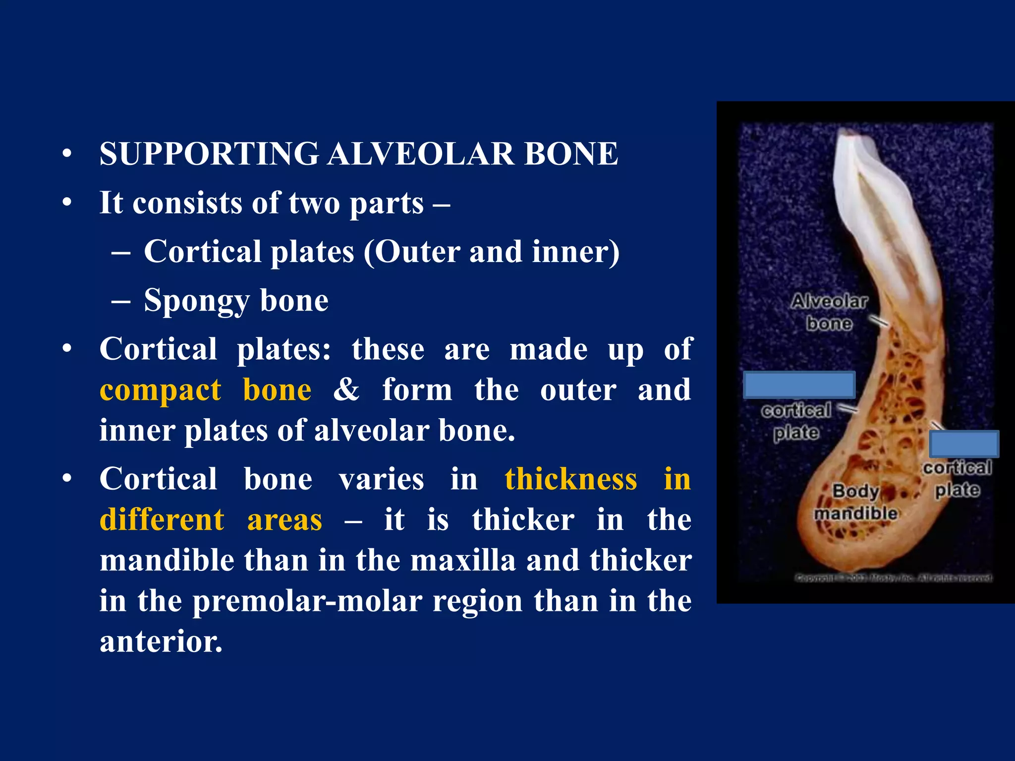
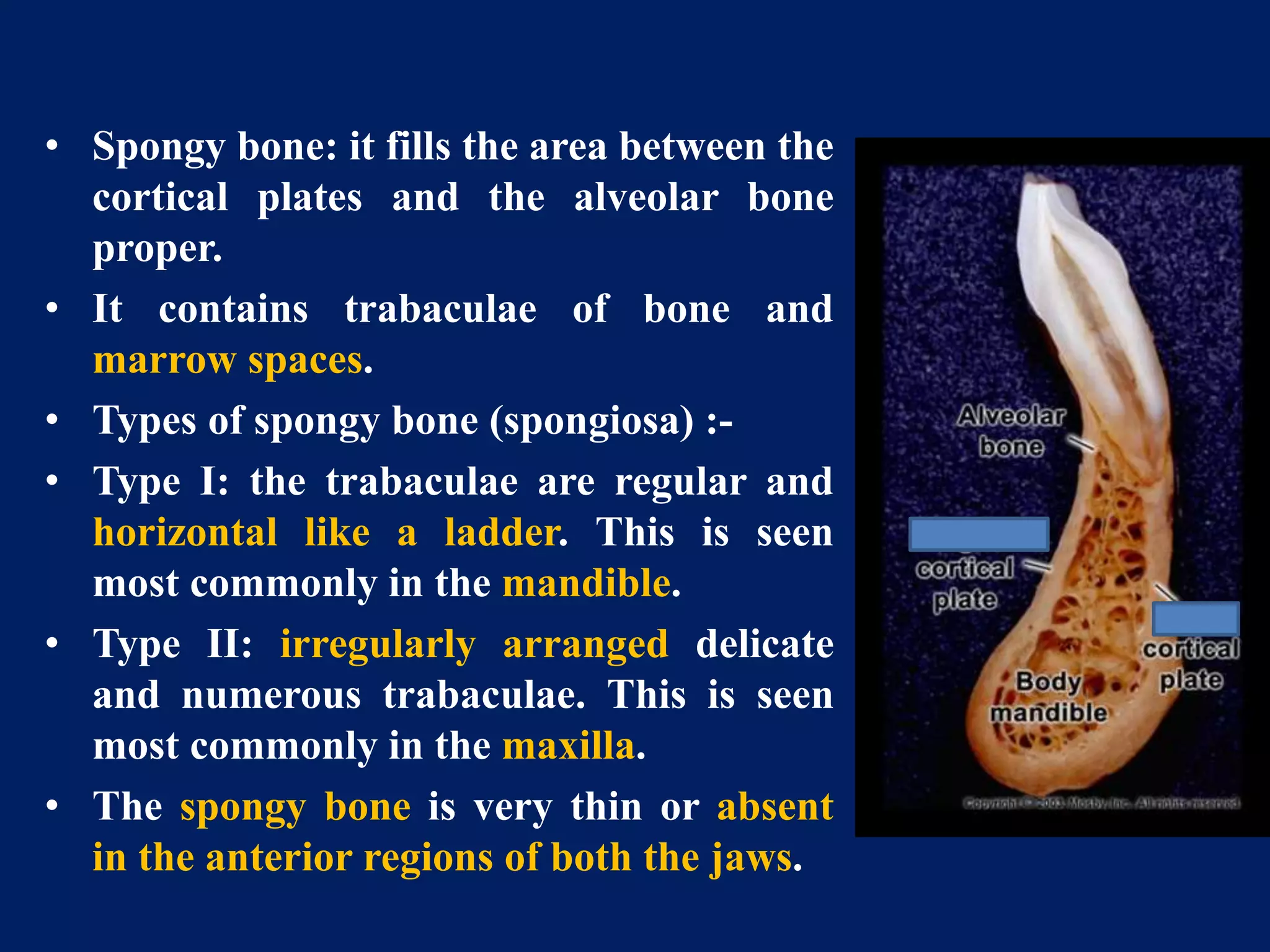
The document discusses diseases of bone, including developmental disorders. It describes four developmental disorders:
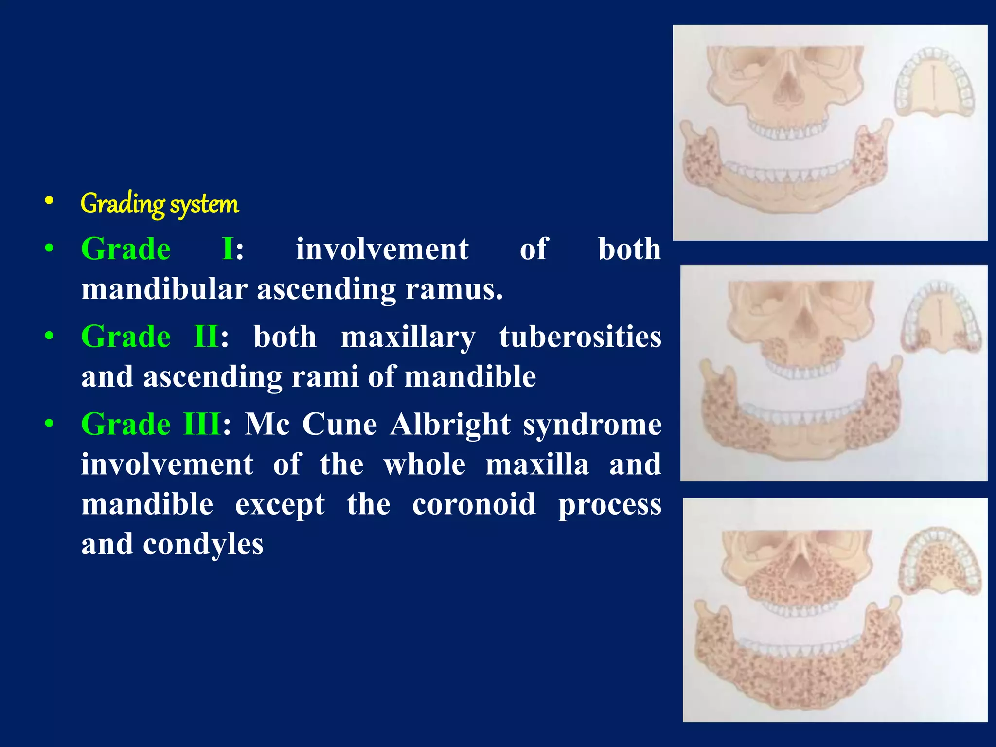
1. Cherubism - A rare jaw condition seen in childhood characterized by bilateral swelling of the jaws due to fibrous dysplasia-like lesions. It is caused by a genetic mutation and lesions typically regress after puberty.
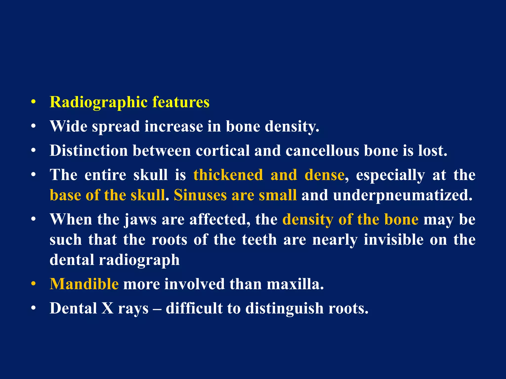
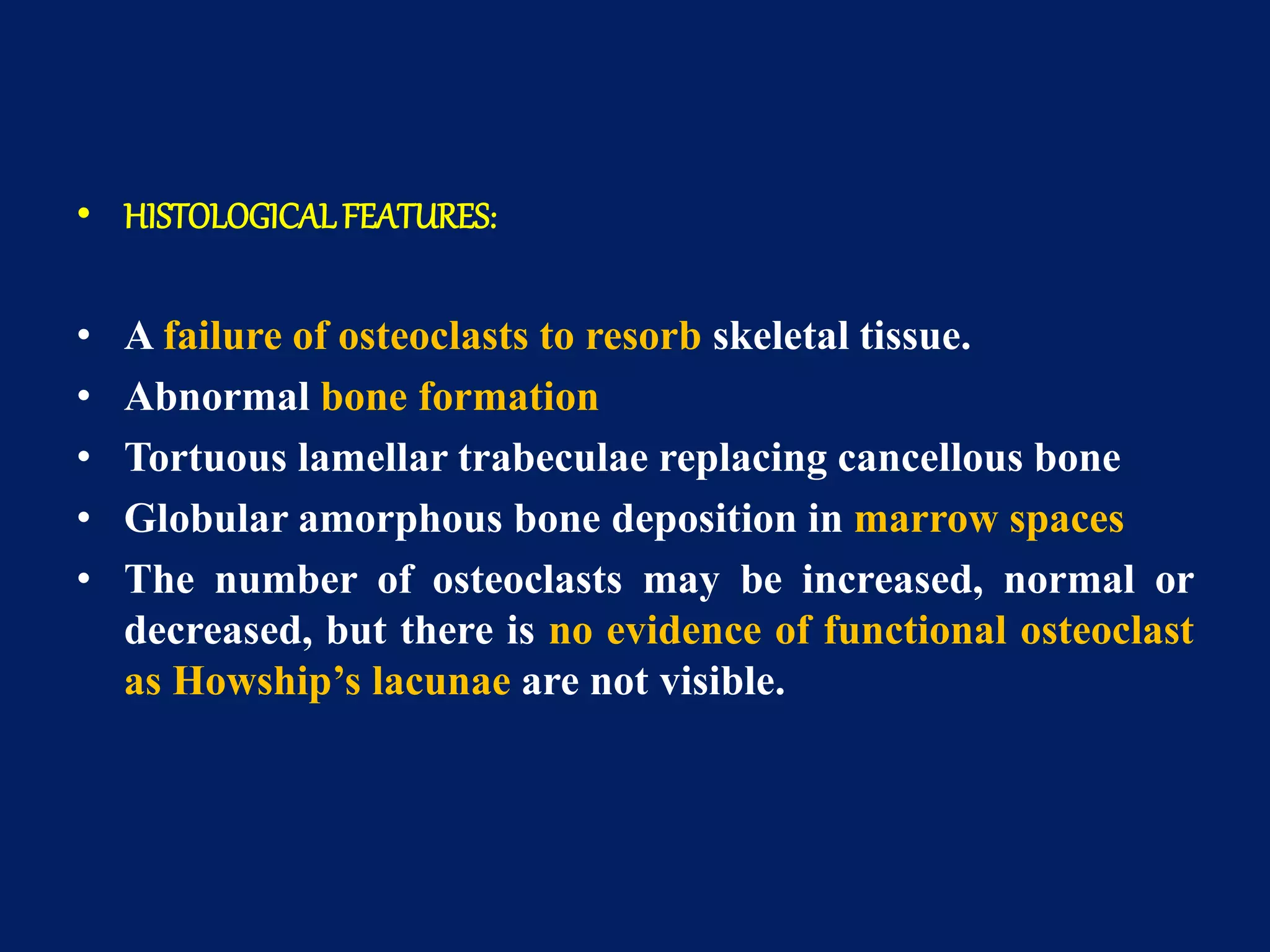
2. Osteopetrosis - A rare disease caused by a defect in bone remodeling resulting in abnormally dense bone that is fragile. It can cause anemia, fractures, and nerve compression.
3. Osteogenesis imperfecta - The most common inherited bone disorder caused by collagen defects, characterized by bone fragility and fractures that easily heal. Other features include blue sclera
Introduction
Epidemiology
Etiology
Manifestations
TNM staging
Squamous cell carcinoma is defined as malignant epithelial neoplasm exhibiting squamous differentiation as characterised by the formation of keratin and/or the presence of intercellular bridges.
( Pindborg et al, 1997).
Introduction
Epidemiology
Etiology
Manifestations
TNM staging
Squamous cell carcinoma is defined as malignant epithelial neoplasm exhibiting squamous differentiation as characterised by the formation of keratin and/or the presence of intercellular bridges.
( Pindborg et al, 1997).
Avitaminosis is any disease caused by chronic or long-term deficiency or caused by a defect in metabolic conversion, such as tryptophan to niacin. They are designated by the same letter as the vitamin.
Systemic Diseases Manifested in the Jawsvahid199212
Systemic Diseases Manifestation the Jaws based on chapter25
Oral Radiology
P R I N C I P L E S
a n d I N T E R P R E T A T I O N Sixth Edition
White and pharoah
The Indian Dental Academy is the Leader in continuing dental education , training dentists in all aspects of dentistry and
offering a wide range of dental certified courses in different formats.
This ppt gives information about alveolar bone . I am a post graduate student from the department of periodontics and implantology , I've tried to make the topic as easy as possible. Hope it is useful for the ug students.
mandibular premolars, common triats and differences between mandibular first and second premolar. buccal aspect, lingual aspect, mesial aspect, distal aspect, occlusal aspect of mandibular premolars
These lecture slides, by Dr Sidra Arshad, offer a quick overview of physiological basis of a normal electrocardiogram.
Learning objectives:
1. Define an electrocardiogram (ECG) and electrocardiography
2. Describe how dipoles generated by the heart produce the waveforms of the ECG
3. Describe the components of a normal electrocardiogram of a typical bipolar leads (limb II)
4. Differentiate between intervals and segments
5. Enlist some common indications for obtaining an ECG
Study Resources:
1. Chapter 11, Guyton and Hall Textbook of Medical Physiology, 14th edition
2. Chapter 9, Human Physiology - From Cells to Systems, Lauralee Sherwood, 9th edition
3. Chapter 29, Ganong’s Review of Medical Physiology, 26th edition
4. Electrocardiogram, StatPearls - https://www.ncbi.nlm.nih.gov/books/NBK549803/
5. ECG in Medical Practice by ABM Abdullah, 4th edition
6. ECG Basics, http://www.nataliescasebook.com/tag/e-c-g-basics
- Video recording of this lecture in English language: https://youtu.be/lK81BzxMqdo
- Video recording of this lecture in Arabic language: https://youtu.be/Ve4P0COk9OI
- Link to download the book free: https://nephrotube.blogspot.com/p/nephrotube-nephrology-books.html
- Link to NephroTube website: www.NephroTube.com
- Link to NephroTube social media accounts: https://nephrotube.blogspot.com/p/join-nephrotube-on-social-media.html
Title: Sense of Smell
Presenter: Dr. Faiza, Assistant Professor of Physiology
Qualifications:
MBBS (Best Graduate, AIMC Lahore)
FCPS Physiology
ICMT, CHPE, DHPE (STMU)
MPH (GC University, Faisalabad)
MBA (Virtual University of Pakistan)
Learning Objectives:
Describe the primary categories of smells and the concept of odor blindness.
Explain the structure and location of the olfactory membrane and mucosa, including the types and roles of cells involved in olfaction.
Describe the pathway and mechanisms of olfactory signal transmission from the olfactory receptors to the brain.
Illustrate the biochemical cascade triggered by odorant binding to olfactory receptors, including the role of G-proteins and second messengers in generating an action potential.
Identify different types of olfactory disorders such as anosmia, hyposmia, hyperosmia, and dysosmia, including their potential causes.
Key Topics:
Olfactory Genes:
3% of the human genome accounts for olfactory genes.
400 genes for odorant receptors.
Olfactory Membrane:
Located in the superior part of the nasal cavity.
Medially: Folds downward along the superior septum.
Laterally: Folds over the superior turbinate and upper surface of the middle turbinate.
Total surface area: 5-10 square centimeters.
Olfactory Mucosa:
Olfactory Cells: Bipolar nerve cells derived from the CNS (100 million), with 4-25 olfactory cilia per cell.
Sustentacular Cells: Produce mucus and maintain ionic and molecular environment.
Basal Cells: Replace worn-out olfactory cells with an average lifespan of 1-2 months.
Bowman’s Gland: Secretes mucus.
Stimulation of Olfactory Cells:
Odorant dissolves in mucus and attaches to receptors on olfactory cilia.
Involves a cascade effect through G-proteins and second messengers, leading to depolarization and action potential generation in the olfactory nerve.
Quality of a Good Odorant:
Small (3-20 Carbon atoms), volatile, water-soluble, and lipid-soluble.
Facilitated by odorant-binding proteins in mucus.
Membrane Potential and Action Potential:
Resting membrane potential: -55mV.
Action potential frequency in the olfactory nerve increases with odorant strength.
Adaptation Towards the Sense of Smell:
Rapid adaptation within the first second, with further slow adaptation.
Psychological adaptation greater than receptor adaptation, involving feedback inhibition from the central nervous system.
Primary Sensations of Smell:
Camphoraceous, Musky, Floral, Pepperminty, Ethereal, Pungent, Putrid.
Odor Detection Threshold:
Examples: Hydrogen sulfide (0.0005 ppm), Methyl-mercaptan (0.002 ppm).
Some toxic substances are odorless at lethal concentrations.
Characteristics of Smell:
Odor blindness for single substances due to lack of appropriate receptor protein.
Behavioral and emotional influences of smell.
Transmission of Olfactory Signals:
From olfactory cells to glomeruli in the olfactory bulb, involving lateral inhibition.
Primitive, less old, and new olfactory systems with different path
Flu Vaccine Alert in Bangalore Karnatakaaddon Scans
As flu season approaches, health officials in Bangalore, Karnataka, are urging residents to get their flu vaccinations. The seasonal flu, while common, can lead to severe health complications, particularly for vulnerable populations such as young children, the elderly, and those with underlying health conditions.
Dr. Vidisha Kumari, a leading epidemiologist in Bangalore, emphasizes the importance of getting vaccinated. "The flu vaccine is our best defense against the influenza virus. It not only protects individuals but also helps prevent the spread of the virus in our communities," he says.
This year, the flu season is expected to coincide with a potential increase in other respiratory illnesses. The Karnataka Health Department has launched an awareness campaign highlighting the significance of flu vaccinations. They have set up multiple vaccination centers across Bangalore, making it convenient for residents to receive their shots.
To encourage widespread vaccination, the government is also collaborating with local schools, workplaces, and community centers to facilitate vaccination drives. Special attention is being given to ensuring that the vaccine is accessible to all, including marginalized communities who may have limited access to healthcare.
Residents are reminded that the flu vaccine is safe and effective. Common side effects are mild and may include soreness at the injection site, mild fever, or muscle aches. These side effects are generally short-lived and far less severe than the flu itself.
Healthcare providers are also stressing the importance of continuing COVID-19 precautions. Wearing masks, practicing good hand hygiene, and maintaining social distancing are still crucial, especially in crowded places.
Protect yourself and your loved ones by getting vaccinated. Together, we can help keep Bangalore healthy and safe this flu season. For more information on vaccination centers and schedules, residents can visit the Karnataka Health Department’s official website or follow their social media pages.
Stay informed, stay safe, and get your flu shot today!
These simplified slides by Dr. Sidra Arshad present an overview of the non-respiratory functions of the respiratory tract.
Learning objectives:
1. Enlist the non-respiratory functions of the respiratory tract
2. Briefly explain how these functions are carried out
3. Discuss the significance of dead space
4. Differentiate between minute ventilation and alveolar ventilation
5. Describe the cough and sneeze reflexes
Study Resources:
1. Chapter 39, Guyton and Hall Textbook of Medical Physiology, 14th edition
2. Chapter 34, Ganong’s Review of Medical Physiology, 26th edition
3. Chapter 17, Human Physiology by Lauralee Sherwood, 9th edition
4. Non-respiratory functions of the lungs https://academic.oup.com/bjaed/article/13/3/98/278874
HOT NEW PRODUCT! BIG SALES FAST SHIPPING NOW FROM CHINA!! EU KU DB BK substit...GL Anaacs
Contact us if you are interested:
Email / Skype : kefaya1771@gmail.com
Threema: PXHY5PDH
New BATCH Ku !!! MUCH IN DEMAND FAST SALE EVERY BATCH HAPPY GOOD EFFECT BIG BATCH !
Contact me on Threema or skype to start big business!!
Hot-sale products:
NEW HOT EUTYLONE WHITE CRYSTAL!!
5cl-adba precursor (semi finished )
5cl-adba raw materials
ADBB precursor (semi finished )
ADBB raw materials
APVP powder
5fadb/4f-adb
Jwh018 / Jwh210
Eutylone crystal
Protonitazene (hydrochloride) CAS: 119276-01-6
Flubrotizolam CAS: 57801-95-3
Metonitazene CAS: 14680-51-4
Payment terms: Western Union,MoneyGram,Bitcoin or USDT.
Deliver Time: Usually 7-15days
Shipping method: FedEx, TNT, DHL,UPS etc.Our deliveries are 100% safe, fast, reliable and discreet.
Samples will be sent for your evaluation!If you are interested in, please contact me, let's talk details.
We specializes in exporting high quality Research chemical, medical intermediate, Pharmaceutical chemicals and so on. Products are exported to USA, Canada, France, Korea, Japan,Russia, Southeast Asia and other countries.
263778731218 Abortion Clinic /Pills In Harare ,sisternakatoto
263778731218 Abortion Clinic /Pills In Harare ,ABORTION WOMEN’S CLINIC +27730423979 IN women clinic we believe that every woman should be able to make choices in her pregnancy. Our job is to provide compassionate care, safety,affordable and confidential services. That’s why we have won the trust from all generations of women all over the world. we use non surgical method(Abortion pills) to terminate…Dr.LISA +27730423979women Clinic is committed to providing the highest quality of obstetrical and gynecological care to women of all ages. Our dedicated staff aim to treat each patient and her health concerns with compassion and respect.Our dedicated group ABORTION WOMEN’S CLINIC +27730423979 IN women clinic we believe that every woman should be able to make choices in her pregnancy. Our job is to provide compassionate care, safety,affordable and confidential services. That’s why we have won the trust from all generations of women all over the world. we use non surgical method(Abortion pills) to terminate…Dr.LISA +27730423979women Clinic is committed to providing the highest quality of obstetrical and gynecological care to women of all ages. Our dedicated staff aim to treat each patient and her health concerns with compassion and respect.Our dedicated group of receptionists, nurses, and physicians have worked together as a teamof receptionists, nurses, and physicians have worked together as a team wwww.lisywomensclinic.co.za/
Report Back from SGO 2024: What’s the Latest in Cervical Cancer?bkling
Are you curious about what’s new in cervical cancer research or unsure what the findings mean? Join Dr. Emily Ko, a gynecologic oncologist at Penn Medicine, to learn about the latest updates from the Society of Gynecologic Oncology (SGO) 2024 Annual Meeting on Women’s Cancer. Dr. Ko will discuss what the research presented at the conference means for you and answer your questions about the new developments.
Prix Galien International 2024 Forum ProgramLevi Shapiro
June 20, 2024, Prix Galien International and Jerusalem Ethics Forum in ROME. Detailed agenda including panels:
- ADVANCES IN CARDIOLOGY: A NEW PARADIGM IS COMING
- WOMEN’S HEALTH: FERTILITY PRESERVATION
- WHAT’S NEW IN THE TREATMENT OF INFECTIOUS,
ONCOLOGICAL AND INFLAMMATORY SKIN DISEASES?
- ARTIFICIAL INTELLIGENCE AND ETHICS
- GENE THERAPY
- BEYOND BORDERS: GLOBAL INITIATIVES FOR DEMOCRATIZING LIFE SCIENCE TECHNOLOGIES AND PROMOTING ACCESS TO HEALTHCARE
- ETHICAL CHALLENGES IN LIFE SCIENCES
- Prix Galien International Awards Ceremony
Recomendações da OMS sobre cuidados maternos e neonatais para uma experiência pós-natal positiva.
Em consonância com os ODS – Objetivos do Desenvolvimento Sustentável e a Estratégia Global para a Saúde das Mulheres, Crianças e Adolescentes, e aplicando uma abordagem baseada nos direitos humanos, os esforços de cuidados pós-natais devem expandir-se para além da cobertura e da simples sobrevivência, de modo a incluir cuidados de qualidade.
Estas diretrizes visam melhorar a qualidade dos cuidados pós-natais essenciais e de rotina prestados às mulheres e aos recém-nascidos, com o objetivo final de melhorar a saúde e o bem-estar materno e neonatal.
Uma “experiência pós-natal positiva” é um resultado importante para todas as mulheres que dão à luz e para os seus recém-nascidos, estabelecendo as bases para a melhoria da saúde e do bem-estar a curto e longo prazo. Uma experiência pós-natal positiva é definida como aquela em que as mulheres, pessoas que gestam, os recém-nascidos, os casais, os pais, os cuidadores e as famílias recebem informação consistente, garantia e apoio de profissionais de saúde motivados; e onde um sistema de saúde flexível e com recursos reconheça as necessidades das mulheres e dos bebês e respeite o seu contexto cultural.
Estas diretrizes consolidadas apresentam algumas recomendações novas e já bem fundamentadas sobre cuidados pós-natais de rotina para mulheres e neonatos que recebem cuidados no pós-parto em unidades de saúde ou na comunidade, independentemente dos recursos disponíveis.
É fornecido um conjunto abrangente de recomendações para cuidados durante o período puerperal, com ênfase nos cuidados essenciais que todas as mulheres e recém-nascidos devem receber, e com a devida atenção à qualidade dos cuidados; isto é, a entrega e a experiência do cuidado recebido. Estas diretrizes atualizam e ampliam as recomendações da OMS de 2014 sobre cuidados pós-natais da mãe e do recém-nascido e complementam as atuais diretrizes da OMS sobre a gestão de complicações pós-natais.
O estabelecimento da amamentação e o manejo das principais intercorrências é contemplada.
Recomendamos muito.
Vamos discutir essas recomendações no nosso curso de pós-graduação em Aleitamento no Instituto Ciclos.
Esta publicação só está disponível em inglês até o momento.
Prof. Marcus Renato de Carvalho
www.agostodourado.com
micro teaching on communication m.sc nursing.pdfAnurag Sharma
Microteaching is a unique model of practice teaching. It is a viable instrument for the. desired change in the teaching behavior or the behavior potential which, in specified types of real. classroom situations, tends to facilitate the achievement of specified types of objectives.
Explore natural remedies for syphilis treatment in Singapore. Discover alternative therapies, herbal remedies, and lifestyle changes that may complement conventional treatments. Learn about holistic approaches to managing syphilis symptoms and supporting overall health.
2. Introduction
• Bone is a living tissue, which makes up the body
• Skeleton and is one of the hardest structures of the animal
body.
• Bone possesses a certain degree of toughness and elasticity.
• It provides shape and support for the body.
• It also provides site of attachment for tendons and muscles,
which are essential for locomotion.
• It also protects vital organs in the body.
• It also provides site for development and storage for blood
cells.
3.
4.
5.
6. Alveolar bone
• ALVEOLAR BONE PROPER
• A) LAMELLATED BONE:
• It is the outer most part of the
alveolar bone proper.
• Some lamellae of the lamellated
bone are arranged roughly parallel
to the surface of the adjacent
marrow spaces, whereas others
forms haversian system.
7. • BUNDLE BONE
• Bundle bone is the part of alveolar bone, into which the
fiber bundles of the PDL insert.
• Fibrils arranged at right angles are Sharpey’s Fibers
• Bundle bone is formed in areas of recent bone deposition.
• Lines of rest seen in bundle bone.
• Radiographically it is called as Lamina Dura because of
increased radiopacity which is due to the presence of thick
bone without trabeculations.
8. radiographically
The lamina dura (arrows) appears as a thin opaque layer of bone
around teeth and around a recent extraction socket.
9. • SUPPORTING ALVEOLAR BONE
• It consists of two parts –
– Cortical plates (Outer and inner)
– Spongy bone
• Cortical plates: these are made up of
compact bone & form the outer and
inner plates of alveolar bone.
• Cortical bone varies in thickness in
different areas – it is thicker in the
mandible than in the maxilla and thicker
in the premolar-molar region than in the
anterior.
10. • Spongy bone: it fills the area between the
cortical plates and the alveolar bone
proper.
• It contains trabaculae of bone and
marrow spaces.
• Types of spongy bone (spongiosa) :-
• Type I: the trabaculae are regular and
horizontal like a ladder. This is seen
most commonly in the mandible.
• Type II: irregularly arranged delicate
and numerous trabaculae. This is seen
most commonly in the maxilla.
• The spongy bone is very thin or absent
in the anterior regions of both the jaws.
17. CHERUBISM
• Rare developmental jaw condition, first described by Jones
in 1933.
• Jones called it as familial multilocular disease of the jaw.
• Other wise named as Familial fibrous dysplasia of jaws,
disseminated juvenile fibrous dysplasia, familial multilocular
cystic disease of jaws, familial fibrous swelling of jaws
• Transmitted as an autosomal dominant trait with 100%
penetrance in males and 50-70% in females
• Affects more than one quadrant
• Non-neoplastic hereditary bone lesion that is histologically
similar to central giant cell granuloma, affects the jaws of
children bilaterally and symmetrically
18. • Child appears as a plump cheeked angels called “Cherub”
• The jaw lesion remit spontaneously when the child reaches
puberty, leaving some facial deformity and malocclusion
but reason for this remission is still unknown.
• The appearance of people with the disorder is caused by a
loss of bone, which the body replaces with excessive
amounts of fibrous tissue
19. • Pathogenesis
• The gene for cherubism was mapped to chromosome 4p16.
• Mutation were identified in the SH3BP2 gene .
• The protein encoded by this gene is believed to function in
signal transduction pathway and to increase the activity of
osteoclasts and osteoblasts during normal tooth eruption.
• It has been suggested that mutation in the SH3BP2 gene
may led to pathologic activation of osteoclasts and
disruption of jaw morphogenesis.
20. • CLINICAL FEATURES
• Age incidence: normal at the birth and are without any
clinically or radiographically evident disease until 14 months
to 3 yrs of age.
• Sex incidence: males = females
• Site predilection:
• Mostly bilateral involvement
• Mandible affected more commonly than maxilla.
• In maxilla, tuberosity region is affected frequently, resulting in
respiratory obstruction and impairment of vision & hearing.
• The lesion are painless and symmetrical.
21. • Signs& symptoms:
• Begins as painless bilateral expansion of affected bone.
• Skin of upper face is stretched.
• A rim of sclera may be seen beneath the iris, giving a
classical “ eyes upturned to heaven” appearance.
• This feature is due to involvement of the infraorbital rim
and orbital floor that tilts the eyeball upwards, as well as to
stretching of the upper facial skin that pulls the lower lid
downwards.
22. • Progressive, extensive bone involvement causes widening
and distortion of alveoli.
• As a result, developing teeth displaced, fail to erupt.
• Numerous dental abnormalities have been reported, such
as agenesis of the 2nd & 3rd mandibular molars,
displacement of the teeth, premature exfoliation of the
primary teeth, delayed eruption of the permanent teeth,
and transposition and rotation of the teeth.
• The permanent dentition is often defective.
• In sever cases root resorption occurs.
• It is been connected to NOONAN’S SYNDROME.
23. • Grading system
• Grade I: involvement of both
mandibular ascending ramus.
• Grade II: both maxillary tuberosities
and ascending rami of mandible
• Grade III: Mc Cune Albright syndrome
involvement of the whole maxilla and
mandible except the coronoid process
and condyles
24. • RADIOGRAPHIC FEATURES
• Appear as expansile, multilocular radiolucency.
• The presence of numerous unerupted teeth and the
destruction of the alveolar bone may displace the teeth,
producing a radiographic appearance referred as
FLOATING TOOTH SYNDROME.
• With adulthood, the cystic areas in the jaws become re-
ossified, which results in irregular patchy sclerosis.
• There is classic ground glass appearance because of the
small, tightly compressed trabecular pattern.
25.
26. • HISTOLOGICALFEATURES
• Features are similar to giant cell tumors.
• Normal bone is partly replaced by pathologic tissue.
• Contains numerous randomly distributed multinucleated
giant cells and vascular spaces within a fibrous connective
tissue stroma.
• Clusters of giant cells and diffuse background of spindled
mononuclear cells, fresh hemorrhage, and eosinophilic
fibrinous material
• An eosinophilic perivascular cuffing is seen.
• The multinucleated giant cells are positive for tartrate
resistant acid phosphatase which is characteristic of
osteoclasts.
27. Multinucleated giant cells are
scattered in vascular fibrous
stroma. Osteoid and newly
formed bone matrix are visible
Multinucleated giant cells are
scattered around blood vessels
30. • DIFFERENTIAL DIAGNOSIS
– Giant cell granulomas of the jaw
– Osteoclastomas
– Aneurysmal bone cyst
– Fibrous dysplasia
– Hyperparathyroidism
31. • TREATMENT
• With time, especially after puberty, the lesions
regress.
• Before puberty, surgery should be carried out only in
cases of severe functional disturbances – otherwise
rapid regrowth of lesions and worsening deformity.
• Use of calcitonin in severe cases.
• Radiation therapy is contraindicated - risk of
development of postirradiation sarcoma.
• The optimal therapy for cherubism - not determined
32. • Keyfeatures:
• Inherited autosomal dominant trait.
• Jaw swelling appears in infancy.
• Angle regions of mandible affected symmetrically giving
typical chubby face.
• Symmetrical involvement of maxillae also seen in sever
cases.
• Radiographically- multilocular cystic lesion.
• Histologically- consist of giant cells in vascular connective
tissue.
• Lesion regress with skeletal maturation and nornal
contour is restored.
33. OSTEOPETROSIS
• Synonyms
• Albers - Schönberg Disease,
• Marble bone disease,
• Osteosclerosis fragilis generalisata
• Rare disease characterized by excessive density of all bones
with obliteration of marrow cavities.
• Due to defect in remodelling caused by failure of
osteoclastic function.
• Osteoclast number is often increased – but no bone is
resorbed.
• Bone has poor mechanical properties
34. • PATHOGENESIS
• Osteoclasts fail to function normally.
• As a result, bone remodeling is affected.
• Defective bone resorption combined with continued bone
deposition results in thickening of cortical bone and sclerosis
of cancellous bone.
• The exact mechanism is unknown.
• Mutation of key elements req. for osteoclastic activity
• H+ ATPase proton pump acidification of resor. lacunae
• Chloride channel regulation of ionic charge
• Carbonic anhydrase resorption of bone
matrix
35. • Cliniclal features
• Clinical types – infantile, intermidiate and adult osteopetrosis.
• Secondary anemia, neutropenia, with susceptibility to
infections.
• Abnormally dense bone is mechanically weak, so fractures are
common.
• Jaws are composed of dense bone with reduced marrow
spaces.
• There may be delayed eruption of teeth.
• Osteomyelitis is a common complication of tooth extraction.
• Other - Cranial nerve compression, hepatosplenomegaly,
facial deformity and marrow failure.
36. • I. INFANTILE OSTEOPETROSIS
• Malignant osteopetrosis
• Diagnosed in early life
• Autosomal recessive trait.
• Diffusely sclerotic skeleton, marrow failure and signs of
cranial nerve compression present.
• Initial signs – normocytic normochromic anemia and
hepatosplenomegaly, due to compensatory extramedullary
heamatopoiesis.
• Increased susceptibility to infections due to
granulocytopenia.
37. • II. INTERMEDIATE OSTEOPETROSIS
• Also called as benign osteopetrosis
• Diagnosed in adolescence and adulthood
• Affected patients have a short stature and are often
asymptomatic at birth, but frequently exhibit fractures by
the end of their first decade of life.
• Marrow failure and hepatosplenomegaly are rare.
• Some present with cranial nerve deficits, macrocephaly,
mild or moderately severe anemia and ankylosed teeth that
may predispose them to osteomyelitis of the jaws.
38. • III. ADULT OSTEOPETROSIS
• Discovered late in life – milder symptoms.
• Autosomal dominant trait.
• About 40% cases are asymptomatic.
• Axial skeleton shows sclerosis, while long bones show little
or no defects.
• Bone pain is seen
• 10% shows osteomyelitis of mandible
39. • Oral manifestations:
• Facial deformity leading to hypertelorism, snub nose,
frontal bossing etc.
• Delayed tooth eruption and osteomyelitis of jaws.
• Sclerosis of skull bones leads to narrowing of foramina
which causes compression of various cranial nerves –
blindness, deafness, facial paralysis etc.
• The medullary spaces of the jaws are reduced.
• Fracture of jaws during extraction procedure can occur
without undue force, due to fragility of the bone.
40. • Radiographic features
• Wide spread increase in bone density.
• Distinction between cortical and cancellous bone is lost.
• The entire skull is thickened and dense, especially at the
base of the skull. Sinuses are small and underpneumatized.
• When the jaws are affected, the density of the bone may be
such that the roots of the teeth are nearly invisible on the
dental radiograph
• Mandible more involved than maxilla.
• Dental X rays – difficult to distinguish roots.
41.
42. • Laboratoryfindings
• Severe anemia due to displacement of hematopoietic
marrow
• Elevated levels of parathyroid harmone, acid phosphatase,
creatinine kinase
43. • HISTOLOGICALFEATURES:
• A failure of osteoclasts to resorb skeletal tissue.
• Abnormal bone formation
• Tortuous lamellar trabeculae replacing cancellous bone
• Globular amorphous bone deposition in marrow spaces
• The number of osteoclasts may be increased, normal or
decreased, but there is no evidence of functional osteoclast
as Howship’s lacunae are not visible.
44. • Keyfeatures
• Rare genetic defect of osteoclastic activity.
• Bone lack medullary cavities but are fragile.
• Extramedullary haemopoiesis in liver and spleen but
anemia common.
• Osteomyelitis a recognised complication.
46. • Most common type of developmental, inherited bone
disorder, showing both autosomal dominant and recessive
pattern.
• Comprises heterogeneous group of heritable CT disorder
in which bone fragility is the primary feature.
• The basic abnormality is a genetic defect in collagen
maturation / synthesis of type I collagen.
• Mutation of genes – COLIA1 – chromosome 17
COLIA2 – chromosome 7
• It is characterized by impairment of collagen maturation.
• Collagen forms a major portion of bone, dentine, sclerae,
ligaments, and skin, OI demonstrates a variety of changes
that involves these sites.
47. • CLINICALFEATURES
• The chief clinical feature is the extreme fragility and
porosity of the bones, with an attended proneness to
fracture.
• Fractures heals readily but with the same quality of bone.
• AGE: Varies with the type.
• The other characteristic feature of OI is the occurrence of
blue sclera.
• The sclera is abnormally thin, and for this reason the
pigmented choroid shows through and produces the bluish
colour.
48. • Other condition in which blue sclera can be seen
– Osteopetrosis
– Fetal rickets
– Turner syndrome
– Pagets disease
– Marfan syndrome &
– Ehlers-Danlos syndrome.
– Some times in normal infants.
49. • Additional signs & symptoms
• Deafness- due to osteosclerosis.
• Abnormalities of teeth.
• Laxity of ligaments.
• A peculiar shape of the skull.
• Abnormal electrical reaction of the muscles.
• Tendency towards capillary bleeding.
50. • CLINICAL FEATURES: -
• Four types present, each having several subtypes.
• TYPE I Osteogenesis imperfecta:
• Commonest type – autosomal dominant.
• Mild to moderate bone fragility – onset is highly variable –
may be present at birth also.
• Hearing loss develops before 30 years.
• Some patients may show dentinogenesis imperfecta sub
type B.
• Blue sclera is seen.
• Kyphoscoliosis
• Easy bruising
• Short stature
51.
52. • TYPE II Osteogenesis imperfecta:
• Extreme bone fragility with frequent fractures.
• Many patients stillborn – 90% die before 4 weeks of age.
• Blue sclera present.
• Dentinogenesis imperfecta present.
• Hearing loss present.
• Micrognathia
• Short trunk.
53. • TYPE III Osteogenesis imperfecta:
• Moderate to severe bone fragility.
• Blue sclera present in infants but fades by adulthood.
• Mortality rate higher in older children.
• Death from cardiopulmonary complications caused by
kyphoscoliosis (backward & lateral curvature of spine).
• Dentinogenesis imperfecta.
• Short limbs.
• Triangluar face, with frontal bossing
54. • TYPE IV Osteogenesis imperfecta: -
• Mild to moderate bone fragility.
• Sclera pale in early life, but fades in later life.
• Fractures present in 50 % case – frequency of fractures
decreases after puberty.
• Some patients may have dentinogenesis imperfecta, some
may not.
55. • Oral manifestation
• Both the dentitions are affected, and demonstrate blue to
brown translucency.
• Radiographically reveals premature obliteration of pulp.
• Although the altered teeth closely resemble dentinogenesis
imperfecta , the two disease are the result of different
mutations and should be considered as separate processes.
• Head size is large.
• Frontal and temporal bossing is seen.
• Class III malocclusion is seen due to maxillary hypoplasia
rather than mandibular hyperplasia.
• Anterior and posterior cross bite and open bites can be
seen.
• Large numbers of impacted and ectopic teeth can be
reported.
• Unerupted 1st and 2nd molar is very common feature.
56. • Radiographic features
• Osteopenia
• Bowing
• Angulation
• Deformity of long bones
• Multiple fratures
• Wormian bone in the skull.
57.
58. • HISTOLOGICALFEATURES:-
• Anomaly due to abnormal collagen synthesis by abnormal
osteoblasts.
• Mass of cortical and cancellous bone is abnormal and
greatly reduced.
• Cortical bone is extremely thin while the cancellous bone is
delicate and shows micro fractures.
• Osteoblasts are present but bone matrix synthesis is
reduced, for this reason the thickness of long bone is
deficient.
• Bone architecture remains immature throughout life.
59. • Treatment
• No known treatment.
• Only treatment of the infection when they occur.
60. • Keyfeatures
• Thin fragile bones due to inadequate type I collagen.
• Inherited as an autosomal dominant trait.
• Multiple factures typically lead to gross deformities.
• Jaw fractures are uncommon.
62. • Bone defects primarily involve skull and clavicle – defects
seen in other bone also.
• Dental abnormalities common
• Inherited as autosomal dominant trait, but almost 40%
cases show spontaneous mutation.
• It is caused due to defect in CBFA 1 gene also called as
RUNX 2 gene of chromosome 6p21. (Runt-related
transcription factor 2 (RUNX2 )
• RUNX2 is a key transcription factor associated with
osteoblast differentiation
• This gene normally guides osteoblastic differentiation and
appropriate bone formation.
63. • CLINICAL FEATURES
• Age incidence: Children
• Sex incidence: Nil
• Site predilection: Skull, clavicles and jaw bones.
64. • Signs & symptoms:
• Short height with large heads showing pronounced frontal
and parietal bossing.
• Nose is broad with depressed nasal bridge.
• Shoulders narrow and droop excessively.
• Sagittal suture is sunken giving the skull a flat appearance.
• Paranasal sinuses are under developed
• Patients show unusual mobility of shoulders due to absence
/ hypoplasia of clavicles.
65.
66. • Oral manifestations:
• Narrow, high arched palate.
• Increased prevalence of cleft palate.
• Maxilla is underdeveloped and smaller than mandible.
• Prolonged retention of deciduous teeth and delay /
complete failure of eruption of permanent teeth.
• OPG and dental radiographs show multiple impacted and
supernumerary teeth.
• The roots of the teeth are short and thinner than usual,
and might be deformed.
67.
68. • Treatment
• No treatment exists for the skull, clavicular, and other bone
anomalies associated with CCD.
• Most patient function well with out any significant
problem.
69. • Keyfeatures
• Rare genetic disorder causing defective formation of
clavicle, delayed closure of fontanells and other defects.
• Many or most permanent teeth typically remains
embedded in the jaw.
• Many additional unerupted teeth also present.
• Sometimes many dentigerous cysts.
70. • A. DEVELOPMENTAL DISEASES:
– Cheurbism
– Osteopetrosis
– Osteogenesis imperfecta
– Cleiodocrainal dysplasia
• B. ENDOCRINAL DISEASES:
– Hyperparathyroidism
• C. IDIOPATHIC DISEASES:
– Idiopathic osteosclerosis
– Massive osteolysis
– Langerhan’s cell disease (Histiocytosis-
X)
– Paget’s disease
• D. REACTIVE DISEASES:
– Giant cell lesion of bone
– Aneurysmal bone cyst
– Simple / Traumatic bone cyst
• E. FIBRO-OSSEOUS LESIONS:
– (i) Non-neoplastic lesions -
• Fibrous dysplasia
• Cemento-osseous dysplasia
– (ii) Neoplasms –
• Ossifying fibroma
• F. INFLAMMATORY DISEASES: -
– (i) Specific:
• Tuberculosis
• Actinomycosis
– (ii) Non specific
• Osteomyelitis
• Dry socket
• Periapical cyst / abscess /
granuloma
• Osteoradionecrosis
• G. NEOPLASTIC DISEASES: -
– (i) Benign:
• Osteoma
• Osteoid osteoma & osteoblastoma
• Chondroma
• Chondromyxoid fibroma
– (ii) Malignant:
• Osteosarcoma
• Ewing’s sarcoma
• Chondrosarcoma
72. PAGET’S DISEASE OF BONE
• Osteitis deformans
• Characterized by abnormal resorption and deposition of
bone, resulting in distortion and weakening of bone.
• ETIOLOGY
• Unknown
• Inflammatory
• Genetic
• Endocrine factors
• Slow virus infection
• Positive family history
73. • CLINICAL FEATURES
• Age incidence: Middle aged individuals
• Sex incidence: Male to female ratio is 2:1
• Site predilection: Bones of skull, lumbar vertebrae ,pelvis,
femur and tibia.
• Common in England, France and Germany.
• Rare in Middle and Far East Asia and Africa.
• Paget’s disease – Monostotic, Polyostotic
74. • Signs and Symptoms -
• Bone pain – most common, severe bone pain and limitation
of movement, especially of joints
• Affected bone becomes thickened, enlarged and weakened
• Weight bearing bones – bowing deformity
• Skull – progressive increase in the circumference of the
head
75. • Maxilla affected more than mandible.
• Maxilla – enlargement of middle third of face (leontiasis
ossea)
• Nasal obstruction, obliterated sinuses and deviated septum
also occur.
• In dentulous patients spacing of teeth is seen, while
edentulous patients complains of tightness of the dentures.
• Mandible involved rarely – may cause prognathism.
76. • Occurs in 3 phases
1. Intense osteoclastic activity and bone resorption
2. Osteoblastic phase producing woven bone, with
ineffective mineralization
3. Dense cortical and trabecullar bone deposition
• Early phase – decrease radiodensity and alteratrion of
trabecular pattern
• In skull – large circumscribed areas of radiolucency -
Osteoporosis circumscripta
77. • Osteoblastic phase – patchy areas of sclerotic bone -
‘cotton wool ‘ appearance
• Later stage – hypercementosis of teeth
• Bone scintingraphy – extent of involvement
• Manbible - marked uptake involving from condyle to
condyle Black beard or Lincoln’s sign
80. • Histopathology
• Uncontrolled alternating resorption and deposition of bone
• Increased osteoclastic activity in resorptive phase
• Osteoblastic activity with formation of osteoid rims around
bone trabeculae
• Vascular fibrous CT replaces marrow
• Presence of basophilic reversal lines in bone – representing
the resorptive and formative phase “Jigsaw puzzle” or
“Mosaic” appearence
81.
82.
83. • Diagnosis
• High level of serum alkaline phosphatase levels
• Normal blood calcium and phosphorus levels
• Urinary hydroxyproline levels are elevated
• Other sensitive markers of bone resorption –
– N-telopeptides
– C-telopeptides
– Pyridinoline cross-link assays
84. • Treatment
• Rx req if the alk. Phosphatase value increase to 25 to 50%
• Bone pain – NSAIDs
• Antiresorptive therapy
• Use of parathyroid hormone antagonists ie, calcitonin and
biphosphonates – mainstays of therapy
• Biphosphonates are administered for – 2 – 6 months
• Dental complications
• Extraction due to hypercementosis
• Osteomyelitis
• Malignant bone tumor - osteosarcoma
• Benign and malignant giant cell lesions
85. • Keyfeatures
• Person past middle age affected.
• Enlargement of skull, thickening but weakness of long
bone and bone pain.
• Maxilla occasionally, but mandible rarely affected.
• Hypercementosis.
• Radiographically- cotton wool appearance.
• Histologically- mosaic/ jigsaw puzzle pattern.
• Serum alkaline phosphatase upto 250 Bodansky units
(normal – 30 to 40).
86. LANGERHANS CELL DISEASE
• The term HISTOCYTOSIS was introduce as a collective
designation for a spectrum of clinicopathologic disorders
characterized by proliferation of histiocytes like cells.
• It is an idiopathic disease characterized by proliferation of
histiocyte like cells (Langerhan's cells), that are
accompanied by varying numbers of eosinophils,
lymphocytes, plasma cells & multinucleated giant cell.
• Believed to be a non neoplastic process.
• Langerhan’s cells are dendritic, mononuclear cells
normally found in epidermis, mucosa, lymph nodes & bone
marrow. They are known as antigen presenting cells.
87. • TYPES
• 1. Eosinophilic granuloma of bone: solitary / multiple bone
involvement without systemic organ involvement. It causes
localized bone destruction with swelling and often pain.
• 2. Hand-Schüller-Christian disease: Chronic disseminated
disease involving bones, viscera and skin. It shows triad of
lytic skull lesion, exophthalmous, and diabetes insipidus.
• 3. Letter-Siwe disease: acute disseminated disease with
bone, visceral and skin involvement, occurring mainly in
infants.
88. • CLINICAL FEATURES
• Age incidence: Predominantly children below 10 years of
age.
• Sex incidence: Definite male predilection.
• Site predilection: Bones - Skull, ribs, vertebrae, femur and
mandible most frequently.
• Oral – gingiva and lips most commonly.
89. • Signs & symptoms:
• Involved bones manifest dull pain and tenderness.
• Visceral involvement results in decreased or failure of
affected organ.
• Jaw bone involvement results in loosening of teeth which
resembles aggressive periodontitis, and the appearance of
teeth “floating in air” are typical.
• Oral mucosa may show ulcerative / proliferative masses on
gingiva.
90.
91. • RADIOGRAPHIC FEATURES
• Multiple, well / poorly defined punched out radiolucent
areas seen.
• Extensive alveolar bone loss occurs, causing the teeth to
appear as if they are “floating in air”.
92.
93. • HISTOLOGICAL FEATURES
• Lesion shows diffuse infiltration of pale staining,
mononuclear cells containing ill defined cell borders and
vesicular nuclei.
• Darker staining eosinophils, plasma cells and lymphocytes
and multinucleated giant cells also seen.
• Electron microscopy shows rod / racquet shaped
characteristic birbeck granules within cytoplasm of
Langerhan's cells
94.
95. • Immunohistochemical studies are needed to confirm the
diagnosis as these cells cannot be distinguished from
normal histiocytes.
• Langerhan’s cells stain positively for S-100 protein.
96. • DIFFERENTIAL DIAGNOSIS
• Other lesions with multifocal, multilocular radiolucency
are
– Cemento-osseous dysplasia
– Hyperparathyroidism
– Cherubism
– Multiple myeloma
97. • A. DEVELOPMENTAL DISEASES:
– Cheurbism
– Osteopetrosis
– Osteogenesis imperfecta
– Cleiodocrainal dysplasia
• B. ENDOCRINAL DISEASES:
– Hyperparathyroidism
• C. IDIOPATHIC DISEASES:
– Idiopathic osteosclerosis
– Massive osteolysis
– Langerhan’s cell disease (Histiocytosis-
X)
– Paget’s disease
• D. REACTIVE DISEASES:
– Giant cell lesion of bone
– Aneurysmal bone cyst
– Simple / Traumatic bone cyst
• E. FIBRO-OSSEOUS LESIONS:
– (i) Non-neoplastic lesions -
• Fibrous dysplasia
• Cemento-osseous dysplasia
– (ii) Neoplasms –
• Ossifying fibroma
• F. INFLAMMATORY DISEASES: -
– (i) Specific:
• Tuberculosis
• Actinomycosis
– (ii) Non specific
• Osteomyelitis
• Dry socket
• Periapical cyst / abscess /
granuloma
• Osteoradionecrosis
• G. NEOPLASTIC DISEASES: -
– (i) Benign:
• Osteoma
• Osteoid osteoma & osteoblastoma
• Chondroma
• Chondromyxoid fibroma
– (ii) Malignant:
• Osteosarcoma
• Ewing’s sarcoma
• Chondrosarcoma
98. • Reactive lesions
– Central giant cell granuloma
– Aneurysmal bone cyst
– Traumatic bone cyst
99. CENTRALGIANT CELL GRANULOMA
• 1st described by Jaffe – central giant cell reparative
granuloma.
• Reparative – term is omitted.
• Considered to be a non neoplastic lesion.
• Uncommon, Benign and proliferative lesion.
• Extraosseous variant – Peripheral giant cell granuloma
100. • CLINICAL FEATURES
• Age – all ages. < 30 yrs
• Sex – F > M
• Site – Both jaws, Mandible > Maxilla
• Mainly anterior segments of jaw & have a tendency to
cross the midline.
• If small – no signs & symptoms, discovered accidently
• Expansion of cortex and perforation
• Mobility , displacement & root resorption
• Diffuse or regular borders
101. • Signs & symptoms
• Mostly asymptomatic and diagnosed only during routine
radiographic examination.
• Manifest usually as painless expansion of affected bone.
• Some aggressive cases may manifest with pain, paresthesia
and perforation of cortical plate.
• Occasionaly shows ulceration of the mucosal surface.
102. Can be grouped under two types
Non Aggressive Aggressive
Slow growing Fast
No root resorption Present
No cortical perforation Present
Shows new bone formation Extensive bone destruction
Less no. of giant cells More
Nuclei are scattered Nuclei are centered
Nuclei no. 3-10 3 - 30
Less area of haemorrhage More
Less hemosedrin deposits More
103. • RADIOGRAPHIC FEATURES
• Present as well defined, unilocular / multilocular radiolucent
defects, but the margins are usually noncorticated.
• Lesions may vary in size from small unilocular
radiolucencies to large multilocular radiolucencies.
104. • DIFFERENTIAL DIAGNOSIS
• Small unilocular lesions can be confused radiographically
with periapical cyst / granuloma.
• Large multilocular lesions should be differentiated from
other multilocular radiolucencies like– ameloblastoma,
aneurysmal bone cyst, Pindborg tumor etc
105. • HISTOLOGICAL FEATURES
• Few to large number of small / large multinucleated giant
cells seen in a background of ovoid / spindle shaped
mesenchymal cells.
• Giant cells believed to represent osteoclasts, and vary in
size from few to many nuclei.(20 nuclei or more )
• Foci of osteoid and newly formed bone may also be seen.
• Areas of hemorrhage and hemosiderin deposition are
common.
107. ANEURYSMAL BONE CYST
• Primarily seen in long bones or vertebrae, and rarely in
jaws.
• Cause and pathogenesis are not yet clear.
• Controversy – whether it arises de novo or occurs as a
result of some “vascular accident” in a pre-existing lesion.
• Aneurysmal bone cyst is an intraosseous accumulation of
variable sized, blood filled spaces surrounded by cellular
fibrous connective tissue that is often admixed with
trabeculae of reactive woven bone.
108. • Pathogenesis is not clear, but some investigators believe
that it arises from a traumatic event, vascular
malformation, or neoplasm that disrupts the normal
osseous hemodynamics and leads to an enlarging,
hemorrhagic extravassation.
• An aneurysmal bone cyst may form when an area of
hemorrhage maintain connection with the disrupted
feeding vessels, subsequently, giant cell granuloma like
area can develop after loss of connection with the original
vascular source.
109. • CLINICAL FEATURES
• Age incidence: First 3 decades.
• Sex incidence: Mainly females.
• Site predilection: molar regions of mandible & maxilla.
110. • Signs & symptoms:
• Hard, rapidly growing swelling which can cause malocclusion.
• Mobility of teeth
• Migration of teeth
• Root resorption
• Pain is often present.
• Paresthesia is present
• If lesion perforates cortical plates, can cause “egg shell
crackling”.
111. • RADIOLOGICAL FEATURES
• Classically seen as a unilocular, ovoid / fusiform
radiolucency which balloons the cortical plates.
• Teeth displacement and root resorption also observed.
112. • HISTOLOGICAL FEATURES:
• Cyst cavity shows many capillaries and blood filled spaces,
of various sizes separated by delicate loose CT.
• Blood filled spaces are not lined by endothelium.
• Many small multinucleated giant cells and trabeculae of
osteoid / woven bone cab be seen.
113.
114. • Key features
• Rare in jaws
• Jaw lesions are mostly seen in ramus and angle region
• Affected patient usually between 10 and 20 years.
• Unknown etiology.
• Soap- bubble radiolucencies –mistaken – ameloblastoma or
OKC.
• Histologically consist of a mass of blood-filled spaces with
scattered giant cells.
• Treated by curettage, but sometimes recur.
115. SIMPLE BONE CYST
• Synonyms: Solitary / Traumatic / Hemorrhagic bone cyst
• The simple bone cyst is a benign, empty, or fluid containing
cavity within bone that is devoid of an epithelial lining.
• Commonly seen in mandible, rare in maxilla.
• Identical to solitary bone cyst of humerus in children and
adolescents.
116. • PATHOGENESIS
• None of the theories are certain about exact cause.
• First theory – cyst may follow trauma to bone that is
insufficient to cause fracture which results in intra
medullary hemorrhage which fails to organize and repair.
This clot subsequently liquefies - resulting in CYST.
• Recent theory – osteogenic cells fail to differentiate locally
and thus instead of bone, the undifferentiated cells form
synovial tissue.
117. • CLINICAL FEATURES
• Age incidence: Young individuals 10-20 yrs
• Sex incidence: Equal
• Site predilection: Body and symphysis of mandible.
118. • Signs & symptoms:
• Asymptomatic.
• Rarely, swelling, pain & paresthesia may be seen.
• Common in premolar and molar region of mandible
• Half of all patients give a history of trauma to the area.
119. • RADIOGRAPHICAL FEATURES
• Appears as a radiolucency with irregular but well defined
edges and slight cortication.
• When many teeth involved – radiolucency scallops between
roots.
• Teeth involved in lesion – usually vital, no root resorption
seen.
120. • HISTOLOGICAL FEATURES
• Wall shows loose fibrovascular CT.
• Hemorrhage and hemosiderin pigment usually present.
• Multinucleated giant cells scattered within the CT.
• Adjacent bone shows osteoclastic resorption on inner
surface.
121. • A. DEVELOPMENTAL DISEASES:
– Cheurbism
– Osteopetrosis
– Osteogenesis imperfecta
– Cleiodocrainal dysplasia
• B. ENDOCRINAL DISEASES:
– Hyperparathyroidism
• C. IDIOPATHIC DISEASES:
– Idiopathic osteosclerosis
– Massive osteolysis
– Langerhan’s cell disease (Histiocytosis-
X)
– Paget’s disease
• D. REACTIVE DISEASES:
– Giant cell lesion of bone
– Aneurysmal bone cyst
– Simple / Traumatic bone cyst
• E. FIBRO-OSSEOUS LESIONS:
– (i) Non-neoplastic lesions -
• Fibrous dysplasia
• Cemento-osseous dysplasia
– (ii) Neoplasms –
• Ossifying fibroma
• F. INFLAMMATORY DISEASES: -
– (i) Specific:
• Tuberculosis
• Actinomycosis
– (ii) Non specific
• Osteomyelitis
• Dry socket
• Periapical cyst / abscess /
granuloma
• Osteoradionecrosis
• G. NEOPLASTIC DISEASES: -
– (i) Benign:
• Osteoma
• Osteoid osteoma & osteoblastoma
• Chondroma
• Chondromyxoid fibroma
– (ii) Malignant:
• Osteosarcoma
• Ewing’s sarcoma
• Chondrosarcoma
122. • Fibro-osseous lesions are a diverse group of lesions
characterized by replacement of normal bone by a fibrous
tissue containing a newly formed, mineralized product. It
is not a specific diagnosis and described only as a process.
• These lesions include developmental, reactive and even
neoplastic lesions.
• Histologic features can be very similar in lesions of
different etiology and biological behavior.
• Clinical, pathological and radiographic correlation is
required to establish a specific
124. FIBROUS DYSPLASIA
• Condition in which normal medullary bone is gradually
replaced by an abnormal fibrous connective tissue
proliferation.
• This mesenchymal tissue contains varying amounts of
osteoid that presumably arises through metaplasia. The
resultant fibro-osseous tissue is poorly formed and
structurally inadequate.
• The condition tends to stabilize and stops growing as
skeletal maturity is reached.
125. • Definition: fibrous dysplasia is an asymptomatic regional
alteration of bone in which the normal architecture is
replaced by fibrous tissue and non functional trabaculae
like osseous structures: lesions may be monostotic or
polyostotic, with or without associated endocrine
disturbances.
126. • ETIOLOGY
• Unknown
• No heriditary influence
• Result from mutation of GNAS 1 gene (guanine nucleotide
binding protein α-stimulating activity polypeptide 1)
• GNAS1 gene encodes a G protein which stimulates
production of c AMP.
• Mutation result in continuous production of cAMP in
affected tissues.
127. • This result in hyperfunction of endocrine organs, giving
rise to precocious puberty, hyperthyroidism, growth
hormone and cortisol production.
• There is increased proliferation of melanocytes resulting in
large café au lait spots
• cAMP effect on differentiation of osteoblasts leading to
fibrous dysplasia
128. • TYPES OF FIBROUS DYSPLASIA
• 1. Monostotic: Fibrous dysplasia (FD) limited to one single
bone. Accounts for 80% – 85% of all cases.
• 2.Polyostotic: FD affects several bones.
• (a) Jaffe type – severe FD with almost entire skeleton
involved.
• (b) McCune-Albright syndrome – along with polyostotic
FD, multiple cutaneous pigmentations and hyperfunction
of one or more endocrine glands.
129. MONOSTOTIC FIBROUS DYSPLASIA
• CLINICAL FEATURES
• Age incidence: 1st or 2nd decade of life.
• Sex incidence: equal
• Site predilection:
• Maxilla involved more than mandible.
• Maxillary lesions often involve adjacent bones like zygoma,
sphenoid etc (called Craniofacial FD).
130. • Signs & symptoms
• Affected bone / bones show a unilateral painless, gradually
enlarging swelling.
• Slow growing and becomes static with skeletal growth
completion
• Teeth within affected jaws remain firm but may be
displaced by the mass.
131. Polyostotic fibrous dysplasia
• Two varients
– Jaffe- Lichtenstein syndrome
– McCune – Albright syndrome
• Involvement of two or more bones
132. • Clinical features
• In children usually less than 10 years of age
• Male:female ratio 1:3
• May present with facial asymmetry
• Clinical features usually dominated by symptoms related
to long bone lesions – pathological factures with pain and
deformity
133. • Café au lait pigmentation – well defined, generally
unilateral tan macules on the trunk and thighs
• May be congenital
• Oral cavity can be involved
• Margins tipically irrregular (resembles map of coastline of
Maine)
134. • RADIOGRAPHIC FEATURES
• Early stages – mixed radiopaque-radiolucent appearance.
• Later stages show a characteristic “ground glass /orange
peel” appearance of affected bones.
• Lesions not well defined and blend into adjacent bone –
limits of lesion cannot be defined.
135.
136. • Differential diagnosis
• Clinically FD must be differentiated from
– Ossifying fibroma
– Pagets disease
• Though, its radiographic appearance is typically, it must
be distinguished from
– Hyperparathyroidism
– Pagets disease (early stages)
137. • Histological features
• Lesion shows typical irregular, shaped trabeculae of
immature woven bone in a cellular, vascular stroma.
• Theses trabeculae are not connected to each other.
• They often assume curvilinear shape, which have been
linked to Chinese script writing.
• These trabeculae believed to arise due to metaplasia and
are not bordered by osteoblasts.
• The surrounding stroma is highly cellular and vascular.
138. • The lesional bone fuses directly to normal bone at the
periphery of the lesion, so that no capsule or line of
demarcation is involved.
• Jaw & skull lesions tends to be more ossified than other
counterparts in the rest of the skeleton.
• Some jaw lesion which rarely undergo maturation shows
lamellar bone in a cellular connective tissue stroma.
139.
140. CEMENTO-OSSEOUS DYSPLASIAS
• Commonest type of fibro-osseous lesions in head and neck
region, occurring in the tooth bearing area.
• Believed to represent some form of reactive process.
• Histopathological features similar to FD and ossifying fibroma.
• Cemento-osseous dysplasia arises in close approximation to
the periodontal ligament and exhibits histopathological
similarities with the structure. Hence some investigators
have suggested these lesion are of periodontal ligament
origin.
• Other investigators believe it is triggered by local factors
and possibly correlated to hormonal imbalance.
141. • TYPES OF CEMENTO-OSSEOUS DYSPLASIAS
• Based on their clinical and radiological features, grouped
into
– 1.Periapical cemento-osseous dysplasia
– 2.Focal cemento-osseous dysplasia
– 3.Florid cemento-osseous dysplasia
142. PERIAPICAL CEMENTO-OSSEOUS DYSPLASIA
• It a reasonably well-defined clinical-radiologic entity
• CLINICAL FEATURES
• Age incidence: 30 – 50 years
• Sex incidence: marked female predilection (female : male
ratio is 14 : 1)
• Racial predilection: Predominantly blacks
• Site predilection: predominantly mandibular anteriors.
143. • SIGNS & SYMPTOMS:
• Can occur either as solitary lesion, but mostly as multiple
lesions.
• Almost always asymptomatic.
• Teeth associated with the lesions are always vital.
144. • RADIOGRAPHIC FEATURES
• (a) Early stage: well circumscribed radiolucency involving
apices of vital teeth (cannot be distinguished from
periapical granuloma / cyst)
145. • (b) Intermediate stage: as mineralized areas begin to appear
in the lesion, mixed radiopaque radiolucent appearance
seen.
146. • (c) Late stage: the entire lesion becomes radiopaque
surrounded by a narrow radiolucent rim.
147. FOCALCEMENTO-OSSEOUS DYSPLASIA
• Benign cemento-osseous lesion.
• Features intermediate between those of periapical and
florid cementoosseous dysplasia.
• Believed to be the commonest fibroosseous lesion.
148. • CLINICAL FEATURES
• Age incidence: 4th and 5th decades.
• Sex incidence: 80 % cases in females.
• Racial predilection: more in whites.
• Site predilection: mostly posterior mandible.
• Signs & symptoms: asymptomatic, detected during routine
radiographic examination.
149. • RADIOGRAPHIC FEATURES
• Lesion may be completely radiolucent / radiopaque.
• Most commonly, it is mixed radiopaqueradiolucent.
• Borders are usually irregular.
150. FLORIDCEMENTO-OSSEOUSDYSPLASIA
• Widespread disease affecting greater area of jaw bones.
• Secondary infections commonly occur (low grade
osteomyelitis) due to exposure of abnormal mineralized
material to oral cavity.
151. • CLINICAL FEATURES
• Age incidence: 2nd and 3rd decades.
• Sex incidence: predominantly females.
• Racial predilection: 90% cases in black women.
• Site predilection:
• Mostly bilaterally symmetrical
• Either jaw may be involved.
152. • SIGNS & SYMPTOMS:
• Usually asymptomatic.
• Patients may complain of dull pain.
• In some cases, yellowish, avascular bone like material may
be seen exposed to oral cavity.
• Affected jaw bone may show expansion.
153. • RADIOGRAPHIC FEATURES
• Lesions are well defined and radiopaque, often mixed with
areas of less well defined mixed radiopaqueradiolucent
regions.
• In some cases, single / multiple simple bone cysts may be
associated with this disease.
154. • DIFFERENTIAL DIAGNOSIS
• In early stages when the lesion is radiolucent,
– 1. Nevoid basal cell carcinoma syndrome.
– 2.Cherubism
– 3.Multiple myeloma
– 4.Brown’s tumor of hyperparathyroidism.
• In later stages when lesion is mixed,
– 1. Odontoma
– 2.Ossifying fibroma
– 3.Ameloblastic fibro-odontoma
– 4.COC
– 5.CEOT
155. • HISTOLOGICAL FEATURES
• In early stages, lesion shows fibroblastic proliferation
which may contain small areas of osteoid formation.
• No evidence of inflammation.
156. • In later stages, the lesion shows increasing deposition of
bone or cementum like material.
• In the final stages, the entire lesion may be composed of
dense mineralized tissue.
157.
158. OSSIFYINGFIBROMA
• Synonyms: Cementifying fibroma / Cemento-ossifying
fibroma
• Ossifying fibroma (OF) is a well circumscribed, sometimes
encapsulated neoplasm composed of fibrous tissue
containing varying amounts of calcified material.
• This calcified material may be bone, cementum like
spheruls or a mixture of both.
159. • It has been suggested that the origin of the tumor
isodontogenic or from periodontal ligaments.
• But identical tumors have been reported in orbital, frontal,
ethmoid, sphenoid and temporal bone, leaving these prior
theories of origin open to question.
160. • CLINICAL FEATURES
• Age incidence: 3rd and 4th decades.
• Sex incidence: Female to male ratio in 5 : 1
• Site predilection: Mandible involved more frequently than
maxilla.
• Within mandible, premolar – molar area is the commonest
site affected.
161. • SIGNS & SYMPTOMS
• Small lesions are asymptomatic and detected only during
routine radiographic examination.
• Larger lesions may cause painless expansion of involved
bone.
• Expansion of bone can cause facial asymmetry.
• Pain and paresthesia are very rarely noted.
162. `
• RADIOGRAPHIC FEATURES
• Most often lesions are well defined, unilocular.
• Some lesions may be mixed radiopaque-radiolucent
depending on the amount of calcified material present in
the tumor.
• Large lesion may produce root divergence and root
resorption
164. • HISTOLOGICAL FEATURES
• Most tumors are well circumscribed masses composed of
fibrous tissue and containing calcified material.
• The calcified material may be in the form of irregular
trabeculae of osteoid or basophilic, globular calcifications
resembling cementum.
• Many times both are present in the same lesion.
165.
166. JUVENILE OSSIFYING FIBROMA
• Synonym: Aggressive ossifying fibroma
• Uncommon lesion of bone.
• Differentiated from ossifying fibroma on the basis of age
incidence, site predilection and clinical behavior.
• However, histologically the distinction from OF is not so
clear.
• Two patterns recognized – trabecular and psammomatoid.
167. • CLINICAL FEATURES
• Age incidence: Patients younger than 15 years of age.
• Sex incidence: Equal.
• Site predilection:
• Most commonly involves orbital and frontal bones.
• Maxilla is involved more commonly.
168. • SIGNS & SYMPTOMS:
• Most tumors show rapid growth.
• In such cases, pain and paresthesia may be noted.
• Psammomatoid variant frequently appears outside the
jaws, mostly arising in the orbital and frontal bone and
paranasal sinuses.
• Cortical expansion and facial asymmetry is seen with jaw
lesions.
• Orbital and sinus involvement may cause exophthalmus,
proptosis and nasal obstruction.
169.
170. • RADIOGRAPHIC FEATURES
• Can be radiolucent or mixed radiopaque-radiolucent
depending on amount of calcified material present within
the tumor.
• Lesion may be well demarcated or may show invasion into
surrounding bone.
171. • HISTOLOGICAL FEATURES
• 1. TRABECULAR J.O.F:
• Both patterns of JOF well circumscribed but not
encapsulated.
• Tumor composed of fibrocellular CT, areas of nuclear
crowding, heamorage and occasional multinucleated giant
cells.
• Mineralized component shows irregular strands of osteoid
lined by plump osteoblasts.
172.
173. • 2. PSAMMOMATOID JOF:
• The stroma is similar to trabecular JOF.
• The mineralized material is composed of concentric,
lamellated and spherical ossicles.
• These ossicles vary in size and typically have basophilic
centres with eosinophilic osteoid rims.
174.
175. • TREATMENT
• For smaller lesions, complete local excision or curettage
appears adequate.
• Rapidly growing lesion, wider resection may be required.
• Recurrence rate is about 30% to 58%.
176. • A. DEVELOPMENTAL DISEASES:
– Cheurbism
– Osteopetrosis
– Osteogenesis imperfecta
– Cleiodocrainal dysplasia
• B. ENDOCRINAL DISEASES:
– Hyperparathyroidism
• C. IDIOPATHIC DISEASES:
– Idiopathic osteosclerosis
– Massive osteolysis
– Langerhan’s cell disease (Histiocytosis-
X)
– Paget’s disease
• D. REACTIVE DISEASES:
– Giant cell lesion of bone
– Aneurysmal bone cyst
– Simple / Traumatic bone cyst
• E. FIBRO-OSSEOUS LESIONS:
– (i) Non-neoplastic lesions -
• Fibrous dysplasia
• Cemento-osseous dysplasia
– (ii) Neoplasms –
• Ossifying fibroma
• F. INFLAMMATORY DISEASES: -
– (i) Specific:
• Tuberculosis
• Actinomycosis
– (ii) Non specific
• Osteomyelitis
• Dry socket
• Periapical cyst / abscess /
granuloma
• Osteoradionecrosis
• G. NEOPLASTIC DISEASES: -
– (i) Benign:
• Osteoma
• Osteoid osteoma & osteoblastoma
• Chondroma
• Chondromyxoid fibroma
– (ii) Malignant:
• Osteosarcoma
• Ewing’s sarcoma
• Chondrosarcoma
177. • A. DEVELOPMENTAL DISEASES:
– Cheurbism
– Osteopetrosis
– Osteogenesis imperfecta
– Cleiodocrainal dysplasia
• B. ENDOCRINAL DISEASES:
– Hyperparathyroidism
• C. IDIOPATHIC DISEASES:
– Idiopathic osteosclerosis
– Massive osteolysis
– Langerhan’s cell disease (Histiocytosis-
X)
– Paget’s disease
• D. REACTIVE DISEASES:
– Giant cell lesion of bone
– Aneurysmal bone cyst
– Simple / Traumatic bone cyst
• E. FIBRO-OSSEOUS LESIONS:
– (i) Non-neoplastic lesions -
• Fibrous dysplasia
• Cemento-osseous dysplasia
– (ii) Neoplasms –
• Ossifying fibroma
• F. INFLAMMATORY DISEASES: -
– (i) Specific:
• Tuberculosis
• Actinomycosis
– (ii) Non specific
• Osteomyelitis
• Dry socket
• Periapical cyst / abscess /
granuloma
• Osteoradionecrosis
• G. NEOPLASTIC DISEASES: -
– (i) Benign:
• Osteoma
• Osteoid osteoma & osteoblastoma
• Chondroma
• Chondromyxoid fibroma
– (ii) Malignant:
• Osteosarcoma
• Ewing’s sarcoma
• Chondrosarcoma
179. OSTEOMA
• Benign tumors composed of mature compact / cancellous
bone.
• Most commonly occur in craniofacial skeleton – rare in
other parts of body.
• Palatal and mandibular tori are not considered as
osteomas although they are histologically identical.
180. • TYPES OF OSTEOMA
• I. Depending on location:
– Periosteal
– Endosteal
• II. Depending on type of bone:
– Compact
– Cancellous
181. • CLINICAL FEATURES
• Age incidence: Young adults.
• Sex incidence: Nil
• Site predilection:
• Mandible affected more commonly than maxilla.
• In mandible – body / condyle.
• Body of mandible – posterior to premolars on lingual
surface.
182. • SIGNS & SYMPTOMS
• 1. Periosteal – slowly growing polypoid / sessile mass,
usually solitary.
• 2.Endosteal – usually seen in condyles, cause progressive
shift in patient’s occlusion towards unaffected side. Other
signs include facial pain swelling and limited mouth
opening.
183. • RADIOGRAPHIC FEATURES
• Endosteal osteomas appear as radiopaque sclerotic masses.
• Periosteal osteomas may appear as uniform ‘opaque mass
or sclerotic periphery with central trabecular pattern.
185. • HISTOLOGICAL FEATURES
• 1. Compact Osteoma:
• Normal appearing mature compact bone showing minimal
marrow tissue.
• 2. Cancellous osteoma:
• Trabeculae of bone and fibrofatty marrow.
• Significant osteoblastic activity may be seen
188. OSTEOSARCOMA
• Osteogenic sarcoma
• Malignancy of mesenchymal cells that have the ability to
produce osteoid or immature bone.
• Commonest malignancy arising within the bone along with
hematopoietic neoplasms.
• Majority arise from within the bone (intramedullary),
some may be peripheral (juxtacortical)
189.
190. • CLINICAL FEATURES
• Age incidence: 3rd and 4th decades.
• Sex incidence: Commoner in males.
• Site predilection: Long bones and Upper / Lower jaws.
• Signs & symptoms:
• Swelling and pain - commonest symptoms.
• Pathologic fracture
• Loosening of teeth, paresthesia and nasal obstruction
(in case of maxillary tumors) may also be noted.
191.
192. • RADIOGRAPHIC FEATURES
• Radiographic features vary from densely sclerotic
• Mixed radiopacity – radiolucency (mottled)
• The characteristic “sunburst” appearance can be noted in
about 25% of jaw tumors.
• Produced by osteophytic bone production.
196. • HISTOLOGICAL FEATURES
• Essentially – osteoid production by malignant
mesenchymal cells.
• In addition to osteoid, chondroid and fibrous material also
seen many times.
• Tumor cells may vary from spindle shaped to highly
pleomorphic types.
197. • Osteosarcomas can be classified on the basis of relative
amounts of chondroid / osteoid / fibers produced by tumor
into:
– Osteoblastic
– Chondroblastic
– Fibroblastic
200. CHONDROMA
• Benign tumors composed of mature hyaline cartilage.
• Common bone tumor, occurring mostly in short bones of
hands and feet.
• Occur very rarely in jaw bones.
• Jaw tumors occur usually in anterior maxilla of adult
patients.
201. • HISTOLOGICAL FEATURES
• Very difficult to differentiate between a benign chondroma
and well differentiated, low grade chondrosarcoma.
• Chondroma composed of mature hyaline cartilage.
• However, a diagnosis of chondroma for jaw lesion is rarely
given as most of them are malignant.
202. CHONDROSARCOMA
• Malignant tumor characterized by cartilage formation, but
not bone, by the tumor cells.
• They comprise about 10% of all primary bone tumors of
skeleton, but occur very rarely in the jaws.
203. • CLINICAL FEATURES
• Age incidence: Occurs in a wide age range. Average age
incidence is 3rd decade.
• Sex incidence: Slightly more in males.
• Site predilection: Involves maxilla and mandible with
equal frequency.
• SIGNS & SYMPTOMS:
• Manifests usually as a painless swelling.
• There may be separation and loosening of teeth also. Pain
is not usually a feature of this tumor, as in case of
osteosarcoma.
• Maxillary tumors – nasal obstruction or epitaxis.
204.
205. • RADIOGRAPHIC FEATURES
• Usually seen as poorly defined radiolucency with variable
amounts of radiopaque foci (caused by calcification of
cartilage matrix).
• If tumor penetrates cortex – sunburst appearance.
207. • HISTOLOGICAL FEATURES
• Composed of cartilage showing varying degrees of
maturation and cellularity.
• Proliferation of atypical chondrocytes and cartilage that
permeate medullary spaces
• Chondrocytic atypia takes the form of binucleation and
multinucleation, nuclear pleomorphism and
hyperchromatism, and more than one chondrocyte within
a lacuna;
• Lobular growth pattern seen with centre of lobule showing
greatest maturation and periphery showing immature
cartilage along with a stroma of round / spindle cells.
208.
209.
210. EWING’S SARCOMA
• Primary malignant tumor of bone.
• Histogenesis is uncertain.
• Comprises 6 % – 10 % of all primary bone tumors.
• Earlier believed to arise from endothelial cells,
hematopoietic cells or undifferentiated mesenchymal cells.
• Now – possibly neuroectodermal origin.
211. • CLINICAL FEATURES
• Age incidence: Children & adolescents.
• Sex incidence: more than 60% cases occur in males.
• Racial incidence: Predominantly in whites.
• Site predilection:
– Primarily affects femur and pelvic bones.
– Jaw tumors very rare.
212. • Signs & symptoms
• Pain and swelling are the commonest manifestations.
• Pain is usually intermittent and can be dull or severe.
• Other signs include paresthesia and tooth mobility.
• General signs – fever & elevated ESR (can be mistaken for
osteomyelitis).
213. • RADIOGRAPHIC FEATURES
• Seen as irregular, radiolucent lesion with poorly defined
margins.
• Cortical destruction or expansion is not usually seen.
214. • HISTOLOGICAL FEATURES
• Composed of small round cells with indistinct cell outlines
but well defined nuclear boundary.
• Tumor cells proliferate in sheets without any pattern.
• Large areas of necrosis and hemorrhage also seen.
• Diagnosis difficult, as it is similar to lymphomas, small cell
osteosarcoma, embryonal rhabdomyosarcoma etc.
215.
216. Marfan syndrome
• Synonyms: marfan-Achardnsyndrome, arachnodactyly
• Autosomal dominent heritable genetic defect
• There is a defect in the connective tissue protein – fibrillin.
• FBN1 gene present on chromosome 15 is mutated
• Famous personality with marfan syndrome is former US
president Abraham Lincoln.
• Effected systems are musculoskeletal, cardiac and ocular.
217. • CLINICAL FEATURES
• Incidence: 1 in 5000 to 1 in 10000
• Multiple skeletal deformities – arachnodactyly,
dolichostenomelia and thoracolumbar scoliosis.
• Shape of skull is long and narrow.
• Other features
– Hyperextensibility of joints with habitual dislocations
– Kyphosis and flat feet
– Aortic dilation, aortic regurgitation, aneurysms mitral
prolapse
– Myopia, cataracts, retinal detachment
218.
219. • ORAL MANIFESTATION:
– High arched palateal vault (prevalant and constant
finding)
– Bifid uvula
– Malocclusion
– Multiple odontogenic cysts in maxilla and mandible
– TMJ dysarthrosis
220. • RADIOGRAPHIC FEATURES:
– Skull radiographs may demonstrate high arched palate,
increased skull height, enlarged frontal sinus.
• TREATMENT AND PROGNOSIS:
– No specific treatment
– Management of cardiovascular manifestations can
decrease the mortality and morbidity of the effected
person.
221. Craniofacial dysostosis
• Inherited as an autosomal dominant triat
• In some cases affected individuals have no family history of
disease
• Disorders characterised by distinctive malformations of
the skull and facial region
222. • CLINICAL FEATURES
• It is a rare genetic disorder that may be evident at birth or
during infancy.
• The fibrous joints between certain bones of the skull close
prematurely
• Facial abnormalities typically include unusual bulging or
protrusion of the eyeballs due to shallow eye cavities.
• Premature closing of suture may result in brachycephalic
that is short head.
• In extreme cases patient may demonstrate clover leaf skull
223. • ORAL MANIFESTATION
• Maxillary hypoplasia which shortened anteroposterior
dimensions of maxillary arch.
• Facial angle is exaggerated and the patient nose is
prominent and pointed resembling parrot beak.
• Dental arch which is reduced and this gives an appearance
of high arched palate.
• Unilateral or bilateral crossbite is evident with open bite
and crowding in mandibular teeth.
• Shovel shaped maxillary incisors cleft lip and palate are
also evident
224. • RADIOGRAFHIC FEATURES
• Digital marking in skull as a result of increased
intracranial pressure from early synostosis of cranial
sutures.
• Malformation of calvarium, flattened mandibular angle,
conical teeth and partial anodontia.
225. • MANAGEMENT
• Midfacial advancement
• Craniectomy – it is needed to alleviate the raise
intracranial pressure
• Fronto orbital advancement – this is done to correct ocular
defect
226. Crouzon syndrome
• Autosomal dominent, 50% due to spontaneous mutation,
complete penetrance, variable expresivity.
• Due to mutation of FGFR-2 gene
• Common findings:
– Craniosynostosis
– Hypertelorism
– Exopthalmos
– Midface hypoplasia
– Mandibular prognathism
– Parrot beaked nose.
– No syndactyly or cervical fusion
– Cognitive function normal to severe mental retardation
227. • Coronal and sagittal sutures are most commonly involved
• Cloverleaf skull is rare and occurs in most severly affected
individuals.
• hydrocephalus
228.
229. Treacher collins syndrome
• Mandibulofacial dysostosis
• Autosomal dominant, 40% will have family history, other
60% new mutations.
• TCOF1 gene found on chromosome 5q
• Malformation of 1st branchial arches
• Otologic: malformed ossicles, auricular deformity, aural
atresia
• 50% will have hearing impairment and middle ear
malformations.
• Preauricular fistulas, mandibular and malar hypoplasia,
antimangoloid palpebral fissures, coloboma of the lower
eyelids, may have cleft lip and palate, normal IQ
230.
231.
232. Aperts syndrome
• Autosomal dominant, most cases due to spontaneous mutation.
• Due to mutation of FGFR-2 gene
• Common findings
– Craniosynostosis (premature fusion of the cranial sutures)
– Severe symmetrical syndactyly
– Low set ears
– Cognitive function normal to severe mental retardation
– Eyes: down slanting palpebrael fissures, hypertelorism, exopthalmos
– Midface hypoplasia
– Mandibular prognatism
– Possible cleft palate
– nose>: parrot beaked nose, possible choanal atresis
– Syndactyly and cervical fusion
233.
234. Pierre robin syndrome
• Triad of:
– Retrognathia
– Glossoptosis
– Cleft palate
• Pathology: due to retrognathia which prevents descent of
the tongue into the oral cavity; prevents secondary palate
fusion.
• Glossoptosis is a medical condition and abnormality which
refer to the downward displacement or retraction of the
tongue
235.
236. • CLINICAL FEATURES
• 1 in 8500 live birth
• Male to female ratio 1:1
• Mandible has small body obtuse gonial angles and
posteriorly located condyles
• Macroglossia ankyloglossia
• Glossoptosis causes severe respiratory and feeding
difficulties
• Cleft palate occasionally bifid uvula
• Musculoskeletal and CNS defects
237. • TREATMENT
• Multidiciplanary approach
• Based on severity of airway compromise and feeding
difficulty
• Severe respiratory difficulty causes failure to thrive
• Surgical intervention