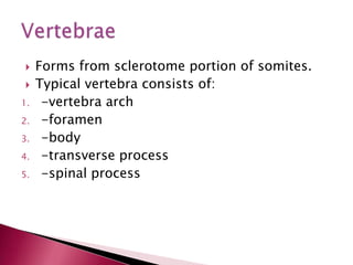
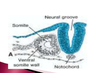
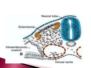
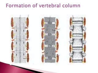
GROUP 3







INTRODUCTION
When sclerotome surrounds the neural tube,
notochord develops.
The position of sclerotomes is changed by
growth of the surrounding structures and not
by its migration.
Pax 1-gene play an important role in
development of vertebral column.


1.
2.
3.

4.
5.

Forms from sclerotome portion of somites.
Typical vertebra consists of:
-vertebra arch
-foramen
-body
-transverse process
-spinal process






During 4th week, sclerotome cells from
around the notochord and spinal cord
migrate to opposing sclerotome on the other
side of the neural tube.
As development continues, sclerotome
portion of somites undergoes
RESEGMENTATION. This forms vertebra.
Patterning and shapes of vertebrae is
regulated by Hox genes.






Mesenchymal cells between cephalic and
caudal parts of original sclerotome fill the
space between the two vertebra bodies
forming the intervertebral disc.
Notochord regresses in the vertebra body.
Notochord enlarges and persists in the
intervertebral disc region.
This contributes to the NUCLEUS PULPOSUS
which is later surrounded by the ANNULUS
FIBROSUS. This gives rise to the intervertebral
disc.








Resegmentation causes myotomes to bridge
the IV disc. This gives the capacity to move
the spine.
Intersegmented arteries pass midway over
vertebral bodies.
Spinal nerves lie IV disc and leave through IV
foramina.
As the vertebrae form, two primary curves of
the spine are established: THORACIC and
SACRAL curvatures.


Later two secondary curves are established:
CERVICAL and LUMBAR curvatures.
SCOLIOSIS
-two vertebrae fuse asymmetrically or half a
vertebra missing.

KLIPPEL-FEIL SEQUENCE
-fewer than normal cervical vertebrae
-vertebrae fuse with abnormal shape
-associated with other defects.

CLEFT VERTEBRA
- imperfect fusion of vertebral arch.
1.
May involve only vertebral arches leaving the
spinal cord intact. In this case, bony defect is
covered by skin and no neurological deficit occur.
2.
Spina Bifida Cystica
-neural tube fails to close
-vertebral arch fails to form
-neurological deficit depend on the extent of the
lesion
-occurrence is 1 in a 1000 births
-can be detected by ultrasound and prevented
administering

folic acid to mother before conception.
 If neural tissue is exposed, amniocentesis can
be used






Bony part of the rib is derived from sclerotome
cells.
COSTAL CARTILAGE: formed by sclerotome
cells that migrate across the lateral somatic
frontier in the adjacent lateral plate mesoderm.
STERNUM: development in the parietal layer of
lateral plate mesoderm in the ventral body wall.
-two sternal bands form in the parietal layer of
the lateral plate mesoderm on the other side of
the midline and these fuse to form
cartilaginous models of the MANUBRIUM,
STERNEBRAE and XIPHOID PROCESS.
RIB DEFECTS
1. Extra rib/Accessory rib
-usually in the lumbar or cervical region

Cervical ribs
-attaches to 7th cervical vertebra
-iminges on the brachial plexus and subclavian
artery
resulting in degrees of anaesthesia in the limb.
-may be unilateral or bilateral

Lumbar ribs
-most common but cause no problem
2. Fused rib
-occurs posteriorly
-two ribs arise to form a single vertebrae
-associated with hemivertebrae (results from
failure of one of the centres to appear leading to
failure of half of the vertebrae to form)
STERNUM
1. Cleft sternum: arises when sternal band fail to
grow together in the midline.
2. Hypoplastic ossification centres, premature
fusion of sternal segment- particularly in infants
with heart disease; multiple manubrial
ossification(common with down’s syndrome)


PECTUS EXCAVATUM



PECTUS CARINATUM

-depressed sternum sunken posteriorly
-most common thoracic wall defect seen by
paediatricians.
-due to overgrowth of costal cartilage that displace
the lower sternum posteriorly or abnormality of
ventral body wall closure.
-flattening of chest bilaterally with projecting
sternum.
-projection of sternum resembles the keel of a boat.
-it has the same cause as P. excavatum.
Skeletal system develops from mesenchyme
 Flat bones e.g. scapula, ribs, sternum)
undergo MEMBRANOUS OSSIFICATION.
 Most bones undergo ENDOCHONDRIAL
OSSIFICATION and form hyaline cartilage.
 Ossification centres appear and the
cartilage gradually ossifies.
NB: vertebrae are modified long bones.


Embro

  • 1.
  • 2.
        INTRODUCTION When sclerotome surroundsthe neural tube, notochord develops. The position of sclerotomes is changed by growth of the surrounding structures and not by its migration. Pax 1-gene play an important role in development of vertebral column.
  • 3.
      1. 2. 3. 4. 5. Forms from sclerotomeportion of somites. Typical vertebra consists of: -vertebra arch -foramen -body -transverse process -spinal process
  • 4.
       During 4th week,sclerotome cells from around the notochord and spinal cord migrate to opposing sclerotome on the other side of the neural tube. As development continues, sclerotome portion of somites undergoes RESEGMENTATION. This forms vertebra. Patterning and shapes of vertebrae is regulated by Hox genes.
  • 8.
       Mesenchymal cells betweencephalic and caudal parts of original sclerotome fill the space between the two vertebra bodies forming the intervertebral disc. Notochord regresses in the vertebra body. Notochord enlarges and persists in the intervertebral disc region. This contributes to the NUCLEUS PULPOSUS which is later surrounded by the ANNULUS FIBROSUS. This gives rise to the intervertebral disc.
  • 9.
        Resegmentation causes myotomesto bridge the IV disc. This gives the capacity to move the spine. Intersegmented arteries pass midway over vertebral bodies. Spinal nerves lie IV disc and leave through IV foramina. As the vertebrae form, two primary curves of the spine are established: THORACIC and SACRAL curvatures.
  • 10.
     Later two secondarycurves are established: CERVICAL and LUMBAR curvatures.
  • 11.
    SCOLIOSIS -two vertebrae fuseasymmetrically or half a vertebra missing.  KLIPPEL-FEIL SEQUENCE -fewer than normal cervical vertebrae -vertebrae fuse with abnormal shape -associated with other defects. 
  • 12.
    CLEFT VERTEBRA - imperfectfusion of vertebral arch. 1. May involve only vertebral arches leaving the spinal cord intact. In this case, bony defect is covered by skin and no neurological deficit occur. 2. Spina Bifida Cystica -neural tube fails to close -vertebral arch fails to form -neurological deficit depend on the extent of the lesion -occurrence is 1 in a 1000 births -can be detected by ultrasound and prevented administering 
  • 13.
    folic acid tomother before conception.  If neural tissue is exposed, amniocentesis can be used
  • 14.
       Bony part ofthe rib is derived from sclerotome cells. COSTAL CARTILAGE: formed by sclerotome cells that migrate across the lateral somatic frontier in the adjacent lateral plate mesoderm. STERNUM: development in the parietal layer of lateral plate mesoderm in the ventral body wall. -two sternal bands form in the parietal layer of the lateral plate mesoderm on the other side of the midline and these fuse to form cartilaginous models of the MANUBRIUM, STERNEBRAE and XIPHOID PROCESS.
  • 15.
    RIB DEFECTS 1. Extrarib/Accessory rib -usually in the lumbar or cervical region  Cervical ribs -attaches to 7th cervical vertebra -iminges on the brachial plexus and subclavian artery resulting in degrees of anaesthesia in the limb. -may be unilateral or bilateral  Lumbar ribs -most common but cause no problem
  • 16.
    2. Fused rib -occursposteriorly -two ribs arise to form a single vertebrae -associated with hemivertebrae (results from failure of one of the centres to appear leading to failure of half of the vertebrae to form) STERNUM 1. Cleft sternum: arises when sternal band fail to grow together in the midline. 2. Hypoplastic ossification centres, premature fusion of sternal segment- particularly in infants with heart disease; multiple manubrial ossification(common with down’s syndrome)
  • 17.
     PECTUS EXCAVATUM  PECTUS CARINATUM -depressedsternum sunken posteriorly -most common thoracic wall defect seen by paediatricians. -due to overgrowth of costal cartilage that displace the lower sternum posteriorly or abnormality of ventral body wall closure. -flattening of chest bilaterally with projecting sternum. -projection of sternum resembles the keel of a boat. -it has the same cause as P. excavatum.
  • 18.
    Skeletal system developsfrom mesenchyme  Flat bones e.g. scapula, ribs, sternum) undergo MEMBRANOUS OSSIFICATION.  Most bones undergo ENDOCHONDRIAL OSSIFICATION and form hyaline cartilage.  Ossification centres appear and the cartilage gradually ossifies. NB: vertebrae are modified long bones. 