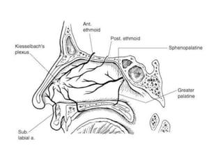
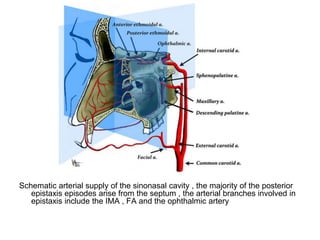
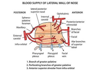
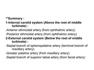
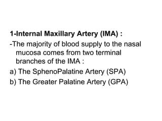
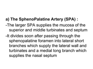
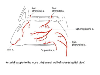
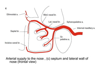
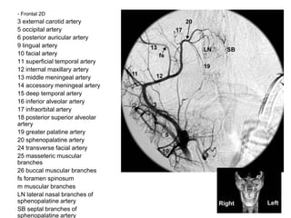
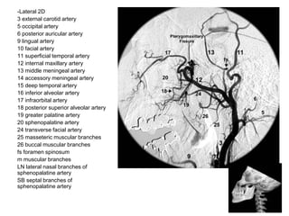
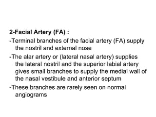
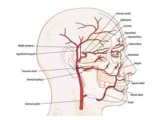
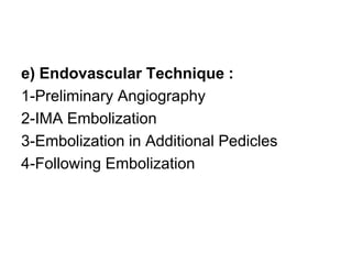
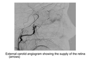
Interventional Neuroradiology
Embolization for Epistaxis
Mohamed M.A. Zaitoun, MD
Interventional Radiology Consultant, Zagazig University Hospitals, Egypt
FINR-Switzerland
zaitoun82@gmail.com
Interventional Radiology Unit,
Zagazig University, Egypt
Knowing as much as possible
about your enemy precedes
successful battle and learning
about the disease process
precedes successful
management.
Embolization for Epistaxis
a) Incidence
b) Causes
c) Vascular Anatomy
d) Indications For Embolization
e) Endovascular Technique
f) Alternative Embolization Techniques
g) Complications of Embolization
h) Outcomes
a) Incidence :
-Epistaxis is common , at least 60 % of us will
have episodes of nose bleeding at some time
-These occur with peak incidence in children (<10
years) and older adults (>50 years)
-Only 6 % of episodes require medical treatment
and these are usually in the older age group
-Bleeding can arise from veins , arteries or
arterialized veins (e.g. associated with a
vascular malforamtion or an arteriovenous
shunt)
-Most episodes are idiopathic , though
epistaxis in adults is frequently associated
with systemic hypertension and other
medical comorbidities
-Two different scenarios can be
differentiated for idiopathic epistaxis ,
these are commonly termed anterior or
posterior nasal bleeding and the latter is
more likely to arise from arteries :
Anterior
Epistaxis
Posterior
Epistaxis
1-Incidence More common
(90%)
Less common
(10%)
2-Blood flow Out from the nose Back into throat
3-Age Children or young
adults
40 years
4-Severity Less severe Severe
5-Common site Little’s area Woodruff’s plexus
6-Common cause Trauma Spontaneous ,
Hypertension or
arteriosclerosis
7-Treatment Usually controlled
by local pressure
or anterior pack
Requires
hospitalization ,
posterior nasal
Anterior Epistaxis Posterior Epistaxis
b) Causes :
1-Idiopathic
2-Trauma
3-Tumors
4-Hereditary Hemorrhagic Telangiectasia
5-Coagulopathies , Blood Dyscrasias &
Anticoagulant Medication
1-Idiopathic :
-This is the commonest form of epistaxis , affecting 70-90
% of all patients
-No underlying structural cause is found but factors
considered to trigger spontaneous epistaxis include
allergies , infections , cold weather and high atmospheric
humidity
-If affects men and women equally
-In adults , it is associated with systemic hypertension ,
atherosclerosis , hypercholesterolemia , smoking , liver
disease and excess alcohol intake
2-Trauma :
-Acute bleeding following trauma is caused by direct vessel
injury and is usually associated with facial fractures
-Delayed bleeding may occur from pseudoaneurysms and
false aneurysms in the nose or paranasal sinuses
-Hemorrhage from posttraumatic aneurysms of the
cavernous carotid artery (which may also cause a fistula)
should be considered in the differential of posttraumatic
epistaxis
-Surgical trauma e.g. biopsy of a neoplasm may precipitate
epistaxis
3-Tumors :
-Nasal and paranasal sinus tumors may present with
epistaxis
-Primary tumors include carcinomas (squamous ,
adenocarcinoma , adenoid cystic carcinoma) , olfactory
neuroblastoma , lymphoma , papillary angiomas &
angiomatous polyps
-It is a common symptom of patients with juvenile
angiofibroma
-Secondary tumors include melanoma , renal cell
carcinoma and other metastatic tumors which develop or
spread into the nose and paranasal tissues
-A small point , when dealing with aggressive
secondary tumors , is the possibility of
embolization including collateral blood supply ,
thus if the ethmoid arteries are enlarged , it has
been suggested that they should be ligated
before embolization is performed in the internal
maxillary artery (IMA) in case they become the
route of additional collateral blood supply to the
tumor from the ophthalmic artery
4-Hereditary Hemorrhagic Telangiectasia :
(Rendu-Osler-Weber Syndrome)
-Epistaxis is the most common presentation of this
autosomal dominant disease that causes
telangiectasia & AVMs in nose , skin , GIT ,
lungs , liver & brain
-Epistaxis affects 95 % of patients
-It is frequently recurrent with serial episodes
which may be severe in a minority of incidents
5-Coagulopathies , Blood Dyscrasias &
Anticoagulant Medication :
-Abnormal clotting should always be
considered as a possible cause
-It may be due to congenital conditions such
as hemophilia , systemic disease such as
cirrhosis or iatrogenic in patients on
anticoagulant or antiplatelet drugs
c) Vascular Anatomy :
-The blood supply to the nasal cavity is
derived from branches of :
1-Internal Maxillary Artery (IMA)
2-Facial Artery (FA)
3-Internal Carotid Artery (ICA)
4-Ascending Pharyngeal Artery (APA)
Schematic arterial supply of the sinonasal cavity , the majority of the posterior
epistaxis episodes arise from the septum , the arterial branches involved in
epistaxis include the IMA , FA and the ophthalmic artery
**Summary :
1-Internal carotid system (Above the root of middle
turbinate) :
-Anterior ethmoidal artery (from ophthalmic artery)
-Posterior ethmoidal artery (from ophthalmic artery)
2-External carotid system (Below the root of middle
turbinate) :
-Septal branch of sphenopalatine artery (terminal branch of
maxillary artery)
-Greater palatine artery (from maxillary artery)
-Septal branch of superior labial artery (from facial artery)
1-Internal Maxillary Artery (IMA) :
-The majority of blood supply to the nasal
mucosa comes from two terminal
branches of the IMA :
a) The SphenoPalatine Artery (SPA)
b) The Greater Palatine Artery (GPA)
a) The SphenoPalatine Artery (SPA) :
-The larger SPA supplies the mucosa of the
superior and middle turbinates and septum
-It divides soon after passing through the
sphenopalatine foramen into lateral short
branches which supply the lateral wall and
turbinates and a medial long branch which
supplies the nasal septum
b) The Greater Palatine Artery (GPA) :
-The GPA or descending palatine artery arises
from the distal IMA just before this artery enters
the sphenopalatine foramen and becomes the
SPA , it runs inferiorly to the hard palate in the
greater palatine canal
-A smaller companion branch of the IMA , the
lesser palatine artery , parallels its course in a
separate bony canal
-They give branches to supply the palate and
mucosa of the inferior lateral nasal margin
-The GPA is the more anterior branch of the
descending palatine arteries and a terminal
branch runs forwards on the hard palate and
enters the nose through the incisive foramen
where on the anterio-inferior part of the nasal
septum , it anastomoses with the septal branch
of the SPA (i.e. at Kiesselbach’s plexus)
-It also give small posterior branches which supply
the superior pharynx and anastomoses with
terminal branches of the SPA on the posterior
inferior septum and inferior turbinate
Arterial supply to the nose , (a) nasal septum
Arterial supply to the nose , (b) lateral wall of nose (sagittal view)
Arterial supply to the nose , (c) septum and lateral wall of
nose (frontal view)
- Frontal 2D
3 external carotid artery
5 occipital artery
6 posterior auricular artery
9 lingual artery
10 facial artery
11 superficial temporal artery
12 internal maxillary artery
13 middle meningeal artery
14 accessory meningeal artery
15 deep temporal artery
16 inferior alveolar artery
17 infraorbital artery
18 posterior superior alveolar
artery
19 greater palatine artery
20 sphenopalatine artery
24 transverse facial artery
25 masseteric muscular
branches
26 buccal muscular branches
fs foramen spinosum
m muscular branches
LN lateral nasal branches of
sphenopalatine artery
SB septal branches of
sphenopalatine artery
-Lateral 2D
3 external carotid artery
5 occipital artery
6 posterior auricular artery
9 lingual artery
10 facial artery
11 superficial temporal artery
12 internal maxillary artery
13 middle meningeal artery
14 accessory meningeal artery
15 deep temporal artery
16 inferior alveolar artery
17 infraorbital artery
18 posterior superior alveolar artery
19 greater palatine artery
20 sphenopalatine artery
24 transverse facial artery
25 masseteric muscular branches
26 buccal muscular branches
fs foramen spinosum
m muscular branches
LN lateral nasal branches of
sphenopalatine artery
SB septal branches of
sphenopalatine artery
-Coned down lateral 2D
3 external carotid artery
5 occipital artery
6 posterior auricular artery
9 lingual artery
10 facial artery
11 superficial temporal artery
12 internal maxillary artery
13 middle meningeal artery
14 accessory meningeal artery
15 deep temporal artery
16 inferior alveolar artery
17 infraorbital artery
18 posterior superior alveolar artery
19 greater palatine artery
20 sphenopalatine artery
24 transverse facial artery
25 masseteric muscular branches
26 buccal muscular branches
fs foramen spinosum
m muscular branches
LN lateral nasal branches of
sphenopalatine
artery
SB septal branches of sphenopalatine
artery
-Frontal 2D early arterial
3 external carotid artery
5 occipital artery
6 posterior auricular artery
9 lingual artery
10 facial artery
11 superficial temporal artery
12 internal maxillary artery
13 middle meningeal artery
14 accessory meningeal artery
15 deep temporal artery
16 inferior alveolar artery
17 infraorbital artery
18 posterior superior alveolar artery
19 greater palatine artery
20 sphenopalatine artery
24 transverse facial artery
25 masseteric muscular branches
26 buccal muscular branches
fs foramen spinosum
m muscular branches
LN lateral nasal branches of
sphenopalatine artery
SB septal branches of sphenopalatine
artery
PDB posterior directed branches of
distal
internal maxillary artery. These are 1.
artery of foramen rotundum 2. artery
of the pterygoid canal (vidian artery),
and 3.
pharyngeal artery (pterygovaginal
artery)
-later arterial phase
3 external carotid artery
5 occipital artery
6 posterior auricular artery
9 lingual artery
10 facial artery
11 superficial temporal artery
12 internal maxillary artery
13 middle meningeal artery
14 accessory meningeal artery
15 deep temporal artery
16 inferior alveolar artery
17 infraorbital artery
18 posterior superior alveolar artery
19 greater palatine artery
20 sphenopalatine artery
24 transverse facial artery
25 masseteric muscular branches
26 buccal muscular branches
fs foramen spinosum
m muscular branches
LN lateral nasal branches of
sphenopalatine artery
SB septal branches of sphenopalatine
artery
PDB posterior directed branches of
distal
internal maxillary artery. These are 1.
artery of foramen rotundum 2. artery of
the pterygoid canal (vidian artery), and
3.
pharyngeal artery (pterygovaginal
artery)
2-Facial Artery (FA) :
-Terminal branches of the facial artery (FA) supply
the nostril and external nose
-The alar artery or (lateral nasal artery) supplies
the lateral nostril and the superior labial artery
gives small branches to supply the medial wall of
the nasal vestibule and anterior septum
-These branches are rarely seen on normal
angiograms
3-Internal Carotid Artery (ICA) :
-The anterior and posterior ethmoidal arteries are branches
of the ophthalmic artery and pass through the cribriform
plate to anastomose with branches of the SPA and
supply the roof of the nasal cavity
-These vessels are rarely seen on normal angiograms
-Finding prominent ethmoidal arteries usually indicates that
embolization in the distal IMA will fail to control epistaxis
-Small branches of the ICA may also contribute to the nasal
blood supply , these are the capsular branch arteries of
McConnell) , artery of foramen rotundum (from ILT) and
the mandibular artery (from the mandibular vidian trunk)
4-Ascending Pharyngeal Artery (APA) :
-The superior and middle pharyngeal arteries
supply the medial and paramedial nasopharynx
-They therefore border the territory of SPA & GPA
branches
-The main supply to posterior nose is from the
SPA and this is the first target for endovascular
treatment , net choose the FA and rarely the
APA
-The anterior and posterior ethmoidal arteries are
best occluded by surgical ligation
d) Indications For Embolization :
1-Embolization is indicated for intractable epistaxis
2-Failure of preliminary treatment by nasal packing
or cautery , these techniques have a relatively
high failure rate in posterior epistaxis (25-50 %) ,
this isn’t surprising because it is difficult to
tamponade the posterior nasal cavity from an
anterior approach
3-Excessive blood loss with falling haemoglobin
level
4-Prior to surgical ligations , embolization is
obviously best performed before surgical ligation
of the IMA , communication and collaboration
with the surgical team should be ongoing so that
patients are referred for embolization at the
appropriate stage
e) Endovascular Technique :
1-Preliminary Angiography
2-IMA Embolization
3-Embolization in Additional Pedicles
4-Following Embolization
1-Preliminary Angiography :
-A detailed angiogram of the cranial
vasculature should be performed and
include examination of :
a) ICA :
-To exclude an underlying pathology or an
anomalous blood supply to the nose
-This can usually be done with a common
carotid injection using biplane DSA
b) ECA :
-By selective injection to assess for any contribution to the
nose from branches other than those of the distal IMA
(i.e. the transverse artery of the face , the ascending
palatine artery and other FA branches & branches of the
APA)
-Selective injection of the FA may be needed and this is
best imaged with a lateral projection
c) The IMA :
-The IMA is then examined and its distal section is best
imaged using a frontal (Water’s projection)
-The operator looks for anastomoses with the ophthalmic
artery , anterior deep temporal artery or the artery of the
foramen rotundum , bleeding points are generally difficult
to identify
2-IMA Embolization :
-Embolization is performed after selective catheterization of
the distal IMA
-A microcatheter (preferably of the largest usable size , e.g.
0.18-0.27) is advanced via a 5F or 6F guide catheter
placed in the ECA until its tip is in the pterygovaginal
portion of the IMA
-Selective angiography is performed and may or may not
show contrast extravasation
-It should show that the SPA & GPA are filling (it is
generally unnecessary to select the long SPA branch)
-If not , the tip is repositioned or steps taken to counter
vasospasm
-Catheter induced vasospasm can be a problem
and some practitioners advocate its treatment
with sublingual glycerol trinitrate rather than
intra-arterial injection of vasodilators because of
concern that the latter may open anastomotic
connections to ICA or ophthalmic artery
-Embolization particles (PVA or acrylic) are initially
selected in the 150-250 μm range and larger
particles in the 300-600 μm range injected after
an initial reduction in mucosal opacification
-A slow free flow injection of particles is made
under fluoroscopic control
External carotid angiogram of a patient with HHT and multiple episodes of severe epistaxis , the target
artery for embolization is the sphenopalatine artery (double black arrows) and the terminal portion
of the facial artery (double white arrows) , a microcatheter for embolization should be placed distal
to the middle meningeal artery (small black arrowhead) and accessory meningeal artery (small
white arrowhead) , B Selective internal maxillary angiogram showing the position of a
microcatheter (large white arrow) , there are separate mucosal hypervascular areas caused by
telangiectasias (small black arrows) , C Selective facial angiogram that shows the supply to the
nasal cavity
A Angiographic anatomy of the distal IMA , the descending palatine artery (black arrows)
outlines the posterior wall and floor of the maxillary antrum , the infraorbital artery
(double white arrows) enters the orbit through the infraorbital fissure , B Selective
distal IMA angiogram in a lateral view , C Completion angiogram after embolization
with microparticles
Internal carotid angiogram in a patient with recurrent epistaxis , there is a rich collateral
supply to the nasal cavity from the ophthalmic artery (arrows) , B The cause of such a
collateral pathway is previous proximal embolisation of the IMA using coils (arrow)
External carotid angiogram showing the supply of the retina
(arrows)
3-Embolization in Additional Pedicles :
-If there is no clear history of unilateral bleeding
then the contralateral distal IMA should be
treated in the same way
-The question of additional embolization in the FA
is difficult
-Selective ipsilateral FA injection may be helpful to
see if there is a significant blood supply to the
nasal septum but treatment probably adds to the
risk of complications and certainly to the length
of procedures
-For these reasons , many prefer to treat the FA
only if a first treatment session fails to control
bleeding
-The procedure is the same as treating the IMA
-The microcatheter tip should be navigated distally
in order to avoid spill of particles to proximal
branches supplying the submandibular gland
and to the masseter muscle branches (since this
is a cause of postoperative pain)
4-Following Embolization :
-Final control angiography is performed to confirm normal
ICA filling
-It may identify persistent nasal supply from the anterior
and posterior ethmoidal arteries which if present , makes
recurrent or persistent bleeding more likely
-It is an indication to consider surgical ligation of these
arteries
-The nasal packs can be removed immediately or after 2-6
hours
-Postprocedure analgesia is rarely required
-Haemoglobin levels should continue to be monitored until
the patient is stable
f) Alternative Embolization Techniques :
-The standard technique described will need
to be adapted in the following
circumstances :
1-Atypical Bleeding :
-When a tumor or traumatic fistula is
identified as the source of bleeding , the
endovascular treatment technique will then
be tailored appropriately
2-Using of larger Particles :
-Large particles , in the 300-800 μm range ,
are advocated as a way of reducing the
risks of the mucosal ulceration and
inadvertent spread to ICA branches
-There is little supportive evidence for this
approach and large particles are more
likely to clump and obstruct in the
microcatheter
3-Use of Gelfoam Plugs & Coils :
-These are used for occlusion of the proximal IMA and FA
-Successful treatment has been reported using large “plug”
embolization
-However , proximal occlusion which risks generating
collateral blood supply is generally considered to be less
effective than distal embolization
4-Use of Liquid Agents :
-It is unnecessary to use liquid agents for spontaneous
bleeding since they are more likely to spread to adjacent
vessels or cranial nerve blood supply
5-Embolization for Rendu-Osler-Weber Syndrome :
-In these patients , embolization is unlikely to be curative
because the disease is too extensive
-Treatment with particles is performed to induce remission
of episodes of severe epistaxis
-It can be useful on an elective basis (i.e. during intervals
between active bleeding) to reduce the frequency of
episodes
-It should be more aggressive and include the arteries
supplying the nasopharyngeal mucosa , middle
pharyngeal and accessory meningeal arteries but arterial
ligations should be avoided since recurrent bleeding in
the long term is very likely
g) Complications of Embolization :
1-Minor :
-Facial pain , trismus ,headache & muscle
ache (may make eating difficult for a few
days , occur in 80 % of procedures)
2-Major : 0.1-3 %
-Stroke , blindness , necrosis of the mucosa
or skin (most common) and VIIth cranial
nerve palsies
h) Outcomes :
-Stopping bleeding can be achieved in 90-100 %
of procedures , it may restart in a minority of
patients (ca. 10 %)
-Thus , a distinction should be made between
technical success , i.e. stopping bleeding and
achieving stable relief , i.e. long term relief
-No recurrence of bleeding occurred during follow
up in 80-90 % of patients
Embolization for Epistaxis

Embolization for Epistaxis

  • 1.
    Interventional Neuroradiology Embolization forEpistaxis Mohamed M.A. Zaitoun, MD Interventional Radiology Consultant, Zagazig University Hospitals, Egypt FINR-Switzerland zaitoun82@gmail.com Interventional Radiology Unit, Zagazig University, Egypt
  • 3.
    Knowing as muchas possible about your enemy precedes successful battle and learning about the disease process precedes successful management.
  • 4.
    Embolization for Epistaxis a)Incidence b) Causes c) Vascular Anatomy d) Indications For Embolization e) Endovascular Technique f) Alternative Embolization Techniques g) Complications of Embolization h) Outcomes
  • 5.
    a) Incidence : -Epistaxisis common , at least 60 % of us will have episodes of nose bleeding at some time -These occur with peak incidence in children (<10 years) and older adults (>50 years) -Only 6 % of episodes require medical treatment and these are usually in the older age group -Bleeding can arise from veins , arteries or arterialized veins (e.g. associated with a vascular malforamtion or an arteriovenous shunt)
  • 6.
    -Most episodes areidiopathic , though epistaxis in adults is frequently associated with systemic hypertension and other medical comorbidities -Two different scenarios can be differentiated for idiopathic epistaxis , these are commonly termed anterior or posterior nasal bleeding and the latter is more likely to arise from arteries :
  • 7.
    Anterior Epistaxis Posterior Epistaxis 1-Incidence More common (90%) Lesscommon (10%) 2-Blood flow Out from the nose Back into throat 3-Age Children or young adults 40 years 4-Severity Less severe Severe 5-Common site Little’s area Woodruff’s plexus 6-Common cause Trauma Spontaneous , Hypertension or arteriosclerosis 7-Treatment Usually controlled by local pressure or anterior pack Requires hospitalization , posterior nasal
  • 8.
  • 10.
    b) Causes : 1-Idiopathic 2-Trauma 3-Tumors 4-HereditaryHemorrhagic Telangiectasia 5-Coagulopathies , Blood Dyscrasias & Anticoagulant Medication
  • 11.
    1-Idiopathic : -This isthe commonest form of epistaxis , affecting 70-90 % of all patients -No underlying structural cause is found but factors considered to trigger spontaneous epistaxis include allergies , infections , cold weather and high atmospheric humidity -If affects men and women equally -In adults , it is associated with systemic hypertension , atherosclerosis , hypercholesterolemia , smoking , liver disease and excess alcohol intake
  • 12.
    2-Trauma : -Acute bleedingfollowing trauma is caused by direct vessel injury and is usually associated with facial fractures -Delayed bleeding may occur from pseudoaneurysms and false aneurysms in the nose or paranasal sinuses -Hemorrhage from posttraumatic aneurysms of the cavernous carotid artery (which may also cause a fistula) should be considered in the differential of posttraumatic epistaxis -Surgical trauma e.g. biopsy of a neoplasm may precipitate epistaxis
  • 13.
    3-Tumors : -Nasal andparanasal sinus tumors may present with epistaxis -Primary tumors include carcinomas (squamous , adenocarcinoma , adenoid cystic carcinoma) , olfactory neuroblastoma , lymphoma , papillary angiomas & angiomatous polyps -It is a common symptom of patients with juvenile angiofibroma -Secondary tumors include melanoma , renal cell carcinoma and other metastatic tumors which develop or spread into the nose and paranasal tissues
  • 14.
    -A small point, when dealing with aggressive secondary tumors , is the possibility of embolization including collateral blood supply , thus if the ethmoid arteries are enlarged , it has been suggested that they should be ligated before embolization is performed in the internal maxillary artery (IMA) in case they become the route of additional collateral blood supply to the tumor from the ophthalmic artery
  • 15.
    4-Hereditary Hemorrhagic Telangiectasia: (Rendu-Osler-Weber Syndrome) -Epistaxis is the most common presentation of this autosomal dominant disease that causes telangiectasia & AVMs in nose , skin , GIT , lungs , liver & brain -Epistaxis affects 95 % of patients -It is frequently recurrent with serial episodes which may be severe in a minority of incidents
  • 16.
    5-Coagulopathies , BloodDyscrasias & Anticoagulant Medication : -Abnormal clotting should always be considered as a possible cause -It may be due to congenital conditions such as hemophilia , systemic disease such as cirrhosis or iatrogenic in patients on anticoagulant or antiplatelet drugs
  • 17.
    c) Vascular Anatomy: -The blood supply to the nasal cavity is derived from branches of : 1-Internal Maxillary Artery (IMA) 2-Facial Artery (FA) 3-Internal Carotid Artery (ICA) 4-Ascending Pharyngeal Artery (APA)
  • 18.
    Schematic arterial supplyof the sinonasal cavity , the majority of the posterior epistaxis episodes arise from the septum , the arterial branches involved in epistaxis include the IMA , FA and the ophthalmic artery
  • 20.
    **Summary : 1-Internal carotidsystem (Above the root of middle turbinate) : -Anterior ethmoidal artery (from ophthalmic artery) -Posterior ethmoidal artery (from ophthalmic artery) 2-External carotid system (Below the root of middle turbinate) : -Septal branch of sphenopalatine artery (terminal branch of maxillary artery) -Greater palatine artery (from maxillary artery) -Septal branch of superior labial artery (from facial artery)
  • 21.
    1-Internal Maxillary Artery(IMA) : -The majority of blood supply to the nasal mucosa comes from two terminal branches of the IMA : a) The SphenoPalatine Artery (SPA) b) The Greater Palatine Artery (GPA)
  • 22.
    a) The SphenoPalatineArtery (SPA) : -The larger SPA supplies the mucosa of the superior and middle turbinates and septum -It divides soon after passing through the sphenopalatine foramen into lateral short branches which supply the lateral wall and turbinates and a medial long branch which supplies the nasal septum
  • 23.
    b) The GreaterPalatine Artery (GPA) : -The GPA or descending palatine artery arises from the distal IMA just before this artery enters the sphenopalatine foramen and becomes the SPA , it runs inferiorly to the hard palate in the greater palatine canal -A smaller companion branch of the IMA , the lesser palatine artery , parallels its course in a separate bony canal -They give branches to supply the palate and mucosa of the inferior lateral nasal margin
  • 24.
    -The GPA isthe more anterior branch of the descending palatine arteries and a terminal branch runs forwards on the hard palate and enters the nose through the incisive foramen where on the anterio-inferior part of the nasal septum , it anastomoses with the septal branch of the SPA (i.e. at Kiesselbach’s plexus) -It also give small posterior branches which supply the superior pharynx and anastomoses with terminal branches of the SPA on the posterior inferior septum and inferior turbinate
  • 26.
    Arterial supply tothe nose , (a) nasal septum
  • 27.
    Arterial supply tothe nose , (b) lateral wall of nose (sagittal view)
  • 28.
    Arterial supply tothe nose , (c) septum and lateral wall of nose (frontal view)
  • 29.
    - Frontal 2D 3external carotid artery 5 occipital artery 6 posterior auricular artery 9 lingual artery 10 facial artery 11 superficial temporal artery 12 internal maxillary artery 13 middle meningeal artery 14 accessory meningeal artery 15 deep temporal artery 16 inferior alveolar artery 17 infraorbital artery 18 posterior superior alveolar artery 19 greater palatine artery 20 sphenopalatine artery 24 transverse facial artery 25 masseteric muscular branches 26 buccal muscular branches fs foramen spinosum m muscular branches LN lateral nasal branches of sphenopalatine artery SB septal branches of sphenopalatine artery
  • 30.
    -Lateral 2D 3 externalcarotid artery 5 occipital artery 6 posterior auricular artery 9 lingual artery 10 facial artery 11 superficial temporal artery 12 internal maxillary artery 13 middle meningeal artery 14 accessory meningeal artery 15 deep temporal artery 16 inferior alveolar artery 17 infraorbital artery 18 posterior superior alveolar artery 19 greater palatine artery 20 sphenopalatine artery 24 transverse facial artery 25 masseteric muscular branches 26 buccal muscular branches fs foramen spinosum m muscular branches LN lateral nasal branches of sphenopalatine artery SB septal branches of sphenopalatine artery
  • 31.
    -Coned down lateral2D 3 external carotid artery 5 occipital artery 6 posterior auricular artery 9 lingual artery 10 facial artery 11 superficial temporal artery 12 internal maxillary artery 13 middle meningeal artery 14 accessory meningeal artery 15 deep temporal artery 16 inferior alveolar artery 17 infraorbital artery 18 posterior superior alveolar artery 19 greater palatine artery 20 sphenopalatine artery 24 transverse facial artery 25 masseteric muscular branches 26 buccal muscular branches fs foramen spinosum m muscular branches LN lateral nasal branches of sphenopalatine artery SB septal branches of sphenopalatine artery
  • 32.
    -Frontal 2D earlyarterial 3 external carotid artery 5 occipital artery 6 posterior auricular artery 9 lingual artery 10 facial artery 11 superficial temporal artery 12 internal maxillary artery 13 middle meningeal artery 14 accessory meningeal artery 15 deep temporal artery 16 inferior alveolar artery 17 infraorbital artery 18 posterior superior alveolar artery 19 greater palatine artery 20 sphenopalatine artery 24 transverse facial artery 25 masseteric muscular branches 26 buccal muscular branches fs foramen spinosum m muscular branches LN lateral nasal branches of sphenopalatine artery SB septal branches of sphenopalatine artery PDB posterior directed branches of distal internal maxillary artery. These are 1. artery of foramen rotundum 2. artery of the pterygoid canal (vidian artery), and 3. pharyngeal artery (pterygovaginal artery)
  • 33.
    -later arterial phase 3external carotid artery 5 occipital artery 6 posterior auricular artery 9 lingual artery 10 facial artery 11 superficial temporal artery 12 internal maxillary artery 13 middle meningeal artery 14 accessory meningeal artery 15 deep temporal artery 16 inferior alveolar artery 17 infraorbital artery 18 posterior superior alveolar artery 19 greater palatine artery 20 sphenopalatine artery 24 transverse facial artery 25 masseteric muscular branches 26 buccal muscular branches fs foramen spinosum m muscular branches LN lateral nasal branches of sphenopalatine artery SB septal branches of sphenopalatine artery PDB posterior directed branches of distal internal maxillary artery. These are 1. artery of foramen rotundum 2. artery of the pterygoid canal (vidian artery), and 3. pharyngeal artery (pterygovaginal artery)
  • 34.
    2-Facial Artery (FA): -Terminal branches of the facial artery (FA) supply the nostril and external nose -The alar artery or (lateral nasal artery) supplies the lateral nostril and the superior labial artery gives small branches to supply the medial wall of the nasal vestibule and anterior septum -These branches are rarely seen on normal angiograms
  • 36.
    3-Internal Carotid Artery(ICA) : -The anterior and posterior ethmoidal arteries are branches of the ophthalmic artery and pass through the cribriform plate to anastomose with branches of the SPA and supply the roof of the nasal cavity -These vessels are rarely seen on normal angiograms -Finding prominent ethmoidal arteries usually indicates that embolization in the distal IMA will fail to control epistaxis -Small branches of the ICA may also contribute to the nasal blood supply , these are the capsular branch arteries of McConnell) , artery of foramen rotundum (from ILT) and the mandibular artery (from the mandibular vidian trunk)
  • 38.
    4-Ascending Pharyngeal Artery(APA) : -The superior and middle pharyngeal arteries supply the medial and paramedial nasopharynx -They therefore border the territory of SPA & GPA branches -The main supply to posterior nose is from the SPA and this is the first target for endovascular treatment , net choose the FA and rarely the APA -The anterior and posterior ethmoidal arteries are best occluded by surgical ligation
  • 39.
    d) Indications ForEmbolization : 1-Embolization is indicated for intractable epistaxis 2-Failure of preliminary treatment by nasal packing or cautery , these techniques have a relatively high failure rate in posterior epistaxis (25-50 %) , this isn’t surprising because it is difficult to tamponade the posterior nasal cavity from an anterior approach 3-Excessive blood loss with falling haemoglobin level 4-Prior to surgical ligations , embolization is obviously best performed before surgical ligation of the IMA , communication and collaboration with the surgical team should be ongoing so that patients are referred for embolization at the appropriate stage
  • 40.
    e) Endovascular Technique: 1-Preliminary Angiography 2-IMA Embolization 3-Embolization in Additional Pedicles 4-Following Embolization
  • 41.
    1-Preliminary Angiography : -Adetailed angiogram of the cranial vasculature should be performed and include examination of : a) ICA : -To exclude an underlying pathology or an anomalous blood supply to the nose -This can usually be done with a common carotid injection using biplane DSA
  • 42.
    b) ECA : -Byselective injection to assess for any contribution to the nose from branches other than those of the distal IMA (i.e. the transverse artery of the face , the ascending palatine artery and other FA branches & branches of the APA) -Selective injection of the FA may be needed and this is best imaged with a lateral projection c) The IMA : -The IMA is then examined and its distal section is best imaged using a frontal (Water’s projection) -The operator looks for anastomoses with the ophthalmic artery , anterior deep temporal artery or the artery of the foramen rotundum , bleeding points are generally difficult to identify
  • 43.
    2-IMA Embolization : -Embolizationis performed after selective catheterization of the distal IMA -A microcatheter (preferably of the largest usable size , e.g. 0.18-0.27) is advanced via a 5F or 6F guide catheter placed in the ECA until its tip is in the pterygovaginal portion of the IMA -Selective angiography is performed and may or may not show contrast extravasation -It should show that the SPA & GPA are filling (it is generally unnecessary to select the long SPA branch) -If not , the tip is repositioned or steps taken to counter vasospasm
  • 44.
    -Catheter induced vasospasmcan be a problem and some practitioners advocate its treatment with sublingual glycerol trinitrate rather than intra-arterial injection of vasodilators because of concern that the latter may open anastomotic connections to ICA or ophthalmic artery -Embolization particles (PVA or acrylic) are initially selected in the 150-250 μm range and larger particles in the 300-600 μm range injected after an initial reduction in mucosal opacification -A slow free flow injection of particles is made under fluoroscopic control
  • 45.
    External carotid angiogramof a patient with HHT and multiple episodes of severe epistaxis , the target artery for embolization is the sphenopalatine artery (double black arrows) and the terminal portion of the facial artery (double white arrows) , a microcatheter for embolization should be placed distal to the middle meningeal artery (small black arrowhead) and accessory meningeal artery (small white arrowhead) , B Selective internal maxillary angiogram showing the position of a microcatheter (large white arrow) , there are separate mucosal hypervascular areas caused by telangiectasias (small black arrows) , C Selective facial angiogram that shows the supply to the nasal cavity
  • 46.
    A Angiographic anatomyof the distal IMA , the descending palatine artery (black arrows) outlines the posterior wall and floor of the maxillary antrum , the infraorbital artery (double white arrows) enters the orbit through the infraorbital fissure , B Selective distal IMA angiogram in a lateral view , C Completion angiogram after embolization with microparticles
  • 47.
    Internal carotid angiogramin a patient with recurrent epistaxis , there is a rich collateral supply to the nasal cavity from the ophthalmic artery (arrows) , B The cause of such a collateral pathway is previous proximal embolisation of the IMA using coils (arrow)
  • 48.
    External carotid angiogramshowing the supply of the retina (arrows)
  • 49.
    3-Embolization in AdditionalPedicles : -If there is no clear history of unilateral bleeding then the contralateral distal IMA should be treated in the same way -The question of additional embolization in the FA is difficult -Selective ipsilateral FA injection may be helpful to see if there is a significant blood supply to the nasal septum but treatment probably adds to the risk of complications and certainly to the length of procedures
  • 50.
    -For these reasons, many prefer to treat the FA only if a first treatment session fails to control bleeding -The procedure is the same as treating the IMA -The microcatheter tip should be navigated distally in order to avoid spill of particles to proximal branches supplying the submandibular gland and to the masseter muscle branches (since this is a cause of postoperative pain)
  • 51.
    4-Following Embolization : -Finalcontrol angiography is performed to confirm normal ICA filling -It may identify persistent nasal supply from the anterior and posterior ethmoidal arteries which if present , makes recurrent or persistent bleeding more likely -It is an indication to consider surgical ligation of these arteries -The nasal packs can be removed immediately or after 2-6 hours -Postprocedure analgesia is rarely required -Haemoglobin levels should continue to be monitored until the patient is stable
  • 52.
    f) Alternative EmbolizationTechniques : -The standard technique described will need to be adapted in the following circumstances : 1-Atypical Bleeding : -When a tumor or traumatic fistula is identified as the source of bleeding , the endovascular treatment technique will then be tailored appropriately
  • 53.
    2-Using of largerParticles : -Large particles , in the 300-800 μm range , are advocated as a way of reducing the risks of the mucosal ulceration and inadvertent spread to ICA branches -There is little supportive evidence for this approach and large particles are more likely to clump and obstruct in the microcatheter
  • 54.
    3-Use of GelfoamPlugs & Coils : -These are used for occlusion of the proximal IMA and FA -Successful treatment has been reported using large “plug” embolization -However , proximal occlusion which risks generating collateral blood supply is generally considered to be less effective than distal embolization 4-Use of Liquid Agents : -It is unnecessary to use liquid agents for spontaneous bleeding since they are more likely to spread to adjacent vessels or cranial nerve blood supply
  • 55.
    5-Embolization for Rendu-Osler-WeberSyndrome : -In these patients , embolization is unlikely to be curative because the disease is too extensive -Treatment with particles is performed to induce remission of episodes of severe epistaxis -It can be useful on an elective basis (i.e. during intervals between active bleeding) to reduce the frequency of episodes -It should be more aggressive and include the arteries supplying the nasopharyngeal mucosa , middle pharyngeal and accessory meningeal arteries but arterial ligations should be avoided since recurrent bleeding in the long term is very likely
  • 56.
    g) Complications ofEmbolization : 1-Minor : -Facial pain , trismus ,headache & muscle ache (may make eating difficult for a few days , occur in 80 % of procedures) 2-Major : 0.1-3 % -Stroke , blindness , necrosis of the mucosa or skin (most common) and VIIth cranial nerve palsies
  • 57.
    h) Outcomes : -Stoppingbleeding can be achieved in 90-100 % of procedures , it may restart in a minority of patients (ca. 10 %) -Thus , a distinction should be made between technical success , i.e. stopping bleeding and achieving stable relief , i.e. long term relief -No recurrence of bleeding occurred during follow up in 80-90 % of patients