SlideShare a Scribd company logo
Epistaxis 
Epistaxis is definied as bleeding from nose. The prosaic definition belies the difficulties associated 
with one of the otolaryngological’s most common& most difficult to treat emergencies. 
Vascular anatomy 
The area most frequently implicated in epistaxis is known as Little’s area or kiesselbach plexus.in the 
posterior nasal cavity, the vessels are larger than those in little area & can more easily be traced to 
their external or internal carotid origin. 
External carotid artery: external carotid artery supplies the nasal cavity via facial & maxillary artery. 
Maxillary artery supply is via sphenopalatine & greater palatine artery. 
Internal carotid artery: internal carotid artery contributes the anterior & posterior ethmoidal 
branches of the ophthalmic artery. Middle turbinate marks the watershed between internal & 
external carotid arteries. 
Woodruff’s plexus : a plexus of prominent blood vessels lying just inferior to the posterior end of the 
inferior turbinate. 
Anteior ethmoidal artery: the artery can be ligated as a treatment for epistaxis via an external 
approach(medial canthal) or endoscopical (transethmoidal). 
Sphenopalatine foramen: sphenopalatine foramen is the portal for major arterial supply of the nasal 
cavity. Lateral to the foramen lies the pterygopalatine space. The sphenopalatine foramen transmits 
the sphenopalatine artery & vein, &maxillary division of trigeminal nerve. A small bony projection 
which lies anterior to the foramen, this landmark is called crista ethmoidalis. 
Blood supply of inferior turbinate: artery reaching the inferior turbinate, artery divided into three 
parallel branches which runs in bony tunnels within the substance of the turbinate. These tunnels 
with their periarterial cuffing of fibrous tissue prevent the arterial constriction & may predispose to 
post-operative haemorrhage. 
Attempts to control haemorrhage following turbinectomy should be directed towards the postero-superior 
aspect of the inferior turbinate where the pressure/bipolar to the submucosal segment of 
the artery should prove effective. 
Classification of epistaxis 
Structured clinical classification 
Classification of epistaxis 
Primary No proven causal factor 
Secondary Proven causal factor 
Childhood <16 years
Adult > 16 years 
Anterior epistaxis Bleeding from anterior to pyriform aperture 
Posterior epistaxis Bleeding from posterior to pyriform aperture. 
Adult primary epistaxis 
The condition can occur at any age. Between 7 to 14% of the adults have epistaxis at some time but 
only 6% are seen by otolaryngologist. Most cases are minor, selflimiting but nevertheless a 
significant number require admission to the hospital. Fewer than 10% of the hospitalized patients 
require a general anaesthetic procedure to secure haemostasis. After head & neck cancer, epistaxis 
stands out as a prominent cause of mortality in ENT patients. 
Aetiology 
By definition, the aetiology of primary epistaxis is unknown, but there are clear suggestions may be 
important. 
Chronobiology: the frequency of admission is greatest in the autumn& winter due to fluctuation of 
temperature & humidity. Circadian level shows a biphasic pattern with peaks in the morning& 
evening.(subarachnoid haemorrhage) 
NSAID: specially aspirin via antiplatelet aggregations effect due to altered platelet membrane 
physiology. 
Alcohol : alcohol causes prolongation of the bleeding time despite normal platelet counts & 
coagulation factors acitivity. 
Hypertension: this has long been considered a cause of epitaxis.recently have failed to show a causal 
relationship between hypertension & epistaxis. 
Septal abnormalities: epistaxis & septal abnormalities could be coincidental. There is no direct 
relationship. 
Factors of adult primary epistaxis (summary 
of 
Aetiology) 
Weather Proven association 
NSAID Proven association 
Alcohol Proven association 
Hypertension No association 
Septal deviation No association
Management 
First , the patient must be resuscitated, bleeding slowed, the nasal cavity examined& a treatment 
plan established. 
Resuscitation: 
First aid by pinching the ala nasi (Trotter’s method). 
History & general examination will help in assessing the amount of blood loss. 
In all but the most minor of bleeds, intravenous access is established. 
Baseline blood estimations should be done. 
A detailed history should be taken,looking for predisposing factors. 
Routine coagulation studies in the absence of a positive history are not indicated. 
Assessment : 
The patient should be assessed in a semi-recumbent position & nersing assistance is 
mandatory. 
Everyone should wear protective visors & clothing as blood aerosol contamination is 
common. 
Basic instrument includes; couch or reclining chair, headlight,suction, vasoconstrictor 
solutions, (now widely used cocaine solution) , a packs, tempons & cautery apparatus. 
More specialized centres should have access to rod lens endoscopy instrument& bipolar 
electrodiathermy. 
Direct therapy(bleeding point specific therapies): 
A committed search for the bleeding vessels should be undertaken. Anterior epistaxis is usually very 
straightforward to identify & treat. At present over 90% of cases are controlled with silver nitrate 
cautery. The use of packing for anterior epistaxis is unwarranted & should be discouraged. 
Posterior epistaxis; systematic examination with a headlight will identify most bleeding points. Once 
identified, bleeding points can be directly controlled with bipolar diathermy, chemical 
cautery(difficult in posterior bleeds) or direct pressure from miniature targeted packs. 
Endoscopic control: failure to locate the bleeding point on initial examination is an indication for 
with a rod lens endoscope. Endoscopy identifies the source of the posterior epistaxis in over 90% of 
cases. Monopolar diathermy should not be used in the nasal cavity as blindness due to propagation 
of current. 
Indirect therapies : (failure to find the bleeding point is an indication for 
use of indirect strategies). 
1.Nasal packing: nasal packing can be anteriorly or posteriorly placed.
Anterior nasal pack : 
Ribbon gauze impregnated with petroleum jelly or BIPP(bismuth iodoform paraffin 
paste) is inserted the entire length of the nasal cavity. 
Once inserted , the pack are left in situ for between 24 to 72 hours. 
Rebleeding or continued bleeding is observed upto 40% of cases. 
Complications of packing include sinusitis, septal perforation alar necrosis, hypoxia, 
myocardial infarction. 
Antibiotic cover should be given. 
Modern alternatives on anterior packing include special tampons(merocel& 
kaltostat). 
Persistent or rebleeding is an indication for further examination of the nasal cavity. 
There is on clear, universally agreed definition of failed packing but who continue to bleed should 
proceed to surgical management sooner than later. 
2.Hot water irrigation:irrigation of the nasal cavity with at 500 has been proposed as an alternative 
to packing( success rate similar to anterior packing Or balloon temponade.) mechanism of action; 
reflex vasodilatation & reduction in nasal lumen dimension. 
3.Systemic medical therapy: Tranexamic acid & epsilon aminocaproic acid are systemic inhibitors of 
fibrinolysis. In epistaxis dose of 1.5gm thrice daily. At present these drugs are best reserved as 
adjuvant therapy in recurrent or refractory cases. 
Surgical management 
Surgical management for continued epistaxis consists of : 
Posterior packing 
Ligation techniques 
Septal surgery techniques 
Embolization techniques. 
1.Posterior nasal packs 
Posterior nasal packing can be carried out under local or general anaesthesia. 
Nasophaygeal tamponade is achieved using special gauze packs inserted transorally& positioned by 
means of tapes passed from posterior choana to anterior nares bilaterally. These posterior Bellocq 
pack are secure against anterior gauze packing. 
The securing tapes are tied over gauze piece(padding positioned to protect the columella from 
pressure ncerosis.
An easier alternative is to insert a foley’s catheter(size 12 or 14). The catheter inflated with 15ml of 
water. 
Ligation techniques 
The ligation should be performed as close as possible to likely bleeding point. 
1.Shenopalatine artery 
2.Internal maxillary artery 
3.External carotid artery 
4.Anterior/posterior ethmoidal artery 
1.Endonasal shenopalatine artery ligation(ESPAL) 
Under G/A or L/A,an incision is made approximately 8mm in length, anterior to & under cover of 
the posterior end of the middle turbinate. The incision is carried down upto bone & a mucosal flap is 
elevated posteriorly until neurovascular sleeve arising from the sphenopalatine foramen is 
identified. Its location is signalled by the crista ethmoidalis. Once major vessel identified, it can be 
ligated using haemostatic clips & divided or coagulated using bipolar diathermy. Success rate almost 
100% 
2. Internal maxillary artery ligation 
Internal maxillary ligation was more frequently prior to the development of ESPAL. In sublabial 
approach, an antrostomy is formed taking care to preserve the infrorbital nerve. The mucosa of the 
posterior wall is elevated & a window is made through into pterygopalatine fossa. The branches of 
internal maxillary artery are identified pulsating within the fat of the fossa & are carefully dissected 
out prior to clipping with haemostatic clips. The proximal internal maxillary artery, descending 
palatine, & sphenopalatine branches are clipped & ideally divided. 
An endoscopic variation of this techniques1)uses a middle meatal antrostomy as an instrument port 
2)with 4mm endoscope is inserted through a canine fossa antrostomy. 
Transantral ligation control haemorrhage in 89% of cases. 
3.External carotid artery ligation 
Uder G/A or L/A a skin crease incision or longitudinal incision along parallel with anterior border of 
sternomastoid. Carotid bifurcation is identified, double check for arterial branches , then ligation in 
continuity. Success rate 14-15%. 
4.Anterior or posterior ethmoidal artery ligation 
Ligation of anterior /posterior ethmoidal artery is adjuvant to above procedures or ethmoidal 
fracture. 
Medial canthal incision which is carried down to the bone of the anterior lacrimal crest. Periosteal 
elevator are used to elevate & lateral retract of bulbur fascia into anterior ethmoidal foramen. The
vassel are identified & clipped & divided. The dissection is continued to identify the posterior artery 
which is located approximately 12mm behind. 
Septal surgery 
When epistaxis originates behind a prominent septal deviation or vomeropalatine spur, septoplasty 
or SMR may be required. Some authors have advocated a septal surgery as a primary treatment for 
failed packing. 
The rationale is that by elvationing the mucoperichondrial flap for septoplasty or SMR, the blood 
supply to the septum is interrupted & haemostasis is secured. 
Embolization 
Under L/A, transfemoral seldinger angiography is used to identify the bleeding point & display nasal 
circulation. It is essential to identify the arteriovenous malformation, aneursyms, fistula prior to 
embolization. Once the bleeding vessel identified, a fine catheter is passed into the internal maxillary 
circulation & particles (polyvinyl alcohol,,tungsten, or steel microcoils) are used to embolize the 
vessels. The ipsilateral facial artery is also embolize in order to prevent recirculation 
Complication are skin necrosis, paresthesia, CVA, groin haematoma. Similar efficacy to ligation 
techniques. 
Secondary epistaxis 
Commomly observed in patients with coagulopathy secondary to liver disease, leukemia, 
myelosuppression. In addition, trauma, post-surgery, warfain therapy deserve special care. 
Best clinical practice 
First line: direct therapy (bipolar/cautery,endoscopic if required) 
Second line: indirect therapy (anterior packing) 
Third line: surgical therapy (ESPAL) 
Fourth line: angiography &embolization.

More Related Content

What's hot

Anesthesia types and complications
Anesthesia types and complicationsAnesthesia types and complications
Anesthesia types and complications
kalyan kumar
 
Endoscopic csf leak repaire
Endoscopic csf leak repaireEndoscopic csf leak repaire
Endoscopic csf leak repaire
Dr.Kumaran Bagavathi Ragavan
 
Epidural anesthesia
Epidural anesthesiaEpidural anesthesia
Epidural anesthesia
robert robert
 
BLS AND ALS
BLS AND ALSBLS AND ALS
BLS AND ALS
tariq620
 
Tracheostomy and its post op care last
Tracheostomy and its post op care lastTracheostomy and its post op care last
Tracheostomy and its post op care last
Jamil Kifayatullah
 
Suction
SuctionSuction
Rigid bronchoscopy
Rigid bronchoscopy Rigid bronchoscopy
Rigid bronchoscopy
Santosh Jha
 
Anesthesia machine
Anesthesia machineAnesthesia machine
Anesthesia machine
JudithJustin3
 
Lasers in otolaryngology
Lasers in otolaryngology Lasers in otolaryngology
Lasers in otolaryngology
Dr Krishna Koirala
 
Anaesthetic techniques
Anaesthetic techniquesAnaesthetic techniques
Anaesthetic techniques
Hitendra Prajapati
 
Tracheostomy
TracheostomyTracheostomy
Tracheostomy
Dr Krishna Koirala
 
Tracheostomy
TracheostomyTracheostomy
Tracheostomy
Rachel Jeevakirubai
 
Rehabilitation after laryngectomy
Rehabilitation after laryngectomyRehabilitation after laryngectomy
Rehabilitation after laryngectomy
Md Roohia
 
Laryngoscope
LaryngoscopeLaryngoscope
Laryngoscope
Pratik Kumar
 
Airway management in ED - Basics and advanced
Airway management in ED - Basics and advancedAirway management in ED - Basics and advanced
Airway management in ED - Basics and advanced
Dr.Venugopalan Poovathum Parambil
 
Tracheostomy tubes by Dr.Ashwin Menon
Tracheostomy tubes by Dr.Ashwin MenonTracheostomy tubes by Dr.Ashwin Menon
Tracheostomy tubes by Dr.Ashwin Menon
Dr.Ashwin Menon
 
Maringoplasty
Maringoplasty Maringoplasty
Maringoplasty
Dr Asmatullah Achakzai
 
Epidural anesthesia & analgesia
Epidural anesthesia & analgesiaEpidural anesthesia & analgesia
Epidural anesthesia & analgesia
SRINIVAS UNDURTY
 
TRACHEOSTOMY CARE
TRACHEOSTOMY CARETRACHEOSTOMY CARE
TRACHEOSTOMY CARE
Rommel Luis III Israel
 
Adenoidectomy
AdenoidectomyAdenoidectomy
Adenoidectomy
Samuel Paul Eda
 

What's hot (20)

Anesthesia types and complications
Anesthesia types and complicationsAnesthesia types and complications
Anesthesia types and complications
 
Endoscopic csf leak repaire
Endoscopic csf leak repaireEndoscopic csf leak repaire
Endoscopic csf leak repaire
 
Epidural anesthesia
Epidural anesthesiaEpidural anesthesia
Epidural anesthesia
 
BLS AND ALS
BLS AND ALSBLS AND ALS
BLS AND ALS
 
Tracheostomy and its post op care last
Tracheostomy and its post op care lastTracheostomy and its post op care last
Tracheostomy and its post op care last
 
Suction
SuctionSuction
Suction
 
Rigid bronchoscopy
Rigid bronchoscopy Rigid bronchoscopy
Rigid bronchoscopy
 
Anesthesia machine
Anesthesia machineAnesthesia machine
Anesthesia machine
 
Lasers in otolaryngology
Lasers in otolaryngology Lasers in otolaryngology
Lasers in otolaryngology
 
Anaesthetic techniques
Anaesthetic techniquesAnaesthetic techniques
Anaesthetic techniques
 
Tracheostomy
TracheostomyTracheostomy
Tracheostomy
 
Tracheostomy
TracheostomyTracheostomy
Tracheostomy
 
Rehabilitation after laryngectomy
Rehabilitation after laryngectomyRehabilitation after laryngectomy
Rehabilitation after laryngectomy
 
Laryngoscope
LaryngoscopeLaryngoscope
Laryngoscope
 
Airway management in ED - Basics and advanced
Airway management in ED - Basics and advancedAirway management in ED - Basics and advanced
Airway management in ED - Basics and advanced
 
Tracheostomy tubes by Dr.Ashwin Menon
Tracheostomy tubes by Dr.Ashwin MenonTracheostomy tubes by Dr.Ashwin Menon
Tracheostomy tubes by Dr.Ashwin Menon
 
Maringoplasty
Maringoplasty Maringoplasty
Maringoplasty
 
Epidural anesthesia & analgesia
Epidural anesthesia & analgesiaEpidural anesthesia & analgesia
Epidural anesthesia & analgesia
 
TRACHEOSTOMY CARE
TRACHEOSTOMY CARETRACHEOSTOMY CARE
TRACHEOSTOMY CARE
 
Adenoidectomy
AdenoidectomyAdenoidectomy
Adenoidectomy
 

Similar to Epistaxis

Surgical management of epistaxix
Surgical management of epistaxixSurgical management of epistaxix
Surgical management of epistaxix
Mohammed Raad
 
Epistaxis.pptx
Epistaxis.pptxEpistaxis.pptx
Epistaxis.pptx
Ahlam Alzuway
 
CAP 47. Epistaxis.pdf
CAP 47. Epistaxis.pdfCAP 47. Epistaxis.pdf
CAP 47. Epistaxis.pdf
eduardocueva13
 
EPISTAXIS.pptx
EPISTAXIS.pptxEPISTAXIS.pptx
EPISTAXIS.pptx
ArpitaHalder8
 
Epistaxis.ppt
Epistaxis.pptEpistaxis.ppt
Epistaxis.ppt
JAYABASKAR J
 
Epistaxis
EpistaxisEpistaxis
Epistaxis
sudha shahi
 
Epistaxis
EpistaxisEpistaxis
Epistaxis
Vinay Bhat
 
EPISTAXIS
EPISTAXISEPISTAXIS
EPISTAXIS
Suraj Dhara
 
Management of epistaxis
Management of epistaxisManagement of epistaxis
Management of epistaxis
susritha17
 
Epistaxis
EpistaxisEpistaxis
Epistaxis
Renold S Xavier
 
Epistasis
EpistasisEpistasis
Epistasis
social service
 
EPISTAXIS final.pdf
EPISTAXIS final.pdfEPISTAXIS final.pdf
EPISTAXIS final.pdf
drravdeepsinghchmans
 
Ppt of epistaxis
Ppt of epistaxisPpt of epistaxis
Ppt of epistaxis
ParagPatil79
 
EPISTAXIS ppt
EPISTAXIS ppt EPISTAXIS ppt
EPISTAXIS ppt
Nethaji Perumal
 
EPITAXSIS.pptx
EPITAXSIS.pptxEPITAXSIS.pptx
EPITAXSIS.pptx
SachinDwivedi57
 
Cerebrospinal fluid rhinorrhoea
Cerebrospinal fluid rhinorrhoeaCerebrospinal fluid rhinorrhoea
Cerebrospinal fluid rhinorrhoea
Shekhar Krishna Debnath
 
Cerebrospinal fluid rhinorrhoea
Cerebrospinal fluid rhinorrhoeaCerebrospinal fluid rhinorrhoea
Cerebrospinal fluid rhinorrhoea
Shekhar Krishna Debnath
 
epistaxis.pptxslidesharethankyousomuchii
epistaxis.pptxslidesharethankyousomuchiiepistaxis.pptxslidesharethankyousomuchii
epistaxis.pptxslidesharethankyousomuchii
1901600146
 
Epistaxis
EpistaxisEpistaxis
Epistaxis
LeslinRaj1
 
Epistaxis- Nose Bleed Overview and Managment
Epistaxis- Nose Bleed Overview and ManagmentEpistaxis- Nose Bleed Overview and Managment
Epistaxis- Nose Bleed Overview and Managment
Bernard Racey
 

Similar to Epistaxis (20)

Surgical management of epistaxix
Surgical management of epistaxixSurgical management of epistaxix
Surgical management of epistaxix
 
Epistaxis.pptx
Epistaxis.pptxEpistaxis.pptx
Epistaxis.pptx
 
CAP 47. Epistaxis.pdf
CAP 47. Epistaxis.pdfCAP 47. Epistaxis.pdf
CAP 47. Epistaxis.pdf
 
EPISTAXIS.pptx
EPISTAXIS.pptxEPISTAXIS.pptx
EPISTAXIS.pptx
 
Epistaxis.ppt
Epistaxis.pptEpistaxis.ppt
Epistaxis.ppt
 
Epistaxis
EpistaxisEpistaxis
Epistaxis
 
Epistaxis
EpistaxisEpistaxis
Epistaxis
 
EPISTAXIS
EPISTAXISEPISTAXIS
EPISTAXIS
 
Management of epistaxis
Management of epistaxisManagement of epistaxis
Management of epistaxis
 
Epistaxis
EpistaxisEpistaxis
Epistaxis
 
Epistasis
EpistasisEpistasis
Epistasis
 
EPISTAXIS final.pdf
EPISTAXIS final.pdfEPISTAXIS final.pdf
EPISTAXIS final.pdf
 
Ppt of epistaxis
Ppt of epistaxisPpt of epistaxis
Ppt of epistaxis
 
EPISTAXIS ppt
EPISTAXIS ppt EPISTAXIS ppt
EPISTAXIS ppt
 
EPITAXSIS.pptx
EPITAXSIS.pptxEPITAXSIS.pptx
EPITAXSIS.pptx
 
Cerebrospinal fluid rhinorrhoea
Cerebrospinal fluid rhinorrhoeaCerebrospinal fluid rhinorrhoea
Cerebrospinal fluid rhinorrhoea
 
Cerebrospinal fluid rhinorrhoea
Cerebrospinal fluid rhinorrhoeaCerebrospinal fluid rhinorrhoea
Cerebrospinal fluid rhinorrhoea
 
epistaxis.pptxslidesharethankyousomuchii
epistaxis.pptxslidesharethankyousomuchiiepistaxis.pptxslidesharethankyousomuchii
epistaxis.pptxslidesharethankyousomuchii
 
Epistaxis
EpistaxisEpistaxis
Epistaxis
 
Epistaxis- Nose Bleed Overview and Managment
Epistaxis- Nose Bleed Overview and ManagmentEpistaxis- Nose Bleed Overview and Managment
Epistaxis- Nose Bleed Overview and Managment
 

More from Shekhar Krishna Debnath

Pta(sbo 3)
Pta(sbo 3)Pta(sbo 3)
Vertigo
VertigoVertigo
Stridor vol 1
Stridor vol  1Stridor vol  1
Obstuctive sleep apnoea in children
Obstuctive sleep apnoea in childrenObstuctive sleep apnoea in children
Obstuctive sleep apnoea in children
Shekhar Krishna Debnath
 
Nose
NoseNose
Diseases of the tonsils 2
Diseases of  the tonsils 2Diseases of  the tonsils 2
Diseases of the tonsils 2
Shekhar Krishna Debnath
 
Disease of tonsils
Disease of tonsilsDisease of tonsils
Disease of tonsils
Shekhar Krishna Debnath
 
Corticosteroid in otolaryngology
Corticosteroid in otolaryngologyCorticosteroid in otolaryngology
Corticosteroid in otolaryngology
Shekhar Krishna Debnath
 
Viruses & antiviral agents
Viruses & antiviral agentsViruses & antiviral agents
Viruses & antiviral agents
Shekhar Krishna Debnath
 
Tumours of the head & neck in the childhood
Tumours of the head & neck in the childhoodTumours of the head & neck in the childhood
Tumours of the head & neck in the childhood
Shekhar Krishna Debnath
 
The adenoid & adenoidectomy
The adenoid & adenoidectomyThe adenoid & adenoidectomy
The adenoid & adenoidectomy
Shekhar Krishna Debnath
 
Otitis media with effusion
Otitis media with effusionOtitis media with effusion
Otitis media with effusion
Shekhar Krishna Debnath
 
Gastro oesophageal reflux & aspiration
Gastro oesophageal reflux & aspirationGastro oesophageal reflux & aspiration
Gastro oesophageal reflux & aspiration
Shekhar Krishna Debnath
 
Fungi( all fungal sinusitis & candidiasis)
Fungi( all fungal sinusitis & candidiasis)Fungi( all fungal sinusitis & candidiasis)
Fungi( all fungal sinusitis & candidiasis)
Shekhar Krishna Debnath
 
Diseases of the tonsil
Diseases of the tonsilDiseases of the tonsil
Diseases of the tonsil
Shekhar Krishna Debnath
 
Chronic otitis media in childhood
Chronic otitis media in childhoodChronic otitis media in childhood
Chronic otitis media in childhood
Shekhar Krishna Debnath
 
Branchial arch fistulae, thyroglossal duct anomalies& lymphangioma
Branchial arch fistulae, thyroglossal duct anomalies& lymphangiomaBranchial arch fistulae, thyroglossal duct anomalies& lymphangioma
Branchial arch fistulae, thyroglossal duct anomalies& lymphangioma
Shekhar Krishna Debnath
 
Antimicrobial therapy
Antimicrobial therapyAntimicrobial therapy
Antimicrobial therapy
Shekhar Krishna Debnath
 
Acute otitis media in children
Acute otitis media in childrenAcute otitis media in children
Acute otitis media in children
Shekhar Krishna Debnath
 
Physiology of swallowing
Physiology of swallowingPhysiology of swallowing
Physiology of swallowing
Shekhar Krishna Debnath
 

More from Shekhar Krishna Debnath (20)

Pta(sbo 3)
Pta(sbo 3)Pta(sbo 3)
Pta(sbo 3)
 
Vertigo
VertigoVertigo
Vertigo
 
Stridor vol 1
Stridor vol  1Stridor vol  1
Stridor vol 1
 
Obstuctive sleep apnoea in children
Obstuctive sleep apnoea in childrenObstuctive sleep apnoea in children
Obstuctive sleep apnoea in children
 
Nose
NoseNose
Nose
 
Diseases of the tonsils 2
Diseases of  the tonsils 2Diseases of  the tonsils 2
Diseases of the tonsils 2
 
Disease of tonsils
Disease of tonsilsDisease of tonsils
Disease of tonsils
 
Corticosteroid in otolaryngology
Corticosteroid in otolaryngologyCorticosteroid in otolaryngology
Corticosteroid in otolaryngology
 
Viruses & antiviral agents
Viruses & antiviral agentsViruses & antiviral agents
Viruses & antiviral agents
 
Tumours of the head & neck in the childhood
Tumours of the head & neck in the childhoodTumours of the head & neck in the childhood
Tumours of the head & neck in the childhood
 
The adenoid & adenoidectomy
The adenoid & adenoidectomyThe adenoid & adenoidectomy
The adenoid & adenoidectomy
 
Otitis media with effusion
Otitis media with effusionOtitis media with effusion
Otitis media with effusion
 
Gastro oesophageal reflux & aspiration
Gastro oesophageal reflux & aspirationGastro oesophageal reflux & aspiration
Gastro oesophageal reflux & aspiration
 
Fungi( all fungal sinusitis & candidiasis)
Fungi( all fungal sinusitis & candidiasis)Fungi( all fungal sinusitis & candidiasis)
Fungi( all fungal sinusitis & candidiasis)
 
Diseases of the tonsil
Diseases of the tonsilDiseases of the tonsil
Diseases of the tonsil
 
Chronic otitis media in childhood
Chronic otitis media in childhoodChronic otitis media in childhood
Chronic otitis media in childhood
 
Branchial arch fistulae, thyroglossal duct anomalies& lymphangioma
Branchial arch fistulae, thyroglossal duct anomalies& lymphangiomaBranchial arch fistulae, thyroglossal duct anomalies& lymphangioma
Branchial arch fistulae, thyroglossal duct anomalies& lymphangioma
 
Antimicrobial therapy
Antimicrobial therapyAntimicrobial therapy
Antimicrobial therapy
 
Acute otitis media in children
Acute otitis media in childrenAcute otitis media in children
Acute otitis media in children
 
Physiology of swallowing
Physiology of swallowingPhysiology of swallowing
Physiology of swallowing
 

Epistaxis

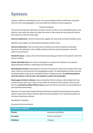
  • 1. Epistaxis Epistaxis is definied as bleeding from nose. The prosaic definition belies the difficulties associated with one of the otolaryngological’s most common& most difficult to treat emergencies. Vascular anatomy The area most frequently implicated in epistaxis is known as Little’s area or kiesselbach plexus.in the posterior nasal cavity, the vessels are larger than those in little area & can more easily be traced to their external or internal carotid origin. External carotid artery: external carotid artery supplies the nasal cavity via facial & maxillary artery. Maxillary artery supply is via sphenopalatine & greater palatine artery. Internal carotid artery: internal carotid artery contributes the anterior & posterior ethmoidal branches of the ophthalmic artery. Middle turbinate marks the watershed between internal & external carotid arteries. Woodruff’s plexus : a plexus of prominent blood vessels lying just inferior to the posterior end of the inferior turbinate. Anteior ethmoidal artery: the artery can be ligated as a treatment for epistaxis via an external approach(medial canthal) or endoscopical (transethmoidal). Sphenopalatine foramen: sphenopalatine foramen is the portal for major arterial supply of the nasal cavity. Lateral to the foramen lies the pterygopalatine space. The sphenopalatine foramen transmits the sphenopalatine artery & vein, &maxillary division of trigeminal nerve. A small bony projection which lies anterior to the foramen, this landmark is called crista ethmoidalis. Blood supply of inferior turbinate: artery reaching the inferior turbinate, artery divided into three parallel branches which runs in bony tunnels within the substance of the turbinate. These tunnels with their periarterial cuffing of fibrous tissue prevent the arterial constriction & may predispose to post-operative haemorrhage. Attempts to control haemorrhage following turbinectomy should be directed towards the postero-superior aspect of the inferior turbinate where the pressure/bipolar to the submucosal segment of the artery should prove effective. Classification of epistaxis Structured clinical classification Classification of epistaxis Primary No proven causal factor Secondary Proven causal factor Childhood <16 years
  • 2. Adult > 16 years Anterior epistaxis Bleeding from anterior to pyriform aperture Posterior epistaxis Bleeding from posterior to pyriform aperture. Adult primary epistaxis The condition can occur at any age. Between 7 to 14% of the adults have epistaxis at some time but only 6% are seen by otolaryngologist. Most cases are minor, selflimiting but nevertheless a significant number require admission to the hospital. Fewer than 10% of the hospitalized patients require a general anaesthetic procedure to secure haemostasis. After head & neck cancer, epistaxis stands out as a prominent cause of mortality in ENT patients. Aetiology By definition, the aetiology of primary epistaxis is unknown, but there are clear suggestions may be important. Chronobiology: the frequency of admission is greatest in the autumn& winter due to fluctuation of temperature & humidity. Circadian level shows a biphasic pattern with peaks in the morning& evening.(subarachnoid haemorrhage) NSAID: specially aspirin via antiplatelet aggregations effect due to altered platelet membrane physiology. Alcohol : alcohol causes prolongation of the bleeding time despite normal platelet counts & coagulation factors acitivity. Hypertension: this has long been considered a cause of epitaxis.recently have failed to show a causal relationship between hypertension & epistaxis. Septal abnormalities: epistaxis & septal abnormalities could be coincidental. There is no direct relationship. Factors of adult primary epistaxis (summary of Aetiology) Weather Proven association NSAID Proven association Alcohol Proven association Hypertension No association Septal deviation No association
  • 3. Management First , the patient must be resuscitated, bleeding slowed, the nasal cavity examined& a treatment plan established. Resuscitation: First aid by pinching the ala nasi (Trotter’s method). History & general examination will help in assessing the amount of blood loss. In all but the most minor of bleeds, intravenous access is established. Baseline blood estimations should be done. A detailed history should be taken,looking for predisposing factors. Routine coagulation studies in the absence of a positive history are not indicated. Assessment : The patient should be assessed in a semi-recumbent position & nersing assistance is mandatory. Everyone should wear protective visors & clothing as blood aerosol contamination is common. Basic instrument includes; couch or reclining chair, headlight,suction, vasoconstrictor solutions, (now widely used cocaine solution) , a packs, tempons & cautery apparatus. More specialized centres should have access to rod lens endoscopy instrument& bipolar electrodiathermy. Direct therapy(bleeding point specific therapies): A committed search for the bleeding vessels should be undertaken. Anterior epistaxis is usually very straightforward to identify & treat. At present over 90% of cases are controlled with silver nitrate cautery. The use of packing for anterior epistaxis is unwarranted & should be discouraged. Posterior epistaxis; systematic examination with a headlight will identify most bleeding points. Once identified, bleeding points can be directly controlled with bipolar diathermy, chemical cautery(difficult in posterior bleeds) or direct pressure from miniature targeted packs. Endoscopic control: failure to locate the bleeding point on initial examination is an indication for with a rod lens endoscope. Endoscopy identifies the source of the posterior epistaxis in over 90% of cases. Monopolar diathermy should not be used in the nasal cavity as blindness due to propagation of current. Indirect therapies : (failure to find the bleeding point is an indication for use of indirect strategies). 1.Nasal packing: nasal packing can be anteriorly or posteriorly placed.
  • 4. Anterior nasal pack : Ribbon gauze impregnated with petroleum jelly or BIPP(bismuth iodoform paraffin paste) is inserted the entire length of the nasal cavity. Once inserted , the pack are left in situ for between 24 to 72 hours. Rebleeding or continued bleeding is observed upto 40% of cases. Complications of packing include sinusitis, septal perforation alar necrosis, hypoxia, myocardial infarction. Antibiotic cover should be given. Modern alternatives on anterior packing include special tampons(merocel& kaltostat). Persistent or rebleeding is an indication for further examination of the nasal cavity. There is on clear, universally agreed definition of failed packing but who continue to bleed should proceed to surgical management sooner than later. 2.Hot water irrigation:irrigation of the nasal cavity with at 500 has been proposed as an alternative to packing( success rate similar to anterior packing Or balloon temponade.) mechanism of action; reflex vasodilatation & reduction in nasal lumen dimension. 3.Systemic medical therapy: Tranexamic acid & epsilon aminocaproic acid are systemic inhibitors of fibrinolysis. In epistaxis dose of 1.5gm thrice daily. At present these drugs are best reserved as adjuvant therapy in recurrent or refractory cases. Surgical management Surgical management for continued epistaxis consists of : Posterior packing Ligation techniques Septal surgery techniques Embolization techniques. 1.Posterior nasal packs Posterior nasal packing can be carried out under local or general anaesthesia. Nasophaygeal tamponade is achieved using special gauze packs inserted transorally& positioned by means of tapes passed from posterior choana to anterior nares bilaterally. These posterior Bellocq pack are secure against anterior gauze packing. The securing tapes are tied over gauze piece(padding positioned to protect the columella from pressure ncerosis.
  • 5. An easier alternative is to insert a foley’s catheter(size 12 or 14). The catheter inflated with 15ml of water. Ligation techniques The ligation should be performed as close as possible to likely bleeding point. 1.Shenopalatine artery 2.Internal maxillary artery 3.External carotid artery 4.Anterior/posterior ethmoidal artery 1.Endonasal shenopalatine artery ligation(ESPAL) Under G/A or L/A,an incision is made approximately 8mm in length, anterior to & under cover of the posterior end of the middle turbinate. The incision is carried down upto bone & a mucosal flap is elevated posteriorly until neurovascular sleeve arising from the sphenopalatine foramen is identified. Its location is signalled by the crista ethmoidalis. Once major vessel identified, it can be ligated using haemostatic clips & divided or coagulated using bipolar diathermy. Success rate almost 100% 2. Internal maxillary artery ligation Internal maxillary ligation was more frequently prior to the development of ESPAL. In sublabial approach, an antrostomy is formed taking care to preserve the infrorbital nerve. The mucosa of the posterior wall is elevated & a window is made through into pterygopalatine fossa. The branches of internal maxillary artery are identified pulsating within the fat of the fossa & are carefully dissected out prior to clipping with haemostatic clips. The proximal internal maxillary artery, descending palatine, & sphenopalatine branches are clipped & ideally divided. An endoscopic variation of this techniques1)uses a middle meatal antrostomy as an instrument port 2)with 4mm endoscope is inserted through a canine fossa antrostomy. Transantral ligation control haemorrhage in 89% of cases. 3.External carotid artery ligation Uder G/A or L/A a skin crease incision or longitudinal incision along parallel with anterior border of sternomastoid. Carotid bifurcation is identified, double check for arterial branches , then ligation in continuity. Success rate 14-15%. 4.Anterior or posterior ethmoidal artery ligation Ligation of anterior /posterior ethmoidal artery is adjuvant to above procedures or ethmoidal fracture. Medial canthal incision which is carried down to the bone of the anterior lacrimal crest. Periosteal elevator are used to elevate & lateral retract of bulbur fascia into anterior ethmoidal foramen. The
  • 6. vassel are identified & clipped & divided. The dissection is continued to identify the posterior artery which is located approximately 12mm behind. Septal surgery When epistaxis originates behind a prominent septal deviation or vomeropalatine spur, septoplasty or SMR may be required. Some authors have advocated a septal surgery as a primary treatment for failed packing. The rationale is that by elvationing the mucoperichondrial flap for septoplasty or SMR, the blood supply to the septum is interrupted & haemostasis is secured. Embolization Under L/A, transfemoral seldinger angiography is used to identify the bleeding point & display nasal circulation. It is essential to identify the arteriovenous malformation, aneursyms, fistula prior to embolization. Once the bleeding vessel identified, a fine catheter is passed into the internal maxillary circulation & particles (polyvinyl alcohol,,tungsten, or steel microcoils) are used to embolize the vessels. The ipsilateral facial artery is also embolize in order to prevent recirculation Complication are skin necrosis, paresthesia, CVA, groin haematoma. Similar efficacy to ligation techniques. Secondary epistaxis Commomly observed in patients with coagulopathy secondary to liver disease, leukemia, myelosuppression. In addition, trauma, post-surgery, warfain therapy deserve special care. Best clinical practice First line: direct therapy (bipolar/cautery,endoscopic if required) Second line: indirect therapy (anterior packing) Third line: surgical therapy (ESPAL) Fourth line: angiography &embolization.
  • 7. vassel are identified & clipped & divided. The dissection is continued to identify the posterior artery which is located approximately 12mm behind. Septal surgery When epistaxis originates behind a prominent septal deviation or vomeropalatine spur, septoplasty or SMR may be required. Some authors have advocated a septal surgery as a primary treatment for failed packing. The rationale is that by elvationing the mucoperichondrial flap for septoplasty or SMR, the blood supply to the septum is interrupted & haemostasis is secured. Embolization Under L/A, transfemoral seldinger angiography is used to identify the bleeding point & display nasal circulation. It is essential to identify the arteriovenous malformation, aneursyms, fistula prior to embolization. Once the bleeding vessel identified, a fine catheter is passed into the internal maxillary circulation & particles (polyvinyl alcohol,,tungsten, or steel microcoils) are used to embolize the vessels. The ipsilateral facial artery is also embolize in order to prevent recirculation Complication are skin necrosis, paresthesia, CVA, groin haematoma. Similar efficacy to ligation techniques. Secondary epistaxis Commomly observed in patients with coagulopathy secondary to liver disease, leukemia, myelosuppression. In addition, trauma, post-surgery, warfain therapy deserve special care. Best clinical practice First line: direct therapy (bipolar/cautery,endoscopic if required) Second line: indirect therapy (anterior packing) Third line: surgical therapy (ESPAL) Fourth line: angiography &embolization.