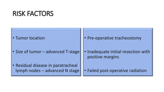
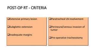
2. INTRODUCTION
• Recurrence of the tumor around the permanent stoma after total
laryngectomy for carcinoma is a serious and fatal complication,
occurring in 1.7 to 15% of the patients.
DEFINITION OF STOMAL RECURRENCE :
Stomal recurrence is a diffuse infiltrate of neoplastic tissue at the
junction of the amputated trachea and skin.
3. INCIDENCE
5-15% in most studies
M:F 93:7
98% of stomal recurrence present within 2 years of initial treatment
Pathogenesis still unknown
4. RISK FACTORS
• Tumor location
• Size of tumor – advanced T-stage
• Residual disease in paratracheal
lymph nodes – advanced N stage
• Pre-operative tracheostomy
• Inadequate initial resection with
positive margins
• Failed post-operative radiation
5. RISK FACTORS contd..
• It is generally associated with subglottic lesions (five fold increased
risk).
• The risk increases with bulky transglottic tumor even in the absence
of subglottic extension
• Pre-operative tracheostomy - most important risk factor.
• The 2-year disease free survival falls to almost 50% in patients with
preoperative tracheostomy compared to patients treated without
tracheostomy
6. TUMOR LOCATION
• Proximity of subglottis to tracheostoma - important risk factor.
• Subglottic tumors are prone to extensive circumferential growth and cartilage
invasion.
• Rubin et al
1)Presence of tumor in subglottis most important factor of recurrence
2)Recurrence rate * Subglottis 14%, *Epiglottis 0.6% * Aryepiglottic fold 1.3%
*Glottis 0.8%
7. TUMOR SIZE – T STAGE
• Increased size of tumor - more likely to develop stomal recurrence
• Rubin et al 1990 - T1 - 0%, T2- 2%, T3 - 9%, T4 - 6%
• Yotakis et al 1996 - T1 - 0%, T2 - 3%, T3 - 1%, T4 - 3%
• T4 statistical significance may be due to extensive spread and
subglottic involvement.
8. ADVANCED NODAL STAGE
• Involvement of paratracheal lymphatics
Welsh et al - radioactive tracers to detect lymphatic drainage
• Sparest on anterior commissure
• Arytenoids follow lymphatics of supraglottis
• Subglottis - 96% involve paratracheal LN
9. • Harris + Butler et al - Clinically undetectable paratracheal LN
metastasis →→50% were positive.
• Weber et al - Subglottic SCC - 52% paratracheal LN metastasis in
absence of cervical metastasis.
• Harrison et al - 65% of subglottic Ca have paratracheal LN metastasis.
Include removal of upper part of manubrium (allow clearance of
LN) and low tracheotomy →→no stomallrecurrence detected..nd
10. PRE-OPERATIVE TRACHEOSTOMY
• Seeding into trachea and periostomal soft tissue
• Keim et al 1965 - Pre-operative tracheotomy recurrence - 41%,
Without tracheotomy-1%
11. SEEDING THROUGH ENDOTRACHEAL INTUBATION
• Malignant cells transferred from laryngeal lesion to trachea via
intubation
Ormerod et al 1953 - endotracheal intubation implanted cells via
tube
Dejong et al 1998 - 51 pts tracheostomy under LA at start of
laryngectomy - 1 recurrence, 63 pts with ET intubation - 1 recurrence
12. PROGNOSIS
Poor with death within 2 years due to
1)Progressive tracheostomal obstruction
2)Hemorrhage caused by erosion of major vessels
Thus focus on prevention and identification of risk factors is the key to
avoid stomal recurrence.
13. CLASSIFICATION OF STOMAL RECURRENCE -
SISSON
• Sisson et al (1976) stomal recurrences post
laryngectomy into 4 types:
• Type I: Localized nodule at the superior
aspect of the laryngostoma without
esophageal involvement.
• Type II: Superior involvement of the
laryngostoma with esophageal
involvement.
• Type III: Inferior involvement of the
laryngostoma, usually with direct
extension to the mediastinum.
• Type IV: Lateral extension, and often
under either of the clavicles.
14. EVALUATION
• Thorough head and neck examination including tracheoscopy and
esophagoscopy.
• To determine the extent of the stomal recurrence. Contrast CT scan of the
chest and neck from skull base, barium swallow, and evaluation under
anesthesia are needed.
• Magnetic resonance imaging has proven useful in assessing prevertebral
musculature involvement.
• If carotid resection is contemplated in select cases, angiography is needed
to determine cerebral crossover flow.
15. IMAGING CONTRAINDICATIONS TO SX
• Overt mediastinal involvement
• Extensive tracheal involvement precluding the possibility of bringing
the residual stump to the skin
• Pre-vertebral musculature and cervical spine involvement
• Massive mediastinal nodal enlargement suggesting metastases
• Great vessel involvement.
16. MANAGEMENT
SURGERY - Stage I, II, and selected cases of stage III stomal
recurrence.
In rare cases, surgery on stage IV stomal recurrence may be indicated
for palliative purposes.
Subglottic tumors or recurrence of previously-treated laryngeal cancer
may require completion laryngectomy and mediastinal dissection.
Mediastinal dissection may be indicated in selected cancers with tracheal
and Level VI node involvement.
Sleeve resection of the trachea or subtotal laryngectomy may also be
17. SURGICAL APPROACH FOR TYPE 1 AND 2
• Watson first described the technique in 1942 and modified by Sisson in 1977
• Extensive removal of tracheostoma , surrounding skin, mediastinal dissection
with removal of manubrium, clavicle heads and resection of involved pharyngo-
esophageal segments with reconstruction using flaps.
• Peri operative mortality 15% - mediastinitis and rupture of great vessels
18. Gluckman et al 1987 - 41 patients underwent surgical treatment
• Type I+II(Sisson 45% survive 2yrs)
• Type III+IV 9% survive 2yrs
• 2 year cause - specific Survival was 24%
19. KEY STEPS IN SURGERY
A 2 cm margin around stoma with lateral cervical extensions and
vertical extension down midline over sternum
Resection involves en bloc removal of all involved tissues between
carotid arteries, anterior to prevertebral fascia, and above innominate
artery with removal of upper mediastinal fat pad and lymph nodes.
Removal of ipsilateral clavicular head (if tumor has predominant side)
and manubrium should be done initially. This maneuver will allow easy
access to the trachea and afford direct vision and protection of the great
vessels.
20. The tumor, involved trachea and resected esophagus (if involved)
are then mobilized from superior to inferior. The esophagus is
mobilized into the thorax as far as possible.
The trachea is transected, Mobilization of stomach and esophagus
is done and the final attachments of the esophagus are divided.
The stomach is mobilized into the neck, and the esophagus is
divided with a GI stapler at the gastroesophageal junction. The
specimen is dissected off.
21. Frozen section margins at the tracheal stump and upper
pharyngoesophageal incision should be sent.
The esophagus is pulled superiorly and mobilized into the neck. 6 to
8 tacking sutures with 2-0 vicryl - placed from the posterior aspect of
the stomach to the prevertebral fascia to maintain its position.
The stomach is sutured to the pharynx in two layers. The mucosa is
closed with 3-0 vicryl and the serosa is sutured to the pharyngeal
musculature with 2-0 or 3-0 vicryl.
It is crucial that the tacking sutures placed to the prevertebral fascia
support the stomach so that the suture line does not have any
tension.
22. If required, the trachea is externalized inferior to the innominate
artery.
A rotational pectoralis myocutaneous flap is harvested.
If required, a small hole can be made in the skin through which the
trachea can be brought and sutured to the skin.
It is important that pectoralis muscle be used to obliterate the dead
space in the mediastinum around the great vessels and trachea.
Large suction drains should be used to drain the right and left neck
and chest. One suction drain should be placed close to the
anastomosis and brought out in a "safe" course over the muscle and
away from the great vessels. This is a safety drain in case the
anastomosis should break down.
A feeding jejunostomy tube is placed while the neck is being closed.
23. CONTRAINDICATIONS TO SURGERY
1.Stomal recurrence classified as stage IV is, in the majority of cases,
inoperable both with regard to a reasonable chance of cure and with
regard to providing safe palliation.
2.Prior mediastinal surgery with a second recurrence at the stoma or
upper mediastinum
3.Patient is medically unfit to tolerate mediastinal surgery.
24. PREVENTION
PRE-OP :
Avoiding preoperative tracheostomy
Pre-operative radiotherapy in high-risk patients
INTRA-OP :
Paratracheal LN dissection in all laryngeal cancers with subglottic extension
Adequate resection in patients with more than 1.5 cm subglottic extension of
growth
POST-OP :
Post-operative radiotherapy in high-risk patients
Post-op RT to the stoma
25. Prevention – contd..
• In the circumstances where preoperative tracheostomy is unavoidable, the interval
between total laryngectomy and tracheostomy should be kept to minimum
• Patients should be advised postoperative radiotherapy to sterilize the area
• The postoperative radiotherapy should be started as early as possible
• Tracheostomy should be done at a higher level
• Tract should be removed at the time of operation, thus creating a new tracheostoma at
the lower level.
• Paratracheal nodes and tissues in tracheo-oesophageal groove should be cleared at the
time of primary surgery
27. TAKE HOME POINTS
• Prevention is the best treatment of stomal recurrence.
• All the risk factors should be evaluated pre-operatively.
• Intensive follow-up should be performed for patients who had preoperative
tracheostomy, paratracheal lymph node metastasis to detect stomal recurrence at an
early stage
• Post-operative radiotherapy should be given in the high risk group.
• Stomal recurrence can be minimized by early detection, comprehensive assessment of
the extent of tumor, timely adjuvant radiation therapy, adequate excision at the time of
primary surgery and radical excision of emergency tracheostomy tract.