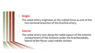
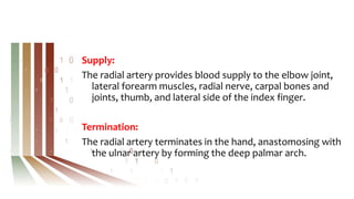
First Master Degree
IR Revision
Mohamed M.A. Zaitoun, MD
Associate Professor of Interventional Radiology
Faculty of Medicine, Zagazig University, Egypt
FINR-Switzerland
Radial Access
Anatomy of the radial artery
Technique of radial access
Anatomy of the radial artery
Origin
Course
Branches
Supply
Termination
Origin:
The radial artery originates at the cubital fossa as one of the
two terminal branches of the brachial artery.
Course:
The radial artery runs along the radial aspect of the anterior
compartment of the forearm under the brachioradialis,
lateral to the flexor carpi radialis tendon.
For the distal section of its course, the radial artery lies on
the surface of the radius.
The radial artery proceeds along the floor of the anatomical
snuff box, passing dorsally around
the scaphoid and trapezium. At the carpal region of the
hand, the radial artery forms the deep palmar arch of the
hand and anastomoses with the ulnar artery.
Branches:
The branches of the radial artery in the forearm include the:
Radial recurrent artery
Palmar carpal branch
Dorsal carpal branch
Muscular branches
Superficial palmar branch
In the hand, the branches are the:
Princeps pollicis artery
Radialis indicis artery
Supply:
The radial artery provides blood supply to the elbow joint,
lateral forearm muscles, radial nerve, carpal bones and
joints, thumb, and lateral side of the index finger.
Termination:
The radial artery terminates in the hand, anastomosing with
the ulnar artery by forming the deep palmar arch.
Technique of radial access
Local anaesthesia
Achieving access
Site of puncture
Cocktail
Local anaesthesia
The local anaesthesia is obtained through the subcutaneous
injection of 2 mL 2% lidocaine and 1 ml of nitroglycerin 1
min before the radial artery puncture, between 1 and 1.5
cm proximal to the styloid process.
Subcutaneously infiltrated nitroglycerin leads to
significant vasodilation of radial artery, this
avoids pre-cannulation spasm of radial artery.
Achieving access
Arterial access may be obtained via either single- or double-
wall puncture technique.
Both techniques are safe and effective and are associated
with low rates of RAO and other complications.
TRA should be achieved using
sonographic guidance with a
single-wall puncture technique
and the Seldinger technique with
a 21-gauge echogenic-tip needle.
The double-wall technique is
associated with a higher first-
pass success rate.
Ultrasound reduces time and number of attempts to achieve
arterial access.
Site of puncture
The radial artery is
accessed 1 to 2 cm
proximal to the radial
styloid process,
i.e. proximal puncture site.
The main advantages are less arterial obstruction and short
hemostasis.
The main disadvantage is the difficulty in cannulation.
Cocktail
After arterial access is achieved, a combination of
medications (radial cocktail) is administered through the
sheath to reduce arterial spasm and vascular tone.
The radial cocktail is a combination of anticoagulants and
spasmolytic:
*Anticoagulant (Heparin) 2500/5000 units
*Spasmolytic:
100-200 µg of nitroglycerin + 2.5 mg of verapamil.
1 milligram (mg) is equal to 1000 micrograms (μg)
Thyroid Ablation
Anatomy of the thyroid gland
Technique of thyroid ablation
Anatomy of the thyroid gland
Gross anatomy
Relations
Arterial supply
Venous drainage
Gross anatomy:
The thyroid extends from C5 to T1 and lies anterior to
the thyroid and cricoid cartilages of the larynx and the
first five or six tracheal rings.
The thyroid is butterfly or "H"-shaped and is composed of
two lobes, each with a superior and inferior pole.
Usually, the superior pole is narrower than the inferior pole
giving a pear-like shape to each lateral lobe.
The lateral lobes are connected in the midline by a narrow
isthmus which is adherent to the 2nd to4th tracheal rings.
Each lobe measures approximately 4 cm in length.
The average weight is 25 g; this is slightly higher in females
and may increase during menstruation and pregnancy.
Relations:
Anteriorly: strap muscles
Posteriorly: thyroid cartilage, cricoid cartilage, trachea
Posteromedially: tracheo-esophageal groove (containing
lymph nodes, recurrent laryngeal nerve, parathyroid
glands)
Posterolaterally: carotid space
Arterial supply:
Superior thyroid artery (from the external carotid artery)
Inferior thyroid artery (from the thyrocervical trunk)
Venous drainage:
Superior thyroid vein (drains to the internal jugular vein)
Middle thyroid vein (drains to the internal jugular vein)
Inferior thyroid vein (drains via plexus to
the brachiocephalic vein)
Technique of thyroid ablation
Patient in supineposition
Mild neckextension
No pre-incision of theskin required
Anaesthesia:perithyroidal LidocaineInjection,conscioussedation
Hydrodissection
Trans-Isthmic Approach
Movingshottechnique/ Pullback technique
Vascularablationtechnique(Artery-firstablation,Marginalvenous
ablation)
TARE (Transarterial radioembolization)
Anatomy of the hepatic artery
Technique of TARE
Anatomy of the hepatic artery
Origin
Course
Branches
Origin:
The common hepatic artery is intermediate in size, commonly
arising as a terminal branch of the celiac artery, which courses
to the right.
Course:
It courses posterior to the parietal peritoneum of the lesser sac,
first passing anteriorly to the pancreas, then coursing
inferiorly towards the first part of the duodenum.
It gives off the right gastric artery, which runs superiorly along
the lower half of the lesser curvature of the stomach to
anastomose with the left gastric artery.
The common hepatic artery then passes slightly superiorly,
where it runs anterior to the portal vein and medial to
the common bile duct after entering the free edge of
the lesser omentum (or hepatoduodenal ligament).
As it courses superiorly towards the porta hepatis, it gives
off the gastroduodenal artery inferiorly, then terminates
as the proper hepatic artery.
Branches:
Right gastric artery
Terminal branches:
Proper hepatic artery (RHA, MHA & LHA)
Gastroduodenal artery
Technique of TARE
Radioembolization is the delivery of radioactive
microspheres to cancers using an endovascular approach.
It is often performed as an outpatient procedure.
Indications
Contraindications
Technique
Indications:
Hepatocellular carcinoma
Hepatic metastases from colorectal carcinoma
Contraindications:
Excessive hepatopulmonary shunting (results in radiation
pneumonitis)
Demonstrable gastrointestinal shunting (results in gastric
ulceration)
Technique:
Typically a transfemoral or transradial intra-arterial catheter
with the tip near the target lesion.
Nuclear medicine specialists prepare the radioactive (e.g.
yttrium-90) microspheres (~32 microns), which when
injected are implanted in the microvascular arterial supply
of the tumor where they become trapped.
The spectrum of yttrium-90 is ~2.7 days, with no remaining
radioactivity after one month.
revision for first master.pptx

revision for first master.pptx

  • 1.
    First Master Degree IRRevision Mohamed M.A. Zaitoun, MD Associate Professor of Interventional Radiology Faculty of Medicine, Zagazig University, Egypt FINR-Switzerland
  • 2.
    Radial Access Anatomy ofthe radial artery Technique of radial access
  • 3.
    Anatomy of theradial artery Origin Course Branches Supply Termination
  • 4.
    Origin: The radial arteryoriginates at the cubital fossa as one of the two terminal branches of the brachial artery. Course: The radial artery runs along the radial aspect of the anterior compartment of the forearm under the brachioradialis, lateral to the flexor carpi radialis tendon.
  • 5.
    For the distalsection of its course, the radial artery lies on the surface of the radius. The radial artery proceeds along the floor of the anatomical snuff box, passing dorsally around the scaphoid and trapezium. At the carpal region of the hand, the radial artery forms the deep palmar arch of the hand and anastomoses with the ulnar artery.
  • 6.
    Branches: The branches ofthe radial artery in the forearm include the: Radial recurrent artery Palmar carpal branch Dorsal carpal branch Muscular branches Superficial palmar branch In the hand, the branches are the: Princeps pollicis artery Radialis indicis artery
  • 7.
    Supply: The radial arteryprovides blood supply to the elbow joint, lateral forearm muscles, radial nerve, carpal bones and joints, thumb, and lateral side of the index finger. Termination: The radial artery terminates in the hand, anastomosing with the ulnar artery by forming the deep palmar arch.
  • 8.
    Technique of radialaccess Local anaesthesia Achieving access Site of puncture Cocktail
  • 9.
    Local anaesthesia The localanaesthesia is obtained through the subcutaneous injection of 2 mL 2% lidocaine and 1 ml of nitroglycerin 1 min before the radial artery puncture, between 1 and 1.5 cm proximal to the styloid process. Subcutaneously infiltrated nitroglycerin leads to significant vasodilation of radial artery, this avoids pre-cannulation spasm of radial artery.
  • 10.
    Achieving access Arterial accessmay be obtained via either single- or double- wall puncture technique. Both techniques are safe and effective and are associated with low rates of RAO and other complications.
  • 11.
    TRA should beachieved using sonographic guidance with a single-wall puncture technique and the Seldinger technique with a 21-gauge echogenic-tip needle. The double-wall technique is associated with a higher first- pass success rate.
  • 12.
    Ultrasound reduces timeand number of attempts to achieve arterial access.
  • 13.
    Site of puncture Theradial artery is accessed 1 to 2 cm proximal to the radial styloid process, i.e. proximal puncture site.
  • 14.
    The main advantagesare less arterial obstruction and short hemostasis. The main disadvantage is the difficulty in cannulation.
  • 15.
    Cocktail After arterial accessis achieved, a combination of medications (radial cocktail) is administered through the sheath to reduce arterial spasm and vascular tone. The radial cocktail is a combination of anticoagulants and spasmolytic: *Anticoagulant (Heparin) 2500/5000 units *Spasmolytic: 100-200 µg of nitroglycerin + 2.5 mg of verapamil.
  • 16.
    1 milligram (mg)is equal to 1000 micrograms (μg)
  • 18.
    Thyroid Ablation Anatomy ofthe thyroid gland Technique of thyroid ablation
  • 19.
    Anatomy of thethyroid gland Gross anatomy Relations Arterial supply Venous drainage
  • 20.
    Gross anatomy: The thyroidextends from C5 to T1 and lies anterior to the thyroid and cricoid cartilages of the larynx and the first five or six tracheal rings. The thyroid is butterfly or "H"-shaped and is composed of two lobes, each with a superior and inferior pole. Usually, the superior pole is narrower than the inferior pole giving a pear-like shape to each lateral lobe.
  • 21.
    The lateral lobesare connected in the midline by a narrow isthmus which is adherent to the 2nd to4th tracheal rings. Each lobe measures approximately 4 cm in length. The average weight is 25 g; this is slightly higher in females and may increase during menstruation and pregnancy.
  • 22.
    Relations: Anteriorly: strap muscles Posteriorly:thyroid cartilage, cricoid cartilage, trachea Posteromedially: tracheo-esophageal groove (containing lymph nodes, recurrent laryngeal nerve, parathyroid glands) Posterolaterally: carotid space
  • 23.
    Arterial supply: Superior thyroidartery (from the external carotid artery) Inferior thyroid artery (from the thyrocervical trunk) Venous drainage: Superior thyroid vein (drains to the internal jugular vein) Middle thyroid vein (drains to the internal jugular vein) Inferior thyroid vein (drains via plexus to the brachiocephalic vein)
  • 24.
    Technique of thyroidablation Patient in supineposition Mild neckextension No pre-incision of theskin required Anaesthesia:perithyroidal LidocaineInjection,conscioussedation Hydrodissection Trans-Isthmic Approach Movingshottechnique/ Pullback technique Vascularablationtechnique(Artery-firstablation,Marginalvenous ablation)
  • 25.
    TARE (Transarterial radioembolization) Anatomyof the hepatic artery Technique of TARE
  • 26.
    Anatomy of thehepatic artery Origin Course Branches
  • 27.
    Origin: The common hepaticartery is intermediate in size, commonly arising as a terminal branch of the celiac artery, which courses to the right. Course: It courses posterior to the parietal peritoneum of the lesser sac, first passing anteriorly to the pancreas, then coursing inferiorly towards the first part of the duodenum. It gives off the right gastric artery, which runs superiorly along the lower half of the lesser curvature of the stomach to anastomose with the left gastric artery.
  • 28.
    The common hepaticartery then passes slightly superiorly, where it runs anterior to the portal vein and medial to the common bile duct after entering the free edge of the lesser omentum (or hepatoduodenal ligament). As it courses superiorly towards the porta hepatis, it gives off the gastroduodenal artery inferiorly, then terminates as the proper hepatic artery.
  • 29.
    Branches: Right gastric artery Terminalbranches: Proper hepatic artery (RHA, MHA & LHA) Gastroduodenal artery
  • 30.
    Technique of TARE Radioembolizationis the delivery of radioactive microspheres to cancers using an endovascular approach. It is often performed as an outpatient procedure. Indications Contraindications Technique
  • 31.
    Indications: Hepatocellular carcinoma Hepatic metastasesfrom colorectal carcinoma Contraindications: Excessive hepatopulmonary shunting (results in radiation pneumonitis) Demonstrable gastrointestinal shunting (results in gastric ulceration)
  • 32.
    Technique: Typically a transfemoralor transradial intra-arterial catheter with the tip near the target lesion. Nuclear medicine specialists prepare the radioactive (e.g. yttrium-90) microspheres (~32 microns), which when injected are implanted in the microvascular arterial supply of the tumor where they become trapped. The spectrum of yttrium-90 is ~2.7 days, with no remaining radioactivity after one month.