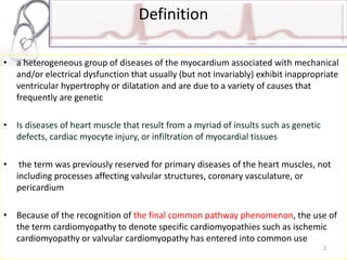
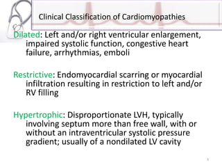
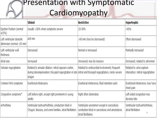
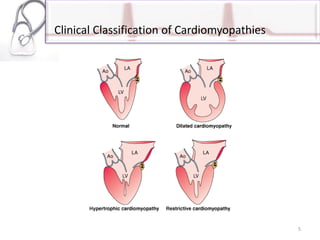
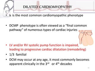
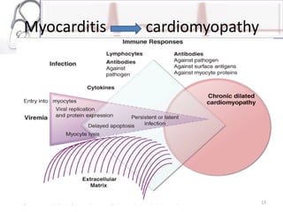
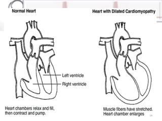
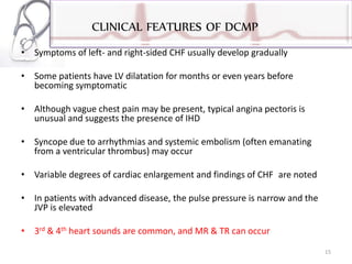
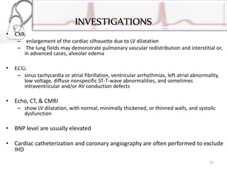
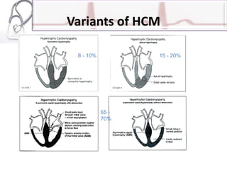
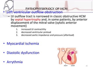
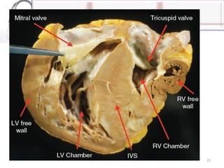
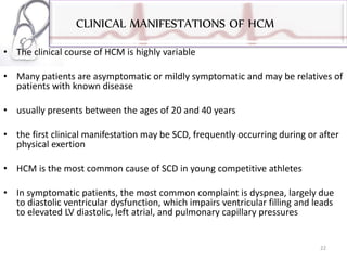
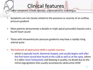
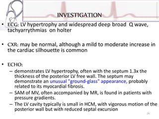
2. Definition
• a heterogeneous group of diseases of the myocardium associated with mechanical
and/or electrical dysfunction that usually (but not invariably) exhibit inappropriate
ventricular hypertrophy or dilatation and are due to a variety of causes that
frequently are genetic
• Is diseases of heart muscle that result from a myriad of insults such as genetic
defects, cardiac myocyte injury, or infiltration of myocardial tissues
• the term was previously reserved for primary diseases of the heart muscles, not
including processes affecting valvular structures, coronary vasculature, or
pericardium
• Because of the recognition of the final common pathway phenomenon, the use of
the term cardiomyopathy to denote specific cardiomyopathies such as ischemic
cardiomyopathy or valvular cardiomyopathy has entered into common use
2
3. Clinical Classification of Cardiomyopathies
Dilated: Left and/or right ventricular enlargement,
impaired systolic function, congestive heart
failure, arrhythmias, emboli
Restrictive: Endomyocardial scarring or myocardial
infiltration resulting in restriction to left and/or
RV filling
Hypertrophic: Disproportionate LVH, typically
involving septum more than free wall, with or
without an intraventricular systolic pressure
gradient; usually of a nondilated LV cavity
3
6. General presentation
• exertional intolerance with breathlessness or fatigue
• Progressive shortness of breath may occur during routine daily
activity, and may manifest as dyspnea or cough in the supine
position
• peripheral edema may not appear despite severe fluid retention,
particularly in younger patients
• atrioventricular valve regurgitation
• typical and atypical chest pain
• atrial and ventricular tachyarrhythmias
• embolic events
6
7. Clinical Evaluation
• Thorough history and physical examination to identify
cardiac and noncardiac disorders
• Detailed family history
• History of alcohol, illicit drugs, chemotherapy or
radiation therapy
• Assessment of ability to perform routine and desired
activities
• Assessment of volume status, orthostatic BP, BMI
7
9. Laboratory evaluation
• Electrocardiogram
• Chest radiograph
• Two-dimensional and Doppler echocardiogram
• Chemistry:
• Na, K, Ca, Mg, FBG,Creatinine, BUN, Albumin, total protein
LFT, Lipid profile, TSH, Serum iron, transferrin saturation,
U/A, Creatine kinase
• Hematology:
• Hgb, WBC count with differential, including eosinophils, ESR
9
10. Initial Evaluation Only in Patients Selected for Possible
Specific Diagnosis
• Titers for infection in presence of clinical suspicion:
– Acute viral (coxsackie virus, echovirus, influenza virus)
– Human immunodeficiency virus
– Chagas' disease
– Lyme disease
– toxoplasmosis
• Catheterization with coronary angiography in patients
with angina who are candidates for intervention
• Serologies for active rheumatologic disease
• Endomyocardial biopsy including sample for electron
microscopy when suspecting specific diagnosis with
therapeutic implications
• Screening for sleep-disordered breathing 10
11. DILATED CARDIOMYOPATHY
• It is the most common cardiomyopathic phenotype
• DCMP phenotype is often viewed as a “final common
pathway” of numerous types of cardiac injuries
• LV and/or RV systolic pump function is impaired,
leading to progressive cardiac dilatation (remodeling)
• 1/3 familial
• DCM may occur at any age, it most commonly becomes
apparent clinically in the 3rd or 4th decades
11
12. Etiology
• Could be idiopathic(primary) or secondary
• Secondary:
– Post infectious (viral myocarditis, HIV, parasitic, bacterial, spirochetal)
– Toxic (alcohol, chemotherapeutics, heavy metals)
– Metabolic (nutritional deficency, endocrinopatheis, electrolyte
deficiencies( Ca,Mg, Phosphate), hemochromatosis)
– Peripartum cardiomyopathy
• last TM of Px to 6mths post partum,
• ? Cross reaction b/n antiuterine ab against cardiac muscles
• A reversible form of DCM may be found with alcohol
abuse, pregnancy , thyroid disease, cocaine use , and
chronic uncontrolled tachycardia
12
15. CLINICAL FEATURES OF DCMP
• Symptoms of left- and right-sided CHF usually develop gradually
• Some patients have LV dilatation for months or even years before
becoming symptomatic
• Although vague chest pain may be present, typical angina pectoris is
unusual and suggests the presence of IHD
• Syncope due to arrhythmias and systemic embolism (often emanating
from a ventricular thrombus) may occur
• Variable degrees of cardiac enlargement and findings of CHF are noted
• In patients with advanced disease, the pulse pressure is narrow and the
JVP is elevated
• 3rd & 4th heart sounds are common, and MR & TR can occur
15
16. INVESTIGATIONS
• CXR:
– enlargement of the cardiac silhouette due to LV dilatation
– The lung fields may demonstrate pulmonary vascular redistribution and interstitial or,
in advanced cases, alveolar edema
• ECG:
– sinus tachycardia or atrial fibrillation, ventricular arrhythmias, left atrial abnormality,
low voltage, diffuse nonspecific ST-T-wave abnormalities, and sometimes
intraventricular and/or AV conduction defects
• Echo, CT, & CMRI
– show LV dilatation, with normal, minimally thickened, or thinned walls, and systolic
dysfunction
• BNP level are usually elevated
• Cardiac catheterization and coronary angiography are often performed to exclude
IHD
16
17. TREATEMENT OF DCMP
• Most patients pursue an inexorably downhill course, and the majority,
particularly those >55 years, die within 4 years of the onset of symptoms
• Spontaneous improvement or stabilization occurs in about one-quarter of
patients
• Patients of African ancestry are more likely to suffer more rapidly
progressive CHF and death than Caucasians
• Death is due to either progressive HF or ventricular tachy- or
bradyarrhythmia; SCD is a constant threat
• Systemic embolization is a concern, and patients should be considered for
chronic anticoagulation
• Standard therapy of HF is indicated
• Treatement of the underlying etiology whenever possible
17
18. HYPERTROPHIC CARDIOMYOPATHY
• HCM is characterized by a thickened but nondilated left
ventricle in the absence of another cardiac or systemic
condition capable of producing the magnitude of hypertrophy
• LV hypertrophy is asymmetric, often with preferential
hypertrophy of the interventricular septum
• the most common of the genetic cardiovascular diseases, is
caused by a multitude of mutations in genes encoding
proteins of the cardiac sarcomere, 50% with AD transmission
• It is found in about 1 in 500 of the general population
18
22. CLINICAL MANIFESTATIONS OF HCM
• The clinical course of HCM is highly variable
• Many patients are asymptomatic or mildly symptomatic and may be relatives of
patients with known disease
• usually presents between the ages of 20 and 40 years
• the first clinical manifestation may be SCD, frequently occurring during or after
physical exertion
• HCM is the most common cause of SCD in young competitive athletes
• In symptomatic patients, the most common complaint is dyspnea, largely due
to diastolic ventricular dysfunction, which impairs ventricular filling and leads
to elevated LV diastolic, left atrial, and pulmonary capillary pressures
22
23. Clinical features
• Other symptoms include syncope, angina pectoris, and fatigue.
• Symptoms are not closely related to the presence or severity of an outflow
pressure gradient
• Most patients demonstrate a double or triple apical precordial impulse and a
fourth heart sound
• Those with intraventricular pressure gradients may have a rapidly rising
arterial pulse
• The hallmark of obstructive HCM is systolic murmur
– which is typically harsh, diamond-shaped, and usually begins well after
the first heart sound best heard at the LLSB as well as at the apex, where
it is often more holosystolic and blowing in quality, no doubt due to the
mitral regurgitation that usually accompanies obstructive HCM
23
24. INVESTIGATION
• ECG: LV hypertrophy and widespread deep broad Q wave,
tachyarrythmias on holter
• CXR: may be normal, although a mild to moderate increase in
the cardiac silhouette is common
• ECHO:
– demonstrates LV hypertrophy, often with the septum 1.3x the
thickness of the posterior LV free wall. The septum may
demonstrate an unusual "ground-glass" appearance, probably
related to its myocardial fibrosis.
– SAM of MV, often accompanied by MR, is found in patients with
pressure gradients.
– The LV cavity typically is small in HCM, with vigorous motion of the
posterior wall but with reduced septal excursion
24
25. TREATEMENT OF HCM
• Since SCD often occurs during or just after physical exertion,
competitive sports and very strenuous activities should be proscribed
• Dehydration should be avoided, and diuretics used with caution In
congested patients
• Beta- blockers drugs and verapamil
– decrease HR and increase the length of time for diastolic filling, as well
as to decrease the inotropic state
• Amiodarone reduces frequency of arrythmias
• diuretics, nitrates, dihydropyridine calcium blockers, vasodilators,
and -adrenergic agonists are best avoided, particularly in patients
with known LV outflow tract pressure gradients
25
26. TREATMENT OF HCM
• Alcohol ingestion may produce sufficient vasodilatation
to exacerbate an outflow pressure gradient
• Atrial fibrillation is poorly tolerated, and a strong effort
should be made to restore and then maintain sinus
rhythm and slow rate
• Surgical myotomy/myectomy and alcohol septal
ablation can also reduce obstruction and improve
symptoms.
• The insertion of an ICD should be considered in
patients with a high-risk profile for SCD 26
27. RESTRICTIVE CARDIOMYOPATHY
• The hallmark of the restrictive cardiomyopathies (RCMs) is
abnormal diastolic function ; the ventricular walls are excessively
rigid and impede ventricular filling
• Partial obliteration of the ventricular cavity by fibrous tissue and
thrombus contributes to the abnormally increased resistance to
ventricular filling
• In late stages systolic function is also impaired
• Myocardial fibrosis, hypertrophy, or infiltration due to a variety of
causes is responsible
Thromboembolic complications are frequent in such patients
27
28. Cause of restrictive cardiomyopathy
include
Idiopathic
Amyloidosis
Sarcoidosis
Hemochromatosis
Chemotherapy or radiation
Hypereosinophilic syndrome
Endomyocardial fibrosis
Long-term chloroquine therapy
In addition, secondary restrictive physiology can
develop during the advanced stages of hypertrophic,
dilated, hypertensive, or ischemic heart disease
28
29. CLINICAL FEATURES
• exercise intolerance and dyspnea are usually prominent
• commonly have dependent edema, ascites, and an
enlarged, tender, and often pulsatile liver
• Kussmaul’s sign is positive
• heart sounds may be distant, and third and fourth heart
sounds are common
• Unlike constrictve pericarditis the apex impulse is usually
easily palpable, and mitral regurgitation is more common
29
30. INVESTIGATION AND TREATEMENT OF RCMS
• ECG often shows low-voltage, nonspecific ST-T-wave abnormalities and
various arrhythmias
• Echocardiography, CTI, and CMRI typically reveal symmetrically thickened
LV walls and normal or slightly reduced ventricular volumes and systolic
function; the atria are usually dilated
• Differentiation of RCM from constrictive pericarditis is of importance
because the latter is often curable by surgery
• Management is usually disappointing, except for hemochromatosis and
Fabry's disease
• Chronic anticoagulation is often recommended to reduce the risk of
embolization from the heart
30